Noma Hidetaka, Yasuda Kanako, Mimura Tatsuya, Suganuma Noboru, Shimura Masahiko
Hachioji Medical Center, Department of Ophthalmology, Tokyo Medical University, Tokyo 193-0998, Japan.
Department of Ophthalmology, Teikyo University School of Medicine, Tokyo 173-8606, Japan.
J Clin Med. 2020 Dec 26;10(1):58. doi: 10.3390/jcm10010058.
To investigate the relationship between retinal blood flow, presence or absence of recurrence of macular edema, and levels of cytokines, after intravitreal ranibizumab injection (IRI) in patients with branch retinal vein occlusion (BRVO).
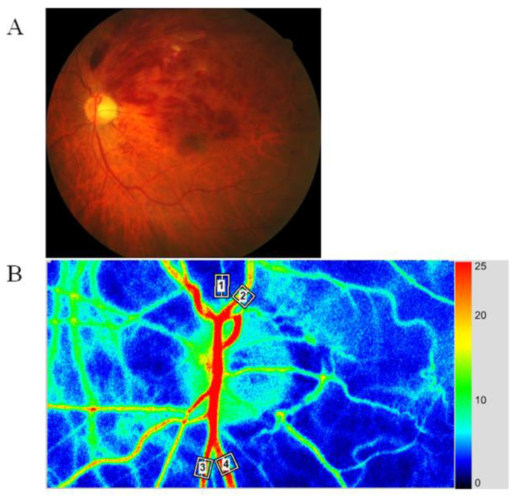
In 47 patients with BRVO and macular edema, we used laser speckle flowgraphy (LSFG) to measure the relative flow volume (RFV) of the retinal arteries and veins passing through the optic disc in the occluded and non-occluded regions of the retina before and after IRI. Aqueous humor samples were obtained at the time of IRI. Levels of vascular endothelial growth factor (VEGF), soluble VEGF receptor (sVEGFR)-1, sVEGFR-2, placental growth factor (PlGF), platelet-derived growth factor (PDGF)-AA, soluble intercellular adhesion molecule (sICAM)-1, monocyte chemoattractant protein 1 (MCP-1), interleukin (IL)-6, IL-8, IL-12 (p70), IL-13 and interferon-inducible 10-kDa protein (IP-10) were measured by the suspension array method. Patients were categorized into two groups on the basis of whether or not macular edema recurred at 2 months after IRI: the nonrecurrent group, = 24; and the recurrent group, = 23.
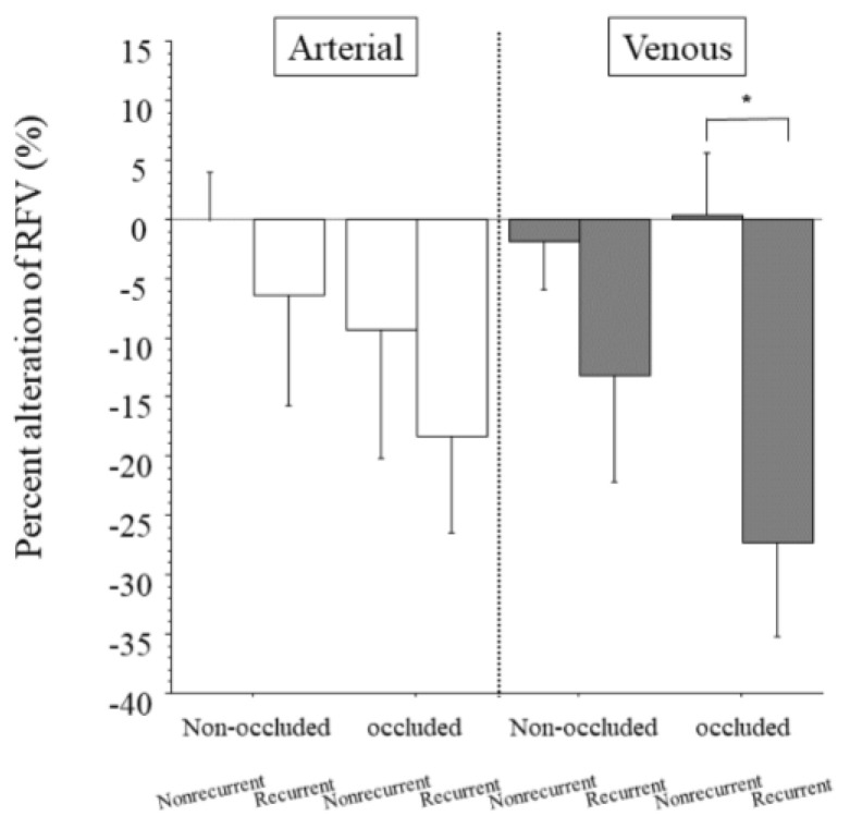
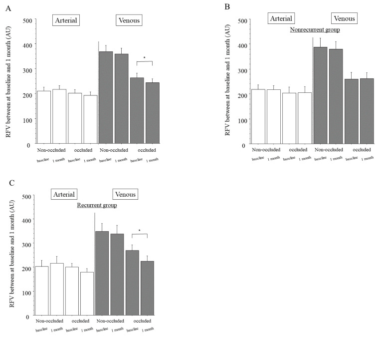
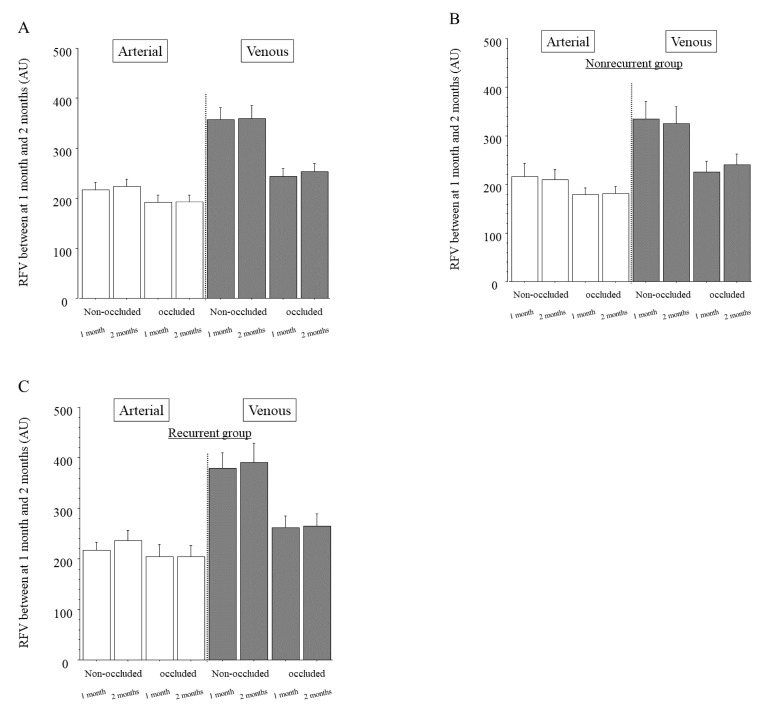
In the veins of the occluded region, RFV showed a significant difference between baseline and 1 month after IRI ( < 0.001) in the recurrent group and the percent change of RFV showed a significant difference between the recurrent and nonrecurrent groups ( = 0.005). Furthermore, we found a significant negative correlation between RFV in the veins of the occluded region and aqueous levels of MCP-1, IL-8 and IP-10 at baseline ( = 0.029, = 0.035, and = 0.039, respectively). In the recurrent group, the arteries and veins of the non-occluded and occluded regions showed no significant association between RFV and the aqueous levels of any factors.
These findings suggested that a decrease in RFV in the veins of the occluded region might be associated with the recurrence of macular edema and that the recurrence might depend on the change in RFV in the veins of the occluded region rather than the levels of cytokines.
研究视网膜分支静脉阻塞(BRVO)患者玻璃体内注射雷珠单抗(IRI)后视网膜血流、黄斑水肿复发与否及细胞因子水平之间的关系。
对47例BRVO合并黄斑水肿患者,在IRI前后,使用激光散斑血流图(LSFG)测量视网膜阻塞区和非阻塞区穿过视盘的视网膜动脉和静脉的相对血流量(RFV)。在IRI时采集房水样本。采用悬浮阵列法测量血管内皮生长因子(VEGF)、可溶性VEGF受体(sVEGFR)-1、sVEGFR-2、胎盘生长因子(PlGF)、血小板衍生生长因子(PDGF)-AA、可溶性细胞间黏附分子(sICAM)-1、单核细胞趋化蛋白1(MCP-1)、白细胞介素(IL)-6、IL-8、IL-12(p70)、IL-13和干扰素诱导10 kDa蛋白(IP-10)的水平。根据IRI后2个月黄斑水肿是否复发,将患者分为两组:未复发组,n = 24;复发组,n = 23。
在复发组中,阻塞区静脉的RFV在基线和IRI后1个月之间存在显著差异(P < 0.001),且RFV的变化百分比在复发组和未复发组之间存在显著差异(P = 0.005)。此外,我们发现基线时阻塞区静脉的RFV与房水中MCP-1、IL-8和IP-10水平之间存在显著负相关(分别为P = 0.029、P = 0.035和P = 0.039)。在复发组中,非阻塞区和阻塞区的动脉和静脉的RFV与任何因素的房水水平之间均无显著关联。
这些发现表明,阻塞区静脉RFV的降低可能与黄斑水肿的复发有关,且复发可能取决于阻塞区静脉RFV的变化而非细胞因子水平。