Department of Surgery, Baba Memorial Hospital, Sakai City, Osaka, Japan.
Department of Gastroenterology, Baba Memorial Hospital, Sakai City, Osaka, Japan.
Am J Case Rep. 2021 Jan 9;22:e927849. doi: 10.12659/AJCR.927849.
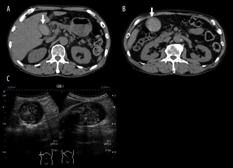
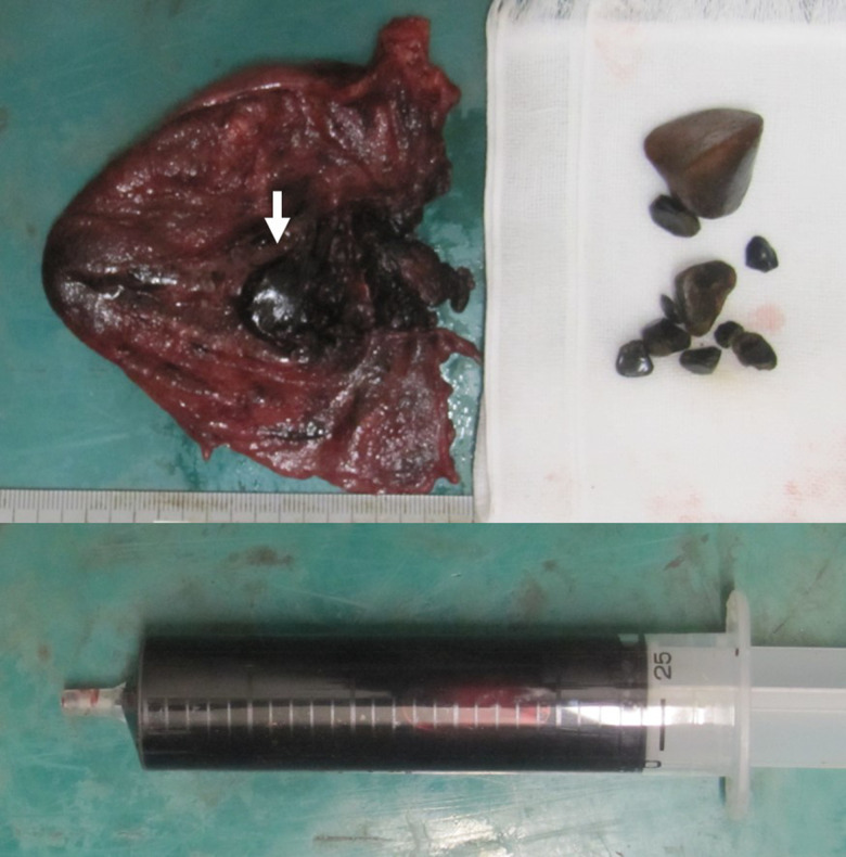
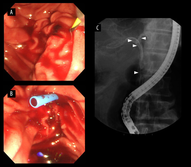
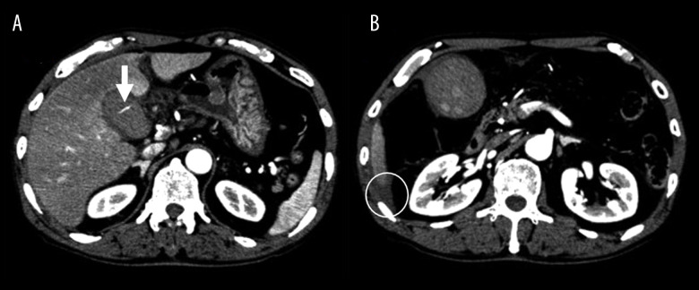
BACKGROUND Hemorrhagic cholecystitis is a rare disease which can be fatal in some cases. Hemorrhagic cholecystitis can sometimes be confused with common biliary diagnoses, as its symptoms imitate other hepatobiliary diseases. We report a case of hemorrhagic cholecystitis with hemobilia caused by the administration of anticoagulant agents. CASE REPORT A 70-year-old man was admitted with abdominal distention and pain. Ultrasound (US) and computed tomography (CT) showed a distended and wall-thickened gallbladder with hyperdense materials. Based on these findings and the laboratory data, the patient was diagnosed with acute cholecystitis with cholangitis. Because the patient's hemodynamics were stable, endoscopic retrograde cholangiopancreatography (ERCP) was performed first to improve the bile flow. The results of ERCP showed blood from the common bile duct by cannulation, which was suspected to reflect hemorrhagic cholecystitis. As the abdominal symptom and CT findings worsened on the day after ERCP, emergency laparoscopic cholecystectomy was performed. An examination of the specimen revealed ulcer formation on the mucosal side of the gallbladder. The patient was discharged 6 days after the operation without any surgical complications. CONCLUSIONS ERCP and early laparoscopic cholecystectomy were performed for a patient with hemorrhagic cholecystitis and hemobilia. Early diagnosis and treatment can lead to good outcomes in patients with hemorrhagic cholecystitis. Since the number of patients who are taking antithrombotic agents is increasing, hemorrhagic cholecystitis should be considered when any unusual imaging findings associated with cholecystitis are observed.
出血性胆囊炎是一种罕见的疾病,在某些情况下可能致命。出血性胆囊炎的症状有时与常见的胆道诊断相混淆,因为其症状模仿其他肝胆疾病。我们报告一例因抗凝剂治疗引起的出血性胆囊炎伴胆血症。
一名 70 岁男性因腹胀和腹痛入院。超声(US)和计算机断层扫描(CT)显示胆囊扩张,壁增厚,伴有高密度物质。根据这些发现和实验室数据,患者被诊断为急性胆囊炎伴胆管炎。由于患者血流动力学稳定,首先进行内镜逆行胰胆管造影(ERCP)以改善胆汁流动。ERCP 的结果显示通过插管从胆总管中抽出血液,这被怀疑反映了出血性胆囊炎。由于 ERCP 后第二天腹部症状和 CT 发现恶化,紧急进行腹腔镜胆囊切除术。标本检查显示胆囊黏膜侧有溃疡形成。患者在手术后 6 天出院,没有任何手术并发症。
对出血性胆囊炎伴胆血症患者进行 ERCP 和早期腹腔镜胆囊切除术。早期诊断和治疗可以导致出血性胆囊炎患者获得良好的结果。由于服用抗血栓药物的患者数量增加,当观察到任何与胆囊炎相关的异常影像学发现时,应考虑出血性胆囊炎。