• 文献检索
  • 文档翻译
  • 深度研究
  • 学术资讯
  • Suppr Zotero 插件Zotero 插件
  • 邀请有礼
  • 套餐&价格
  • 历史记录
应用&插件
Suppr Zotero 插件Zotero 插件浏览器插件Mac 客户端Windows 客户端微信小程序
定价
高级版会员购买积分包购买API积分包
服务
文献检索文档翻译深度研究API 文档MCP 服务
关于我们
关于 Suppr公司介绍联系我们用户协议隐私条款
关注我们

Suppr 超能文献

核心技术专利:CN118964589B侵权必究
粤ICP备2023148730 号-1Suppr @ 2026

文献检索

告别复杂PubMed语法,用中文像聊天一样搜索,搜遍4000万医学文献。AI智能推荐,让科研检索更轻松。

立即免费搜索

文件翻译

保留排版,准确专业,支持PDF/Word/PPT等文件格式,支持 12+语言互译。

免费翻译文档

深度研究

AI帮你快速写综述,25分钟生成高质量综述,智能提取关键信息,辅助科研写作。

立即免费体验

肺部空洞:严重 COVID-19 肺部疾病的一种被低估的晚期并发症。

Pulmonary cavitation: an under-recognized late complication of severe COVID-19 lung disease.

机构信息

Department of Pulmonology, Respiratory Institute, Cleveland Clinic Abu Dhabi, Abu Dhabi, United Arab Emirates.

Department of Infectious Diseases, Medical Specialties Institute, Cleveland Clinic Abu Dhabi, Abu Dhabi, United Arab Emirates.

出版信息

BMC Pulm Med. 2021 Jan 12;21(1):24. doi: 10.1186/s12890-020-01379-1.

DOI:10.1186/s12890-020-01379-1
PMID:33435949
原文链接:https://pmc.ncbi.nlm.nih.gov/articles/PMC7803002/
Abstract

BACKGROUND

Pulmonary radiological findings of the novel coronavirus disease 2019 (COVID-19) have been well documented and range from scattered ground-glass infiltrates in milder cases to confluent ground-glass change, dense consolidation, and crazy paving in the critically ill. However, lung cavitation has not been commonly described in these patients. The objective of this study was to assess the incidence of pulmonary cavitation in patients with COVID-19 and describe its characteristics and evolution.

METHODS

We conducted a retrospective review of all patients admitted to our institution with COVID-19 and reviewed electronic medical records and imaging to identify patients who developed pulmonary cavitation.

RESULTS

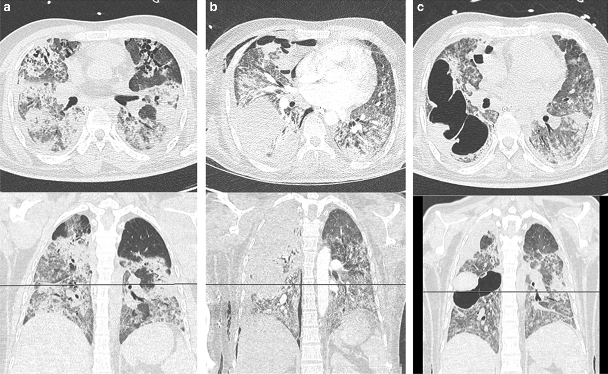
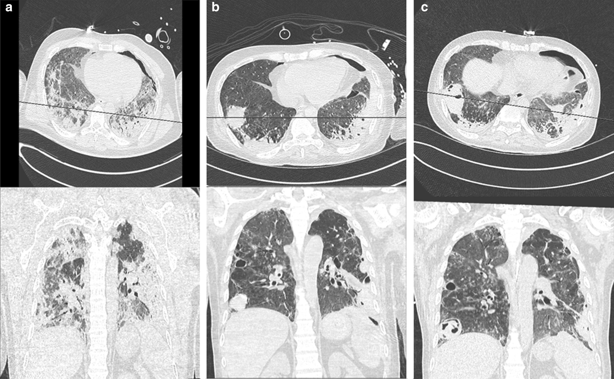
Twelve out of 689 (1.7%) patients admitted to our institution with COVID-19 developed pulmonary cavitation, comprising 3.3% (n = 12/359) of patients who developed COVID-19 pneumonia, and 11% (n = 12/110) of those admitted to the intensive care unit. We describe the imaging characteristics of the cavitation and present the clinical, pharmacological, laboratory, and microbiological parameters for these patients. In this cohort six patients have died, and six discharged home.

CONCLUSION

Cavitary lung disease in patients with severe COVID-19 disease is not uncommon, and is associated with a high level of morbidity and mortality.

摘要

背景

新型冠状病毒病 2019(COVID-19)的肺部放射学表现已有详细记录,范围从轻度病例的散在磨玻璃影到重症患者的融合性磨玻璃影、致密实变和铺路石征。然而,这些患者中并未常描述肺空洞。本研究旨在评估 COVID-19 患者肺空洞的发生率,并描述其特征和演变。

方法

我们对我院收治的所有 COVID-19 患者进行了回顾性分析,并查阅了电子病历和影像学资料,以确定发生肺空洞的患者。

结果

我院收治的 689 例 COVID-19 患者中有 12 例(1.7%)发生肺空洞,其中 3.3%(n=12/359)的 COVID-19 肺炎患者和 11%(n=12/110)的重症监护病房患者发生肺空洞。我们描述了空洞的影像学特征,并介绍了这些患者的临床、药理学、实验室和微生物学参数。在该队列中,有 6 例患者死亡,6 例出院回家。

结论

重症 COVID-19 患者的空洞性肺病并不少见,与高发病率和死亡率相关。

https://cdn.ncbi.nlm.nih.gov/pmc/blobs/d064/7805052/e9ab3d8f874f/12890_2020_1379_Fig2_HTML.jpg
https://cdn.ncbi.nlm.nih.gov/pmc/blobs/d064/7805052/7343feca0c41/12890_2020_1379_Fig1_HTML.jpg
https://cdn.ncbi.nlm.nih.gov/pmc/blobs/d064/7805052/e9ab3d8f874f/12890_2020_1379_Fig2_HTML.jpg
https://cdn.ncbi.nlm.nih.gov/pmc/blobs/d064/7805052/7343feca0c41/12890_2020_1379_Fig1_HTML.jpg
https://cdn.ncbi.nlm.nih.gov/pmc/blobs/d064/7805052/e9ab3d8f874f/12890_2020_1379_Fig2_HTML.jpg

相似文献

1
Pulmonary cavitation: an under-recognized late complication of severe COVID-19 lung disease.肺部空洞:严重 COVID-19 肺部疾病的一种被低估的晚期并发症。
BMC Pulm Med. 2021 Jan 12;21(1):24. doi: 10.1186/s12890-020-01379-1.
2
Clinical imaging characteristics of inpatients with coronavirus disease-2019 in Heilongjiang Province, China: a retrospective study.中国黑龙江省2019冠状病毒病住院患者的临床影像特征:一项回顾性研究。
Aging (Albany NY). 2020 Jul 20;12(14):13860-13868. doi: 10.18632/aging.103633.
3
[Risk factors of pulmonary cavitation in COVID-19 pneumonia].[新型冠状病毒肺炎肺空洞形成的危险因素]
Khirurgiia (Mosk). 2024(5):36-42. doi: 10.17116/hirurgia202405136.
4
Pulmonary cavitation in follow-up COVID 2019 cases: An etiological perspective.2019冠状病毒病后续病例中的肺空洞形成:病因学视角
Med J Armed Forces India. 2022 Aug 19;80(Suppl 1):S29-37. doi: 10.1016/j.mjafi.2022.06.015.
5
[Spatial and temporal distribution and predictive value of chest CT scoring in patients with COVID-19].新型冠状病毒肺炎患者胸部CT评分的时空分布及预测价值
Zhonghua Jie He He Hu Xi Za Zhi. 2021 Mar 12;44(3):230-236. doi: 10.3760/cma.j.cn112147-20200522-00626.
6
Dandelion and focal crazy paving signs: the lung CT based predictors for evaluation of the severity of coronavirus disease.蒲公英征和局灶性铺路石征:基于肺部 CT 的预测因子用于评估冠状病毒病的严重程度。
Curr Med Res Opin. 2021 Feb;37(2):219-224. doi: 10.1080/03007995.2020.1846173. Epub 2020 Nov 26.
7
Lung cavitation as a consequence of coronavirus-19 pneumonia.新冠肺炎导致的肺空洞。
Eur Rev Med Pharmacol Sci. 2021 Oct;25(19):5936-5941. doi: 10.26355/eurrev_202110_26870.
8
Similarities and Differences of Early Pulmonary CT Features of Pneumonia Caused by SARS-CoV-2, SARS-CoV and MERS-CoV: Comparison Based on a Systemic Review.新型冠状病毒、严重急性呼吸综合征冠状病毒和中东呼吸综合征冠状病毒所致肺炎早期肺部CT特征的异同:基于系统评价的比较
Chin Med Sci J. 2020 Sep 30;35(3):254-261. doi: 10.24920/003727.
9
Imaging and clinical features of patients with 2019 novel coronavirus SARS-CoV-2.新型冠状病毒 2019 年 SARS-CoV-2 患者的影像学和临床特征。
Eur J Nucl Med Mol Imaging. 2020 May;47(5):1275-1280. doi: 10.1007/s00259-020-04735-9. Epub 2020 Feb 28.
10
COVID-19 Infection in Octagenarian Patients: Imaging Aspects and Clinical Correlations.八旬老人感染 COVID-19:影像学表现及临床相关性。
Gerontology. 2022;68(3):261-271. doi: 10.1159/000516166. Epub 2021 Aug 5.

引用本文的文献

1
An Observational Cohort Study of Bronchoalveolar Lavage Fluid Galactomannan and Culture Positivity in Patients Requiring Mechanical Ventilation.一项针对需要机械通气患者的支气管肺泡灌洗液体半乳甘露聚糖及培养阳性率的观察性队列研究。
Open Forum Infect Dis. 2025 Feb 15;12(3):ofaf090. doi: 10.1093/ofid/ofaf090. eCollection 2025 Mar.
2
Impact of underlying diseases and complications on COVID-19 mortality in South Korea: analysis of national health insurance service data.基础疾病及并发症对韩国新冠疫情死亡率的影响:基于国民健康保险服务数据的分析
Arch Public Health. 2025 Jan 23;83(1):20. doi: 10.1186/s13690-025-01509-3.
3

本文引用的文献

1
Chest Imaging Appearance of COVID-19 Infection.新型冠状病毒肺炎感染的胸部影像学表现。
Radiol Cardiothorac Imaging. 2020 Feb 13;2(1):e200028. doi: 10.1148/ryct.2020200028. eCollection 2020 Feb.
2
COVID-19 Infection Presenting with CT Halo Sign.表现为CT晕征的新型冠状病毒肺炎感染
Radiol Cardiothorac Imaging. 2020 Feb 12;2(1):e200026. doi: 10.1148/ryct.2020200026. eCollection 2020 Feb.
3
Spontaneous pneumomediastinum, pneumothorax and subcutaneous emphysema in COVID-19: case report and literature review.新型冠状病毒肺炎并发自发性纵隔气肿、气胸及皮下气肿:病例报告与文献综述
Evaluating the Cavitary Lung Lesions on CT Scan of COVID-19 Patients: A Retrospective Study.
评估新型冠状病毒肺炎患者胸部CT扫描中的肺空洞病变:一项回顾性研究。
J Community Hosp Intern Med Perspect. 2024 Nov 2;14(6):23-29. doi: 10.55729/2000-9666.1411. eCollection 2024.
4
COVID-19 associated lung cavitary lesions: A case series.新型冠状病毒肺炎相关肺空洞性病变:病例系列
Caspian J Intern Med. 2024 Sep 7;15(4):713-728. doi: 10.22088/cjim.15.4.713. eCollection 2024 Fall.
5
Surgical therapy for major lung parenchymal damage from late coronavirus disease complication: case report and literature review.冠状病毒病晚期并发症致严重肺实质损伤的手术治疗:病例报告及文献综述
AME Case Rep. 2023 Sep 1;7:33. doi: 10.21037/acr-22-36. eCollection 2023.
6
Concomitant Pulmonary Aspergillosis Following Severe COVID-19.重症新型冠状病毒肺炎合并肺曲霉病
Cureus. 2023 Jun 28;15(6):e41112. doi: 10.7759/cureus.41112. eCollection 2023 Jun.
7
COVID-19 and Lung Cavitation: A Clue to Pathogenesis?新冠病毒与肺空洞:发病机制的线索?
HCA Healthc J Med. 2022 Aug 29;3(4):253-256. doi: 10.36518/2689-0216.1361. eCollection 2022.
8
The impact of COVID-19 on TB in Iran: An illustrative study.2019年冠状病毒病对伊朗结核病的影响:一项实例研究。
J Clin Tuberc Other Mycobact Dis. 2023 May;31:100357. doi: 10.1016/j.jctube.2023.100357. Epub 2023 Mar 1.
9
COVID-19 and Cavitary Lesion in Lung.新型冠状病毒肺炎与肺部空洞性病变
Cureus. 2023 Jan 23;15(1):e34098. doi: 10.7759/cureus.34098. eCollection 2023 Jan.
10
Cavitary lung lesion as sequelae of mild COVID-19 in a patient with HIV.一名感染艾滋病毒的患者出现轻度新冠后遗症,表现为肺部空洞性病变。
Clin Infect Pract. 2023 Jan;17:100218. doi: 10.1016/j.clinpr.2023.100218. Epub 2023 Jan 18.
Rev Inst Med Trop Sao Paulo. 2020 Oct 9;62:e76. doi: 10.1590/S1678-9946202062076. eCollection 2020.
4
Cavitary pulmonary tuberculosis with COVID-19 coinfection.空洞型肺结核合并新型冠状病毒肺炎感染
IDCases. 2020;22:e00973. doi: 10.1016/j.idcr.2020.e00973. Epub 2020 Sep 28.
5
Similarities and Differences of Early Pulmonary CT Features of Pneumonia Caused by SARS-CoV-2, SARS-CoV and MERS-CoV: Comparison Based on a Systemic Review.新型冠状病毒、严重急性呼吸综合征冠状病毒和中东呼吸综合征冠状病毒所致肺炎早期肺部CT特征的异同:基于系统评价的比较
Chin Med Sci J. 2020 Sep 30;35(3):254-261. doi: 10.24920/003727.
6
A rare case of Pneumopericardium secondary to COVID-19.COVID-19 导致罕见的心包积气病例。
Heart Lung. 2020 Nov-Dec;49(6):679-680. doi: 10.1016/j.hrtlng.2020.08.017. Epub 2020 Aug 21.
7
Tocilizumab in patients with severe COVID-19: a retrospective cohort study.托珠单抗治疗重症新型冠状病毒肺炎患者:一项回顾性队列研究
Lancet Rheumatol. 2020 Aug;2(8):e474-e484. doi: 10.1016/S2665-9913(20)30173-9. Epub 2020 Jun 24.
8
Lung cavitation in COVID-19: co-infection complication or rare evolution?新型冠状病毒肺炎中的肺空洞形成:合并感染并发症还是罕见演变?
Einstein (Sao Paulo). 2020;18:eAI5822. doi: 10.31744/einstein_journal/2020ai5822. Epub 2020 Jul 22.
9
Remdesivir for the Treatment of Covid-19 - Preliminary Report. Reply.瑞德西韦治疗新冠病毒病-初步报告。回复。
N Engl J Med. 2020 Sep 3;383(10):994. doi: 10.1056/NEJMc2022236. Epub 2020 Jul 10.
10
Lung cavitation due to COVID-19 pneumonia.新型冠状病毒肺炎所致的肺空洞形成。
BMJ Case Rep. 2020 Jul 6;13(7):e237245. doi: 10.1136/bcr-2020-237245.