Solanki Anu K, Lali Ferdinand V, Autefage Hélène, Agarwal Shweta, Nommeots-Nomm Amy, Metcalfe Anthony D, Stevens Molly M, Jones Julian R
Department of Materials, Imperial College London, South Kensington, London, SW7 2AZ, UK.
Institute of Biomedical Engineering, Imperial College London, South Kensington, London, SW7 2AZ, UK.
Biomater Res. 2021 Jan 15;25(1):1. doi: 10.1186/s40824-020-00202-6.
Bioactive glasses are traditionally associated with bonding to bone through a hydroxycarbonate apatite (HCA) surface layer but the release of active ions is more important for bone regeneration. They are now being used to deliver ions for soft tissue applications, particularly wound healing. Cobalt is known to simulate hypoxia and provoke angiogenesis. The aim here was to develop new bioactive glass compositions designed to be scaffold materials to locally deliver pro-angiogenic cobalt ions, at a controlled rate, without forming an HCA layer, for wound healing applications.
New melt-derived bioactive glass compositions were designed that had the same network connectivity (mean number of bridging covalent bonds between silica tetrahedra), and therefore similar biodegradation rate, as the original 45S5 Bioglass. The amount of magnesium and cobalt in the glass was varied, with the aim of reducing or removing calcium and phosphate from the compositions. Electrospun poly(ε-caprolactone)/bioactive glass composites were also produced. Glasses were tested for ion release in dissolution studies and their influence on Hypoxia-Inducible Factor 1-alpha (HIF-1α) and expression of Vascular Endothelial Growth Factor (VEGF) from fibroblast cells was investigated.
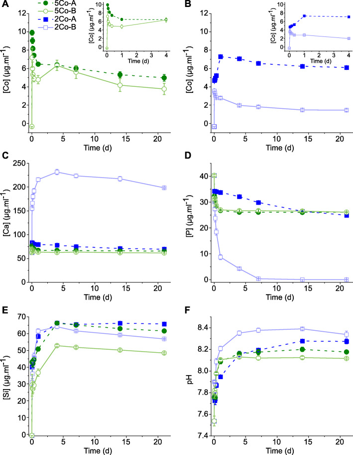
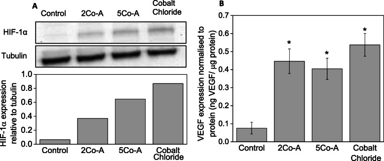
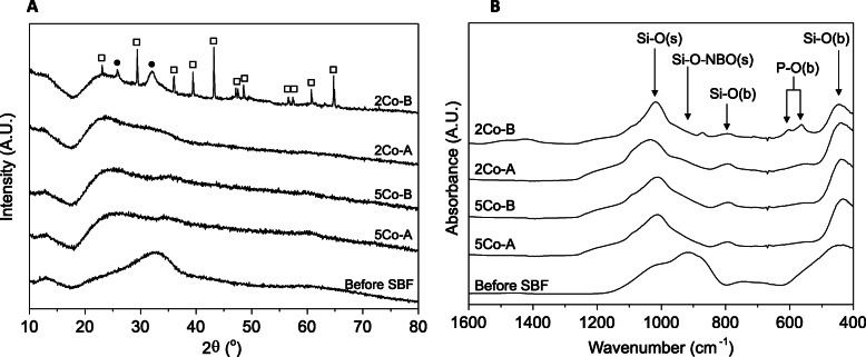
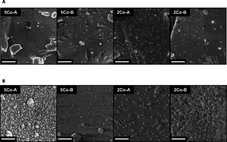
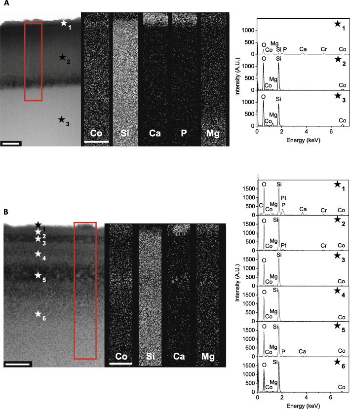
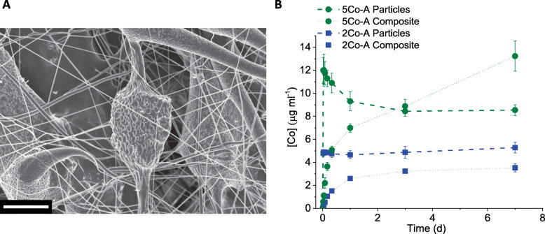
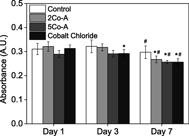
Dissolution tests showed the silica rich layer differed depending on the amount of MgO in the glass, which influenced the delivery of cobalt. The electrospun composites delivered a more sustained ion release relative to glass particles alone. Exposing fibroblasts to conditioned media from these composites did not cause a detrimental effect on metabolic activity but glasses containing cobalt did stabilise HIF-1α and provoked a significantly higher expression of VEGF (not seen in Co-free controls).
The composite fibres containing new bioactive glass compositions delivered cobalt ions at a sustained rate, which could be mediated by the magnesium content of the glass. The dissolution products stabilised HIF-1α and provoked a significantly higher expression of VEGF, suggesting the composites activated the HIF pathway to stimulate angiogenesis.
传统上,生物活性玻璃通过羟基碳酸磷灰石(HCA)表面层与骨结合,但活性离子的释放对骨再生更为重要。目前它们正被用于软组织应用中输送离子,尤其是伤口愈合。已知钴可模拟缺氧并促进血管生成。本研究的目的是开发新的生物活性玻璃组合物,设计成支架材料,以可控速率局部递送促血管生成的钴离子,不形成HCA层,用于伤口愈合应用。
设计了新的熔融衍生生物活性玻璃组合物,其具有与原始45S5生物玻璃相同的网络连接性(硅四面体之间桥连共价键的平均数),因此具有相似的生物降解速率。改变玻璃中镁和钴的含量,目的是减少或去除组合物中的钙和磷。还制备了电纺聚(ε-己内酯)/生物活性玻璃复合材料。在溶解研究中测试了玻璃的离子释放,并研究了它们对缺氧诱导因子1α(HIF-1α)的影响以及对成纤维细胞中血管内皮生长因子(VEGF)表达的影响。
溶解试验表明,富含二氧化硅的层因玻璃中氧化镁的含量而异,这影响了钴的递送。相对于单独的玻璃颗粒,电纺复合材料的离子释放更持久。将成纤维细胞暴露于这些复合材料的条件培养基中对代谢活性没有不利影响,但含钴的玻璃确实使HIF-1α稳定,并引发VEGF的显著更高表达(在无钴对照中未观察到)。
含有新生物活性玻璃组合物的复合纤维以持续速率递送钴离子,这可以由玻璃中的镁含量介导。溶解产物使HIF-1α稳定,并引发VEGF的显著更高表达,表明复合材料激活了HIF途径以刺激血管生成。