• 文献检索
  • 文档翻译
  • 深度研究
  • 学术资讯
  • Suppr Zotero 插件Zotero 插件
  • 邀请有礼
  • 套餐&价格
  • 历史记录
应用&插件
Suppr Zotero 插件Zotero 插件浏览器插件Mac 客户端Windows 客户端微信小程序
定价
高级版会员购买积分包购买API积分包
服务
文献检索文档翻译深度研究API 文档MCP 服务
关于我们
关于 Suppr公司介绍联系我们用户协议隐私条款
关注我们

Suppr 超能文献

核心技术专利:CN118964589B侵权必究
粤ICP备2023148730 号-1Suppr @ 2026

文献检索

告别复杂PubMed语法,用中文像聊天一样搜索,搜遍4000万医学文献。AI智能推荐,让科研检索更轻松。

立即免费搜索

文件翻译

保留排版,准确专业,支持PDF/Word/PPT等文件格式,支持 12+语言互译。

免费翻译文档

深度研究

AI帮你快速写综述,25分钟生成高质量综述,智能提取关键信息,辅助科研写作。

立即免费体验

人工智能量化的COVID-19胸部CT与患者预后的关联。

Association of AI quantified COVID-19 chest CT and patient outcome.

作者信息

Fang Xi, Kruger Uwe, Homayounieh Fatemeh, Chao Hanqing, Zhang Jiajin, Digumarthy Subba R, Arru Chiara D, Kalra Mannudeep K, Yan Pingkun

机构信息

Department of Biomedical Engineering, Center for Biotechnology and Interdisciplinary Studies, Rensselaer Polytechnic Institute, Troy, NY, 12180, USA.

Department of Radiology, Massachusetts General Hospitals, Harvard Medical School, Boston, MA, 02114, USA.

出版信息

Int J Comput Assist Radiol Surg. 2021 Mar;16(3):435-445. doi: 10.1007/s11548-020-02299-5. Epub 2021 Jan 23.

DOI:10.1007/s11548-020-02299-5
PMID:33484428
原文链接:https://pmc.ncbi.nlm.nih.gov/articles/PMC7822756/
Abstract

PURPOSE

Severity scoring is a key step in managing patients with COVID-19 pneumonia. However, manual quantitative analysis by radiologists is a time-consuming task, while qualitative evaluation may be fast but highly subjective. This study aims to develop artificial intelligence (AI)-based methods to quantify disease severity and predict COVID-19 patient outcome.

METHODS

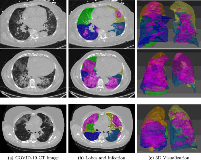
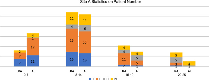
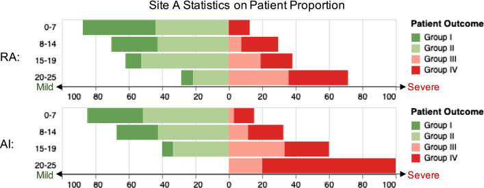
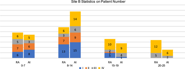
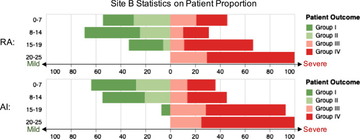
We develop an AI-based framework that employs deep neural networks to efficiently segment lung lobes and pulmonary opacities. The volume ratio of pulmonary opacities inside each lung lobe gives the severity scores of the lobes, which are then used to predict ICU admission and mortality with three different machine learning methods. The developed methods were evaluated on datasets from two hospitals (site A: Firoozgar Hospital, Iran, 105 patients; site B: Massachusetts General Hospital, USA, 88 patients).

RESULTS

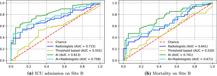
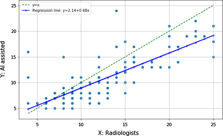
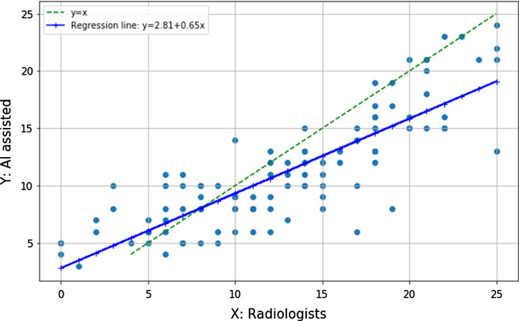
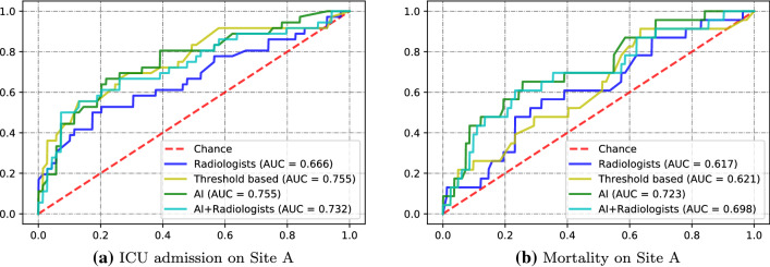
AI-based severity scores are strongly associated with those evaluated by radiologists (Spearman's rank correlation 0.837, [Formula: see text]). Using AI-based scores produced significantly higher ([Formula: see text]) area under the ROC curve (AUC) values. The developed AI method achieved the best performance of AUC = 0.813 (95% CI [0.729, 0.886]) in predicting ICU admission and AUC = 0.741 (95% CI [0.640, 0.837]) in mortality estimation on the two datasets.

CONCLUSIONS

Accurate severity scores can be obtained using the developed AI methods over chest CT images. The computed severity scores achieved better performance than radiologists in predicting COVID-19 patient outcome by consistently quantifying image features. Such developed techniques of severity assessment may be extended to other lung diseases beyond the current pandemic.

摘要

目的

严重程度评分是管理新冠肺炎肺炎患者的关键步骤。然而,放射科医生进行的手动定量分析是一项耗时的任务,而定性评估可能很快但主观性很强。本研究旨在开发基于人工智能(AI)的方法来量化疾病严重程度并预测新冠肺炎患者的预后。

方法

我们开发了一个基于AI的框架,该框架采用深度神经网络来有效地分割肺叶和肺内混浊区域。每个肺叶内肺内混浊区域的体积比给出了各肺叶的严重程度评分,然后使用三种不同的机器学习方法来预测入住重症监护病房(ICU)情况和死亡率。所开发的方法在来自两家医院的数据集上进行了评估(地点A:伊朗菲罗兹加尔医院,105例患者;地点B:美国马萨诸塞州总医院,88例患者)。

结果

基于AI的严重程度评分与放射科医生评估的评分密切相关(斯皮尔曼等级相关系数为0.837,[公式:见原文])。使用基于AI的评分在ROC曲线下面积(AUC)值上显著更高([公式:见原文])。所开发的AI方法在预测ICU入住情况时,在两个数据集上的AUC = 0.813(95%可信区间[0.729, 0.886]),在死亡率估计方面AUC = 0.741(95%可信区间[0.640, 0.837]),表现最佳。

结论

通过所开发的AI方法可以在胸部CT图像上获得准确的严重程度评分。通过持续量化图像特征,计算得到的严重程度评分在预测新冠肺炎患者预后方面比放射科医生表现更好。这种所开发的严重程度评估技术可能会扩展到当前大流行之外的其他肺部疾病。

https://cdn.ncbi.nlm.nih.gov/pmc/blobs/4d31/7822756/65a7c643bcdb/11548_2020_2299_Fig9_HTML.jpg
https://cdn.ncbi.nlm.nih.gov/pmc/blobs/4d31/7822756/8f1aaacf970a/11548_2020_2299_Fig1_HTML.jpg
https://cdn.ncbi.nlm.nih.gov/pmc/blobs/4d31/7822756/5a35b92abf15/11548_2020_2299_Fig2_HTML.jpg
https://cdn.ncbi.nlm.nih.gov/pmc/blobs/4d31/7822756/a8b9befb7e00/11548_2020_2299_Fig3_HTML.jpg
https://cdn.ncbi.nlm.nih.gov/pmc/blobs/4d31/7822756/3d3056dfcae2/11548_2020_2299_Fig4_HTML.jpg
https://cdn.ncbi.nlm.nih.gov/pmc/blobs/4d31/7822756/e6e8c678dd7d/11548_2020_2299_Fig5_HTML.jpg
https://cdn.ncbi.nlm.nih.gov/pmc/blobs/4d31/7822756/ae19e6947bed/11548_2020_2299_Fig6_HTML.jpg
https://cdn.ncbi.nlm.nih.gov/pmc/blobs/4d31/7822756/157a40c3145c/11548_2020_2299_Fig7_HTML.jpg
https://cdn.ncbi.nlm.nih.gov/pmc/blobs/4d31/7822756/e7c2559d651f/11548_2020_2299_Fig8_HTML.jpg
https://cdn.ncbi.nlm.nih.gov/pmc/blobs/4d31/7822756/65a7c643bcdb/11548_2020_2299_Fig9_HTML.jpg

相似文献

1
Association of AI quantified COVID-19 chest CT and patient outcome.人工智能量化的COVID-19胸部CT与患者预后的关联。
Int J Comput Assist Radiol Surg. 2021 Mar;16(3):435-445. doi: 10.1007/s11548-020-02299-5. Epub 2021 Jan 23.
2
Multicenter Assessment of CT Pneumonia Analysis Prototype for Predicting Disease Severity and Patient Outcome.多中心 CT 肺炎分析原型评估:预测疾病严重程度和患者转归。
J Digit Imaging. 2021 Apr;34(2):320-329. doi: 10.1007/s10278-021-00430-9. Epub 2021 Feb 25.
3
From community-acquired pneumonia to COVID-19: a deep learning-based method for quantitative analysis of COVID-19 on thick-section CT scans.从社区获得性肺炎到 COVID-19:一种基于深度学习的 CT 厚层扫描 COVID-19 定量分析方法。
Eur Radiol. 2020 Dec;30(12):6828-6837. doi: 10.1007/s00330-020-07042-x. Epub 2020 Jul 18.
4
Automated Assessment of COVID-19 Reporting and Data System and Chest CT Severity Scores in Patients Suspected of Having COVID-19 Using Artificial Intelligence.利用人工智能对疑似 COVID-19 患者的 COVID-19 报告和数据系统及胸部 CT 严重程度评分进行自动评估。
Radiology. 2021 Jan;298(1):E18-E28. doi: 10.1148/radiol.2020202439. Epub 2020 Jul 30.
5
Artificial intelligence-assisted quantification of COVID-19 pneumonia burden from computed tomography improves prediction of adverse outcomes over visual scoring systems.人工智能辅助 CT 对 COVID-19 肺炎负担的定量评估可提高对不良结局的预测能力,优于视觉评分系统。
Br J Radiol. 2023 Sep;96(1149):20220180. doi: 10.1259/bjr.20220180. Epub 2023 Jun 27.
6
Initial chest radiographs and artificial intelligence (AI) predict clinical outcomes in COVID-19 patients: analysis of 697 Italian patients.初始胸部 X 光片和人工智能 (AI) 预测 COVID-19 患者的临床结局:对 697 例意大利患者的分析。
Eur Radiol. 2021 Mar;31(3):1770-1779. doi: 10.1007/s00330-020-07269-8. Epub 2020 Sep 18.
7
Using artificial intelligence to assist radiologists in distinguishing COVID-19 from other pulmonary infections.利用人工智能辅助放射科医生区分 COVID-19 与其他肺部感染。
J Xray Sci Technol. 2021;29(1):1-17. doi: 10.3233/XST-200735.
8
Artificial intelligence matches subjective severity assessment of pneumonia for prediction of patient outcome and need for mechanical ventilation: a cohort study.人工智能匹配肺炎的主观严重程度评估以预测患者结局和机械通气需求:一项队列研究。
Sci Rep. 2021 Jan 13;11(1):858. doi: 10.1038/s41598-020-79470-0.
9
Analysis of clinical features and imaging signs of COVID-19 with the assistance of artificial intelligence.人工智能辅助分析 COVID-19 的临床特征和影像征象。
Eur Rev Med Pharmacol Sci. 2020 Aug;24(15):8210-8218. doi: 10.26355/eurrev_202008_22510.
10
Prognostication of patients with COVID-19 using artificial intelligence based on chest x-rays and clinical data: a retrospective study.基于胸部 X 光片和临床数据的人工智能预测 COVID-19 患者的预后:一项回顾性研究。
Lancet Digit Health. 2021 May;3(5):e286-e294. doi: 10.1016/S2589-7500(21)00039-X. Epub 2021 Mar 24.

引用本文的文献

1
Human-in-the-Loop-A Deep Learning Strategy in Combination with a Patient-Specific Gaussian Mixture Model Leads to the Fast Characterization of Volumetric Ground-Glass Opacity and Consolidation in the Computed Tomography Scans of COVID-19 Patients.人在回路中——一种结合患者特异性高斯混合模型的深度学习策略可快速表征COVID-19患者计算机断层扫描中的磨玻璃密度影和实变。
J Clin Med. 2024 Sep 4;13(17):5231. doi: 10.3390/jcm13175231.
2
Development of a new prognostic model to predict pneumonia outcome using artificial intelligence-based chest radiograph results.利用人工智能胸片结果开发一种新的预测肺炎结局的预后模型。
Sci Rep. 2024 Jun 22;14(1):14415. doi: 10.1038/s41598-024-65488-1.
3

本文引用的文献

1
Automated Assessment and Tracking of COVID-19 Pulmonary Disease Severity on Chest Radiographs using Convolutional Siamese Neural Networks.使用卷积暹罗神经网络对胸部X光片上的COVID-19肺部疾病严重程度进行自动评估和跟踪。
Radiol Artif Intell. 2020 Jul 22;2(4):e200079. doi: 10.1148/ryai.2020200079. eCollection 2020 Jul.
2
Chest CT Severity Score: An Imaging Tool for Assessing Severe COVID-19.胸部CT严重程度评分:一种评估重症COVID-19的影像学工具。
Radiol Cardiothorac Imaging. 2020 Mar 30;2(2):e200047. doi: 10.1148/ryct.2020200047. eCollection 2020 Apr.
3
Synergistic learning of lung lobe segmentation and hierarchical multi-instance classification for automated severity assessment of COVID-19 in CT images.
Robust Medical Diagnosis: A Novel Two-Phase Deep Learning Framework for Adversarial Proof Disease Detection in Radiology Images.
稳健医学诊断:一种新颖的两阶段深度学习框架,用于放射图像中的对抗性证明疾病检测。
J Imaging Inform Med. 2024 Feb;37(1):308-338. doi: 10.1007/s10278-023-00916-8. Epub 2024 Jan 10.
4
A Multiclass Radiomics Method-Based WHO Severity Scale for Improving COVID-19 Patient Assessment and Disease Characterization From CT Scans.一种基于多分类放射组学方法的世卫组织严重程度评分标准,用于改善 CT 扫描对 COVID-19 患者的评估和疾病特征描述。
Invest Radiol. 2023 Dec 1;58(12):882-893. doi: 10.1097/RLI.0000000000001005. Epub 2023 Jul 27.
5
The Role of Computed Tomography in the Management of Hospitalized Patients With COVID-19.计算机断层扫描在新型冠状病毒肺炎住院患者管理中的作用
Cureus. 2023 Mar 28;15(3):e36821. doi: 10.7759/cureus.36821. eCollection 2023 Mar.
6
Quantification of pulmonary involvement in COVID-19 pneumonia: an upgrade of the software for lung CT segmentation.新型冠状病毒肺炎肺部受累情况的量化:肺CT分割软件的升级
Eur Phys J Plus. 2023;138(4):326. doi: 10.1140/epjp/s13360-023-03896-4. Epub 2023 Apr 11.
7
Chest computed tomography radiomics to predict the outcome for patients with COVID-19 at an early stage.胸部计算机断层扫描放射组学预测 COVID-19 患者早期预后。
Diagn Interv Radiol. 2023 Jan 31;29(1):91-102. doi: 10.5152/dir.2022.21576. Epub 2023 Jan 18.
8
3D CT-Inclusive Deep-Learning Model to Predict Mortality, ICU Admittance, and Intubation in COVID-19 Patients.基于 3D CT 的深度学习模型预测 COVID-19 患者的死亡率、入住 ICU 率和插管率。
J Digit Imaging. 2023 Apr;36(2):603-616. doi: 10.1007/s10278-022-00734-4. Epub 2022 Nov 30.
9
Determination of the Severity and Percentage of COVID-19 Infection through a Hierarchical Deep Learning System.通过分层深度学习系统确定新冠病毒感染的严重程度和百分比
J Pers Med. 2022 Mar 28;12(4):535. doi: 10.3390/jpm12040535.
10
COVID-19 mortality prediction in the intensive care unit with deep learning based on longitudinal chest X-rays and clinical data.基于纵向胸部 X 光片和临床数据的深度学习在重症监护病房中对 COVID-19 死亡率的预测。
Eur Radiol. 2022 Jul;32(7):4446-4456. doi: 10.1007/s00330-022-08588-8. Epub 2022 Feb 19.
用于CT图像中COVID-19自动严重程度评估的肺叶分割与分层多实例分类协同学习
Pattern Recognit. 2021 May;113:107828. doi: 10.1016/j.patcog.2021.107828. Epub 2021 Jan 16.
4
Integrative analysis for COVID-19 patient outcome prediction.COVID-19 患者预后预测的综合分析。
Med Image Anal. 2021 Jan;67:101844. doi: 10.1016/j.media.2020.101844. Epub 2020 Oct 13.
5
Joint prediction and time estimation of COVID-19 developing severe symptoms using chest CT scan.利用胸部 CT 扫描联合预测和时间估计 COVID-19 发展为严重症状。
Med Image Anal. 2021 Jan;67:101824. doi: 10.1016/j.media.2020.101824. Epub 2020 Oct 10.
6
Severity assessment of COVID-19 using CT image features and laboratory indices.基于 CT 图像特征和实验室指标评估 COVID-19 严重程度。
Phys Med Biol. 2021 Jan 26;66(3):035015. doi: 10.1088/1361-6560/abbf9e.
7
Automatic lung segmentation in routine imaging is primarily a data diversity problem, not a methodology problem.常规影像中的自动肺分割主要是一个数据多样性问题,而不是方法学问题。
Eur Radiol Exp. 2020 Aug 20;4(1):50. doi: 10.1186/s41747-020-00173-2.
8
Artificial intelligence for the detection of COVID-19 pneumonia on chest CT using multinational datasets.利用多国数据集的人工智能检测胸部 CT 中的 COVID-19 肺炎。
Nat Commun. 2020 Aug 14;11(1):4080. doi: 10.1038/s41467-020-17971-2.
9
Multi-Organ Segmentation Over Partially Labeled Datasets With Multi-Scale Feature Abstraction.多器官分割在具有多尺度特征抽象的部分标记数据集上。
IEEE Trans Med Imaging. 2020 Nov;39(11):3619-3629. doi: 10.1109/TMI.2020.3001036. Epub 2020 Oct 28.
10
Clinical and Chest Radiography Features Determine Patient Outcomes in Young and Middle-aged Adults with COVID-19.临床和胸部放射摄影特征可预测 COVID-19 中青年患者的结局。
Radiology. 2020 Oct;297(1):E197-E206. doi: 10.1148/radiol.2020201754. Epub 2020 May 14.