• 文献检索
  • 文档翻译
  • 深度研究
  • 学术资讯
  • Suppr Zotero 插件Zotero 插件
  • 邀请有礼
  • 套餐&价格
  • 历史记录
应用&插件
Suppr Zotero 插件Zotero 插件浏览器插件Mac 客户端Windows 客户端微信小程序
定价
高级版会员购买积分包购买API积分包
服务
文献检索文档翻译深度研究API 文档MCP 服务
关于我们
关于 Suppr公司介绍联系我们用户协议隐私条款
关注我们

Suppr 超能文献

核心技术专利:CN118964589B侵权必究
粤ICP备2023148730 号-1Suppr @ 2026

文献检索

告别复杂PubMed语法,用中文像聊天一样搜索,搜遍4000万医学文献。AI智能推荐,让科研检索更轻松。

立即免费搜索

文件翻译

保留排版,准确专业,支持PDF/Word/PPT等文件格式,支持 12+语言互译。

免费翻译文档

深度研究

AI帮你快速写综述,25分钟生成高质量综述,智能提取关键信息,辅助科研写作。

立即免费体验

基于胸部 X 光片和临床数据的人工智能预测 COVID-19 患者的预后:一项回顾性研究。

Prognostication of patients with COVID-19 using artificial intelligence based on chest x-rays and clinical data: a retrospective study.

机构信息

Department of Radiology, Perelman School of Medicine, University of Pennsylvania, Philadelphia, PA, USA.

Department of Diagnostic Imaging, Rhode Island Hospital and Warren Alpert Medical School of Brown University, Providence, RI, USA.

出版信息

Lancet Digit Health. 2021 May;3(5):e286-e294. doi: 10.1016/S2589-7500(21)00039-X. Epub 2021 Mar 24.

DOI:10.1016/S2589-7500(21)00039-X
PMID:33773969
原文链接:https://pmc.ncbi.nlm.nih.gov/articles/PMC7990487/
Abstract

BACKGROUND

Chest x-ray is a relatively accessible, inexpensive, fast imaging modality that might be valuable in the prognostication of patients with COVID-19. We aimed to develop and evaluate an artificial intelligence system using chest x-rays and clinical data to predict disease severity and progression in patients with COVID-19.

METHODS

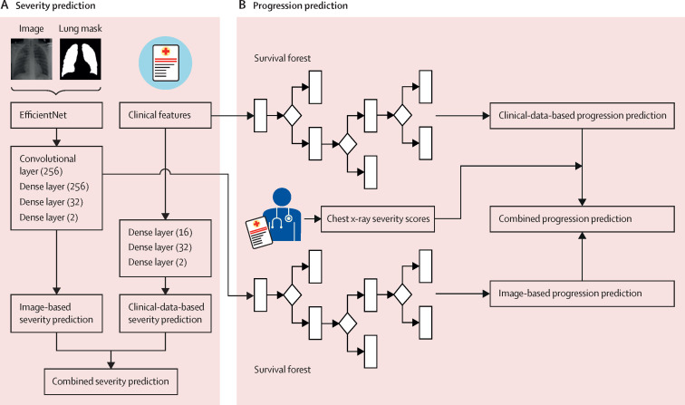
We did a retrospective study in multiple hospitals in the University of Pennsylvania Health System in Philadelphia, PA, USA, and Brown University affiliated hospitals in Providence, RI, USA. Patients who presented to a hospital in the University of Pennsylvania Health System via the emergency department, with a diagnosis of COVID-19 confirmed by RT-PCR and with an available chest x-ray from their initial presentation or admission, were retrospectively identified and randomly divided into training, validation, and test sets (7:1:2). Using the chest x-rays as input to an EfficientNet deep neural network and clinical data, models were trained to predict the binary outcome of disease severity (ie, critical or non-critical). The deep-learning features extracted from the model and clinical data were used to build time-to-event models to predict the risk of disease progression. The models were externally tested on patients who presented to an independent multicentre institution, Brown University affiliated hospitals, and compared with severity scores provided by radiologists.

FINDINGS

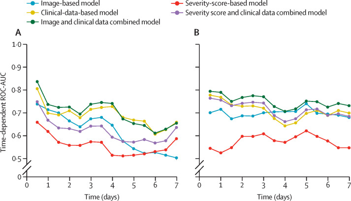
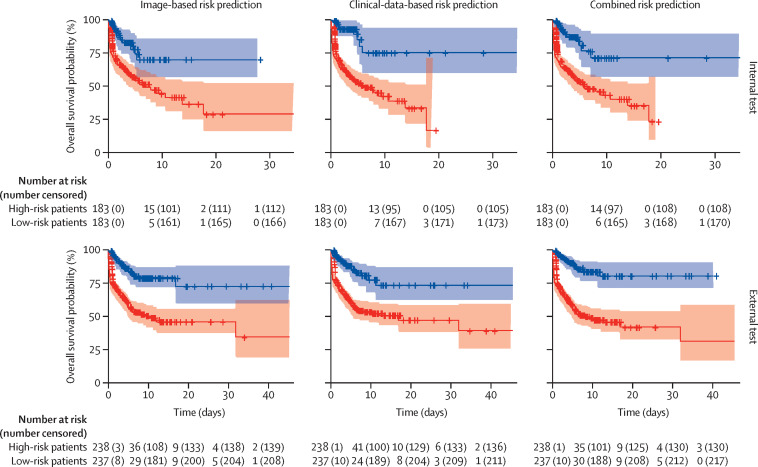
1834 patients who presented via the University of Pennsylvania Health System between March 9 and July 20, 2020, were identified and assigned to the model training (n=1285), validation (n=183), or testing (n=366) sets. 475 patients who presented via the Brown University affiliated hospitals between March 1 and July 18, 2020, were identified for external testing of the models. When chest x-rays were added to clinical data for severity prediction, area under the receiver operating characteristic curve (ROC-AUC) increased from 0·821 (95% CI 0·796-0·828) to 0·846 (0·815-0·852; p<0·0001) on internal testing and 0·731 (0·712-0·738) to 0·792 (0·780-0 ·803; p<0·0001) on external testing. When deep-learning features were added to clinical data for progression prediction, the concordance index (C-index) increased from 0·769 (0·755-0·786) to 0·805 (0·800-0·820; p<0·0001) on internal testing and 0·707 (0·695-0·729) to 0·752 (0·739-0·764; p<0·0001) on external testing. The image and clinical data combined model had significantly better prognostic performance than combined severity scores and clinical data on internal testing (C-index 0·805 vs 0·781; p=0·0002) and external testing (C-index 0·752 vs 0·715; p<0·0001).

INTERPRETATION

In patients with COVID-19, artificial intelligence based on chest x-rays had better prognostic performance than clinical data or radiologist-derived severity scores. Using artificial intelligence, chest x-rays can augment clinical data in predicting the risk of progression to critical illness in patients with COVID-19.

FUNDING

Brown University, Amazon Web Services Diagnostic Development Initiative, Radiological Society of North America, National Cancer Institute and National Institute of Biomedical Imaging and Bioengineering of the National Institutes of Health.

摘要

背景

胸部 X 光是一种相对容易获得、价格低廉、快速的成像方式,可能对预测 COVID-19 患者的预后有价值。我们旨在开发和评估一种人工智能系统,该系统使用胸部 X 光和临床数据来预测 COVID-19 患者的疾病严重程度和进展。

方法

我们在美国宾夕法尼亚大学卫生系统的多个医院和美国罗德岛州普罗维登斯的布朗大学附属医院进行了一项回顾性研究。通过急诊室就诊于宾夕法尼亚大学卫生系统医院、通过 RT-PCR 确诊 COVID-19 且初次就诊或入院时可获得胸部 X 光的患者被回顾性识别并随机分为训练集、验证集和测试集(7:1:2)。使用胸部 X 光作为输入到 EfficientNet 深度神经网络和临床数据,训练模型以预测疾病严重程度(即危急或非危急)的二项结局。从模型和临床数据中提取的深度学习特征用于构建预测疾病进展风险的生存时间模型。该模型在宾夕法尼亚大学卫生系统以外的一家独立多中心机构布朗大学附属医院的患者中进行了外部测试,并与放射科医生提供的严重程度评分进行了比较。

结果

2020 年 3 月 9 日至 7 月 20 日期间,通过宾夕法尼亚大学卫生系统就诊的 1834 名患者被确定并分配到模型训练(n=1285)、验证(n=183)或测试(n=366)组。2020 年 3 月 1 日至 7 月 18 日期间,通过布朗大学附属医院就诊的 475 名患者被确定用于模型的外部测试。当胸部 X 光与临床数据一起用于严重程度预测时,内部测试中曲线下面积(ROC-AUC)从 0.821(95%CI 0.796-0.828)增加到 0.846(0.815-0.852;p<0.0001),外部测试中从 0.731(0.712-0.738)增加到 0.792(0.780-0.803;p<0.0001)。当深度学习特征与临床数据一起用于进展预测时,内部测试中的一致性指数(C-index)从 0.769(0.755-0.786)增加到 0.805(0.800-0.820;p<0.0001),外部测试中从 0.707(0.695-0.729)增加到 0.752(0.739-0.764;p<0.0001)。图像和临床数据联合模型在内部测试(C-index 0.805 与 0.781;p=0.0002)和外部测试(C-index 0.752 与 0.715;p<0.0001)中的预后性能均显著优于联合严重程度评分和临床数据。

解释

在 COVID-19 患者中,基于胸部 X 光的人工智能具有比临床数据或放射科医生严重程度评分更好的预后性能。使用人工智能,胸部 X 光可以增强临床数据,以预测 COVID-19 患者向危急疾病进展的风险。

资助

布朗大学、亚马逊网络服务诊断开发倡议、北美放射学会、美国国立卫生研究院国家癌症研究所和国家生物医学成像和生物工程研究所。

https://cdn.ncbi.nlm.nih.gov/pmc/blobs/53ed/7990487/cd566c6aa8c6/gr3_lrg.jpg
https://cdn.ncbi.nlm.nih.gov/pmc/blobs/53ed/7990487/47a8bf3d7ae5/gr1_lrg.jpg
https://cdn.ncbi.nlm.nih.gov/pmc/blobs/53ed/7990487/f3d37de33da6/gr2_lrg.jpg
https://cdn.ncbi.nlm.nih.gov/pmc/blobs/53ed/7990487/cd566c6aa8c6/gr3_lrg.jpg
https://cdn.ncbi.nlm.nih.gov/pmc/blobs/53ed/7990487/47a8bf3d7ae5/gr1_lrg.jpg
https://cdn.ncbi.nlm.nih.gov/pmc/blobs/53ed/7990487/f3d37de33da6/gr2_lrg.jpg
https://cdn.ncbi.nlm.nih.gov/pmc/blobs/53ed/7990487/cd566c6aa8c6/gr3_lrg.jpg

相似文献

1
Prognostication of patients with COVID-19 using artificial intelligence based on chest x-rays and clinical data: a retrospective study.基于胸部 X 光片和临床数据的人工智能预测 COVID-19 患者的预后:一项回顾性研究。
Lancet Digit Health. 2021 May;3(5):e286-e294. doi: 10.1016/S2589-7500(21)00039-X. Epub 2021 Mar 24.
2
Automated diagnosis and prognosis of COVID-19 pneumonia from initial ER chest X-rays using deep learning.利用深度学习技术从急诊初始胸部 X 光片中自动诊断和预测 COVID-19 肺炎。
BMC Infect Dis. 2022 Jul 21;22(1):637. doi: 10.1186/s12879-022-07617-7.
3
Artificial Intelligence Augmentation of Radiologist Performance in Distinguishing COVID-19 from Pneumonia of Other Origin at Chest CT.人工智能增强放射科医生在胸部 CT 上区分 COVID-19 与其他病因肺炎的性能。
Radiology. 2020 Sep;296(3):E156-E165. doi: 10.1148/radiol.2020201491. Epub 2020 Apr 27.
4
Initial chest radiographs and artificial intelligence (AI) predict clinical outcomes in COVID-19 patients: analysis of 697 Italian patients.初始胸部 X 光片和人工智能 (AI) 预测 COVID-19 患者的临床结局:对 697 例意大利患者的分析。
Eur Radiol. 2021 Mar;31(3):1770-1779. doi: 10.1007/s00330-020-07269-8. Epub 2020 Sep 18.
5
Determination of disease severity in COVID-19 patients using deep learning in chest X-ray images.利用胸部 X 光图像中的深度学习技术确定 COVID-19 患者的疾病严重程度。
Diagn Interv Radiol. 2021 Jan;27(1):20-27. doi: 10.5152/dir.2020.20205.
6
Machine Learning-Based Prediction of COVID-19 Severity and Progression to Critical Illness Using CT Imaging and Clinical Data.基于机器学习的 CT 影像学与临床数据预测 COVID-19 严重程度及向危重症的进展。
Korean J Radiol. 2021 Jul;22(7):1213-1224. doi: 10.3348/kjr.2020.1104. Epub 2021 Mar 9.
7
Effect of a comprehensive deep-learning model on the accuracy of chest x-ray interpretation by radiologists: a retrospective, multireader multicase study.深度学习模型对放射科医师解读胸部 X 光片准确性的影响:一项回顾性、多读者多病例研究。
Lancet Digit Health. 2021 Aug;3(8):e496-e506. doi: 10.1016/S2589-7500(21)00106-0. Epub 2021 Jul 1.
8
Diagnosis of Coronavirus Disease 2019 Pneumonia by Using Chest Radiography: Value of Artificial Intelligence.利用胸部 X 线摄影诊断 2019 年冠状病毒病肺炎:人工智能的价值。
Radiology. 2021 Feb;298(2):E88-E97. doi: 10.1148/radiol.2020202944. Epub 2020 Sep 24.
9
Automated Assessment of COVID-19 Reporting and Data System and Chest CT Severity Scores in Patients Suspected of Having COVID-19 Using Artificial Intelligence.利用人工智能对疑似 COVID-19 患者的 COVID-19 报告和数据系统及胸部 CT 严重程度评分进行自动评估。
Radiology. 2021 Jan;298(1):E18-E28. doi: 10.1148/radiol.2020202439. Epub 2020 Jul 30.
10
3D CT-Inclusive Deep-Learning Model to Predict Mortality, ICU Admittance, and Intubation in COVID-19 Patients.基于 3D CT 的深度学习模型预测 COVID-19 患者的死亡率、入住 ICU 率和插管率。
J Digit Imaging. 2023 Apr;36(2):603-616. doi: 10.1007/s10278-022-00734-4. Epub 2022 Nov 30.

引用本文的文献

1
A bibliometric analysis of clinical studies on artificial intelligence in emergency medicine.急诊医学中人工智能临床研究的文献计量分析
Medicine (Baltimore). 2025 Jul 11;104(28):e43282. doi: 10.1097/MD.0000000000043282.
2
Artificial intelligence in atrial fibrillation: emerging applications, research directions and ethical considerations.人工智能在心房颤动中的应用:新兴应用、研究方向及伦理考量
Front Cardiovasc Med. 2025 Jun 24;12:1596574. doi: 10.3389/fcvm.2025.1596574. eCollection 2025.
3
Early Diagnosis of Pneumonia and Chronic Obstructive Pulmonary Disease with a Smart Stethoscope with Cloud Server-Embedded Machine Learning in the Post-COVID-19 Era.

本文引用的文献

1
COVID-Classifier: an automated machine learning model to assist in the diagnosis of COVID-19 infection in chest X-ray images.COVID-Classifier:一种自动化机器学习模型,可辅助诊断胸部 X 光图像中的 COVID-19 感染。
Sci Rep. 2021 May 10;11(1):9887. doi: 10.1038/s41598-021-88807-2.
2
Automated Assessment and Tracking of COVID-19 Pulmonary Disease Severity on Chest Radiographs using Convolutional Siamese Neural Networks.使用卷积暹罗神经网络对胸部X光片上的COVID-19肺部疾病严重程度进行自动评估和跟踪。
Radiol Artif Intell. 2020 Jul 22;2(4):e200079. doi: 10.1148/ryai.2020200079. eCollection 2020 Jul.
3
AI-driven quantification, staging and outcome prediction of COVID-19 pneumonia.
后新冠疫情时代,利用嵌入云服务器机器学习的智能听诊器对肺炎和慢性阻塞性肺疾病进行早期诊断
Biomedicines. 2025 Feb 4;13(2):354. doi: 10.3390/biomedicines13020354.
4
Generation of short-term follow-up chest CT images using a latent diffusion model in COVID-19.在新冠肺炎中使用潜在扩散模型生成短期随访胸部CT图像
Jpn J Radiol. 2025 Apr;43(4):622-633. doi: 10.1007/s11604-024-01699-w. Epub 2024 Nov 25.
5
Assessing the prognostic utility of clinical and radiomic features for COVID-19 patients admitted to ICU: challenges and lessons learned.评估入住重症监护病房的COVID-19患者临床和影像组学特征的预后效用:挑战与经验教训。
Harv Data Sci Rev. 2024 Winter;6(1). doi: 10.1162/99608f92.9d86a749. Epub 2024 Jan 31.
6
Development of a new prognostic model to predict pneumonia outcome using artificial intelligence-based chest radiograph results.利用人工智能胸片结果开发一种新的预测肺炎结局的预后模型。
Sci Rep. 2024 Jun 22;14(1):14415. doi: 10.1038/s41598-024-65488-1.
7
COVID-19 mortality prediction in Hungarian ICU settings implementing random forest algorithm.在实施随机森林算法的匈牙利 ICU 环境中预测 COVID-19 死亡率。
Sci Rep. 2024 May 24;14(1):11941. doi: 10.1038/s41598-024-62791-9.
8
Multimodal data fusion using sparse canonical correlation analysis and cooperative learning: a COVID-19 cohort study.使用稀疏典型相关分析和协同学习的多模态数据融合:一项新冠肺炎队列研究
NPJ Digit Med. 2024 May 7;7(1):117. doi: 10.1038/s41746-024-01128-2.
9
21st century critical care medicine: An overview.21世纪危重症医学概述
World J Crit Care Med. 2024 Mar 9;13(1):90176. doi: 10.5492/wjccm.v13.i1.90176.
10
Development and Validation of a Robust and Interpretable Early Triaging Support System for Patients Hospitalized With COVID-19: Predictive Algorithm Modeling and Interpretation Study.开发和验证用于 COVID-19 住院患者的强大且可解释的早期分诊支持系统:预测算法建模和解释研究。
J Med Internet Res. 2024 Jan 11;26:e52134. doi: 10.2196/52134.
AI 驱动的 COVID-19 肺炎量化、分期和预后预测。
Med Image Anal. 2021 Jan;67:101860. doi: 10.1016/j.media.2020.101860. Epub 2020 Oct 15.
4
The role of initial chest X-ray in triaging patients with suspected COVID-19 during the pandemic.在疫情期间,初始胸部X光在对疑似COVID-19患者进行分诊中的作用。
Emerg Radiol. 2020 Dec;27(6):617-621. doi: 10.1007/s10140-020-01808-y. Epub 2020 Jun 22.
5
Chest X-ray in new Coronavirus Disease 2019 (COVID-19) infection: findings and correlation with clinical outcome.新型冠状病毒病 2019(COVID-19)感染的胸部 X 线表现:特征与临床转归的相关性。
Radiol Med. 2020 Aug;125(8):730-737. doi: 10.1007/s11547-020-01232-9. Epub 2020 Jun 9.
6
The role of biomarkers in diagnosis of COVID-19 - A systematic review.生物标志物在 COVID-19 诊断中的作用 - 系统评价。
Life Sci. 2020 Aug 1;254:117788. doi: 10.1016/j.lfs.2020.117788. Epub 2020 May 13.
7
Radiographic findings in 240 patients with COVID-19 pneumonia: time-dependence after the onset of symptoms.240 例新冠肺炎患者的放射学表现:症状出现后的时间依赖性。
Eur Radiol. 2020 Nov;30(11):6161-6169. doi: 10.1007/s00330-020-06967-7. Epub 2020 May 30.
8
Clinically Applicable AI System for Accurate Diagnosis, Quantitative Measurements, and Prognosis of COVID-19 Pneumonia Using Computed Tomography.利用计算机断层扫描技术对 COVID-19 肺炎进行准确诊断、定量测量和预后的临床适用人工智能系统。
Cell. 2020 Jun 11;181(6):1423-1433.e11. doi: 10.1016/j.cell.2020.04.045. Epub 2020 May 4.
9
Clinical and Chest Radiography Features Determine Patient Outcomes in Young and Middle-aged Adults with COVID-19.临床和胸部放射摄影特征可预测 COVID-19 中青年患者的结局。
Radiology. 2020 Oct;297(1):E197-E206. doi: 10.1148/radiol.2020201754. Epub 2020 May 14.
10
Development and Validation of a Clinical Risk Score to Predict the Occurrence of Critical Illness in Hospitalized Patients With COVID-19.开发和验证一种临床风险评分,以预测 COVID-19 住院患者发生危重症的情况。
JAMA Intern Med. 2020 Aug 1;180(8):1081-1089. doi: 10.1001/jamainternmed.2020.2033.