Laboratory for Brain Connectomics Imaging, RIKEN Center for Biosystems Dynamics Research, 6-7-3 MI R&D Center 3F, Minatojima-minamimachi, Chuo-ku, Kobe 650-0047, Japan; Department of Neurobiology, Kyoto University Graduate School of Medicine, Kyoto, Japan.
Inserm, Stem Cell and Brain Research Institute U1208, Univ Lyon, Université Claude Bernard Lyon 1, Bron, France.
Neuroimage. 2021 Apr 1;229:117726. doi: 10.1016/j.neuroimage.2021.117726. Epub 2021 Jan 20.
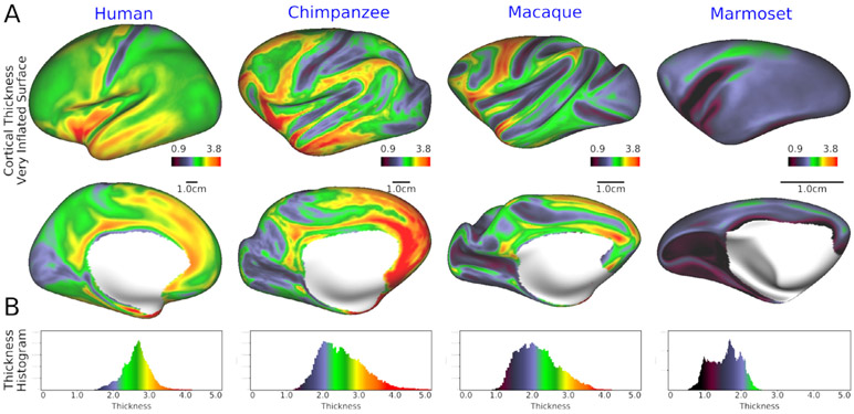
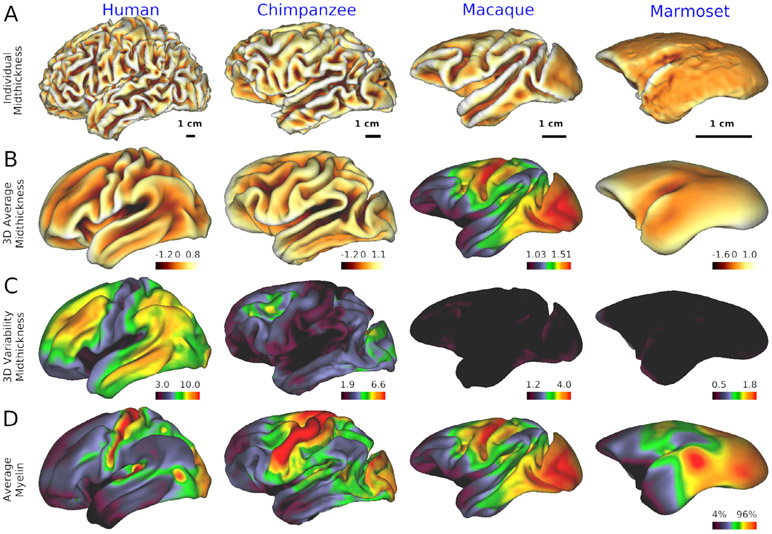
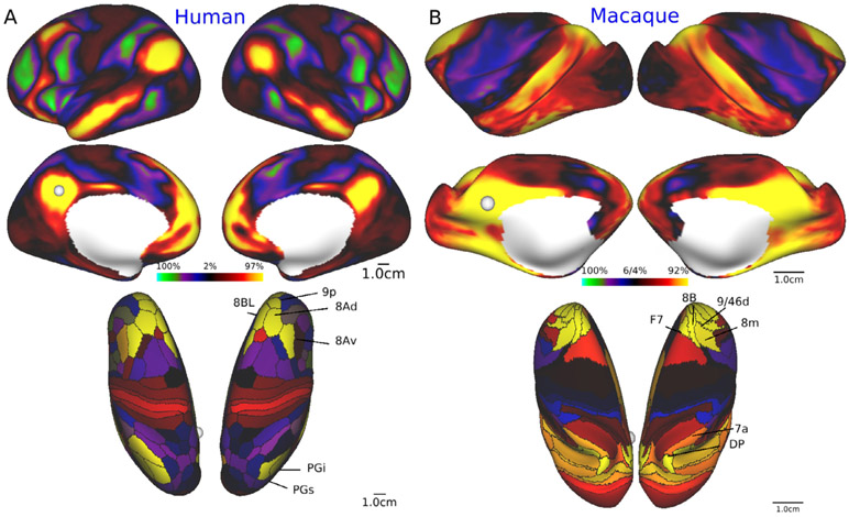
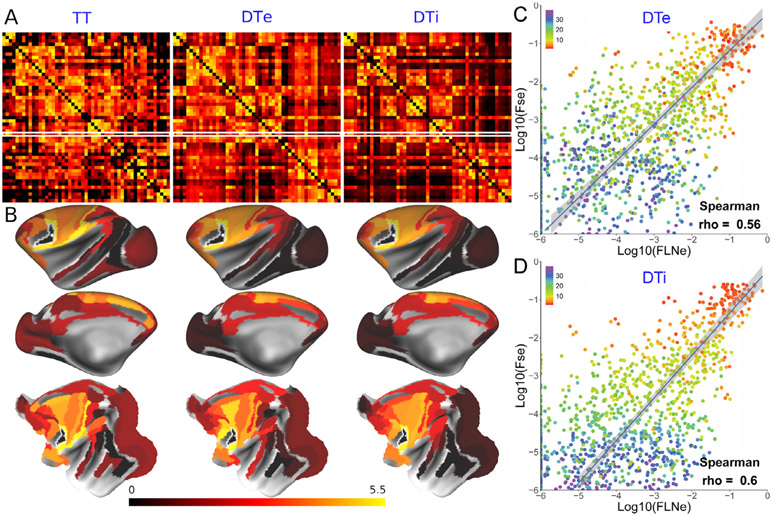
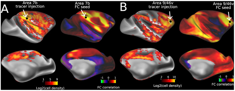
Multi-modal neuroimaging projects such as the Human Connectome Project (HCP) and UK Biobank are advancing our understanding of human brain architecture, function, connectivity, and their variability across individuals using high-quality non-invasive data from many subjects. Such efforts depend upon the accuracy of non-invasive brain imaging measures. However, 'ground truth' validation of connectivity using invasive tracers is not feasible in humans. Studies using nonhuman primates (NHPs) enable comparisons between invasive and non-invasive measures, including exploration of how "functional connectivity" from fMRI and "tractographic connectivity" from diffusion MRI compare with long-distance connections measured using tract tracing. Our NonHuman Primate Neuroimaging & Neuroanatomy Project (NHP_NNP) is an international effort (6 laboratories in 5 countries) to: (i) acquire and analyze high-quality multi-modal brain imaging data of macaque and marmoset monkeys using protocols and methods adapted from the HCP; (ii) acquire quantitative invasive tract-tracing data for cortical and subcortical projections to cortical areas; and (iii) map the distributions of different brain cell types with immunocytochemical stains to better define brain areal boundaries. We are acquiring high-resolution structural, functional, and diffusion MRI data together with behavioral measures from over 100 individual macaques and marmosets in order to generate non-invasive measures of brain architecture such as myelin and cortical thickness maps, as well as functional and diffusion tractography-based connectomes. We are using classical and next-generation anatomical tracers to generate quantitative connectivity maps based on brain-wide counting of labeled cortical and subcortical neurons, providing ground truth measures of connectivity. Advanced statistical modeling techniques address the consistency of both kinds of data across individuals, allowing comparison of tracer-based and non-invasive MRI-based connectivity measures. We aim to develop improved cortical and subcortical areal atlases by combining histological and imaging methods. Finally, we are collecting genetic and sociality-associated behavioral data in all animals in an effort to understand how genetic variation shapes the connectome and behavior.
多模态神经影像学项目,如人类连接组计划(HCP)和英国生物银行,正在使用来自许多个体的高质量非侵入性数据,推进我们对人类大脑结构、功能、连接及其个体变异性的理解。这些努力依赖于非侵入性脑成像测量的准确性。然而,使用侵入性示踪剂对连接进行“真实”验证在人类中是不可行的。使用非人类灵长类动物(NHP)的研究可以在侵入性和非侵入性测量之间进行比较,包括探索功能磁共振成像的“功能连接”和弥散磁共振成像的“轨迹连接”与使用示踪技术测量的远距离连接之间的关系。我们的非人类灵长类动物神经影像学和神经解剖学项目(NHP_NNP)是一项国际合作(来自 5 个国家的 6 个实验室),旨在:(i)采用来自 HCP 的协议和方法,获取和分析猕猴和狨猴的高质量多模态脑成像数据;(ii)获取皮质和皮质下投射到皮质区域的定量侵入性示踪数据;(iii)用免疫细胞化学染色标记不同的脑细胞类型,更好地定义大脑区域边界。我们正在从 100 多只猕猴和狨猴中获取高分辨率结构、功能和弥散 MRI 数据以及行为测量数据,以便生成脑结构的非侵入性测量,如髓鞘和皮质厚度图,以及基于功能和弥散轨迹的连接组。我们正在使用经典和下一代解剖示踪剂,根据大脑中标记的皮质和皮质下神经元的数量生成定量连接图,为连接提供真实的测量值。先进的统计建模技术解决了个体间两种数据的一致性问题,允许比较示踪剂和非侵入性 MRI 连接测量值。我们旨在通过结合组织学和成像方法来开发改进的皮质和皮质下区域图谱。最后,我们正在对所有动物收集遗传和社会性相关的行为数据,以了解遗传变异如何塑造连接组和行为。