• 文献检索
  • 文档翻译
  • 深度研究
  • 学术资讯
  • Suppr Zotero 插件Zotero 插件
  • 邀请有礼
  • 套餐&价格
  • 历史记录
应用&插件
Suppr Zotero 插件Zotero 插件浏览器插件Mac 客户端Windows 客户端微信小程序
定价
高级版会员购买积分包购买API积分包
服务
文献检索文档翻译深度研究API 文档MCP 服务
关于我们
关于 Suppr公司介绍联系我们用户协议隐私条款
关注我们

Suppr 超能文献

核心技术专利:CN118964589B侵权必究
粤ICP备2023148730 号-1Suppr @ 2026

文献检索

告别复杂PubMed语法,用中文像聊天一样搜索,搜遍4000万医学文献。AI智能推荐,让科研检索更轻松。

立即免费搜索

文件翻译

保留排版,准确专业,支持PDF/Word/PPT等文件格式,支持 12+语言互译。

免费翻译文档

深度研究

AI帮你快速写综述,25分钟生成高质量综述,智能提取关键信息,辅助科研写作。

立即免费体验

单核苷酸β-抑制蛋白2变体A248T类似于活化抑制蛋白的动力学特性。

The single nucleotide β -arrestin2 variant, A248T, resembles dynamical properties of activated arrestin.

作者信息

Şensoy Özge

机构信息

Department of Computer Engineering, The School of Engineering and Natural Sciences, İstanbul Medipol University, İstanbul Turkey.

出版信息

Turk J Chem. 2020 Apr 1;44(2):409-420. doi: 10.3906/kim-1910-46. eCollection 2020.

DOI:10.3906/kim-1910-46
PMID:33488166
原文链接:https://pmc.ncbi.nlm.nih.gov/articles/PMC7671214/
Abstract

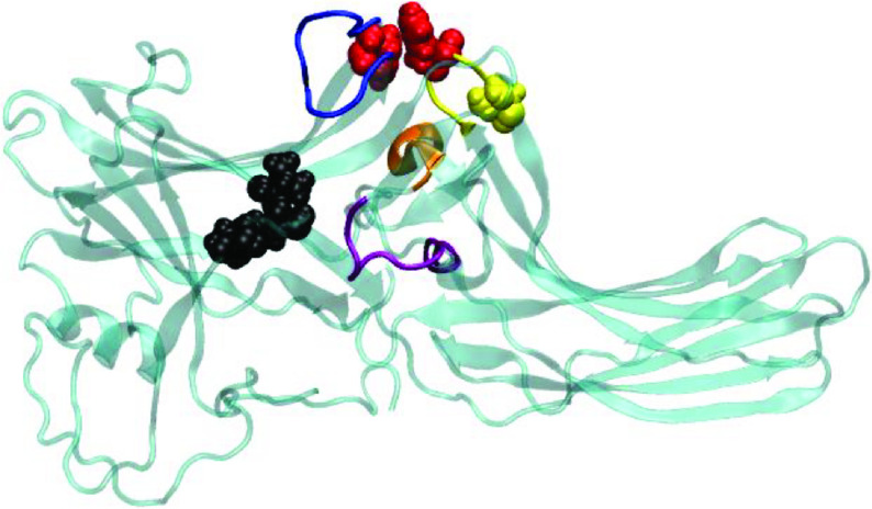
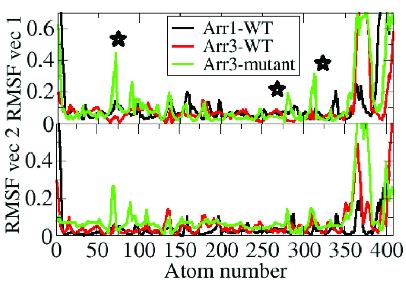
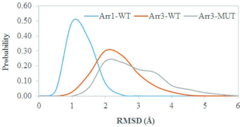
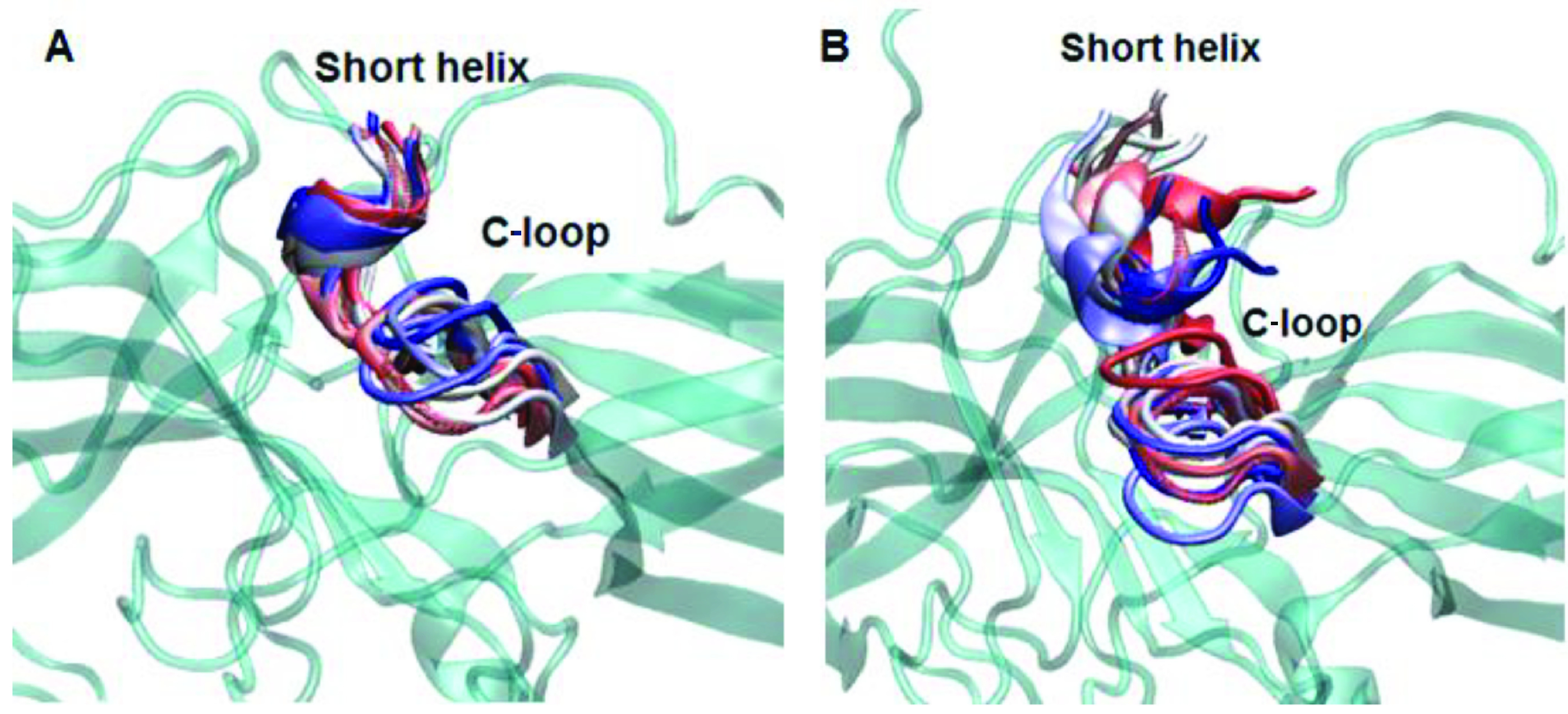
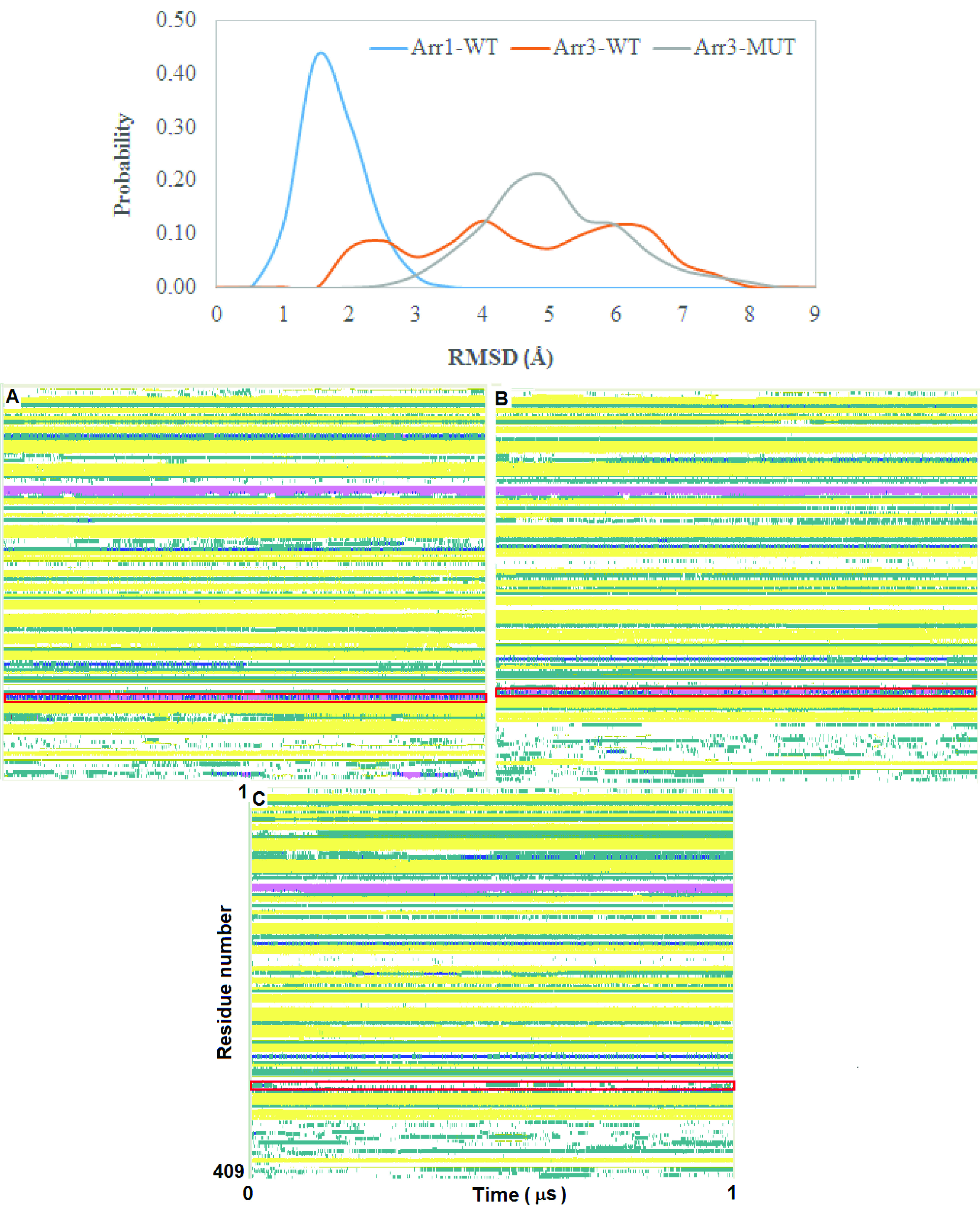
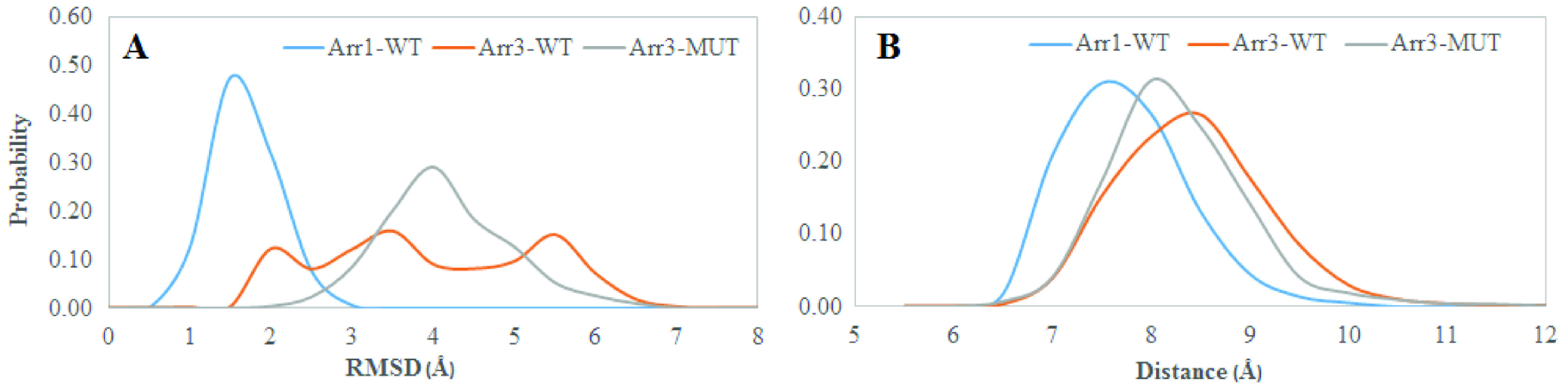
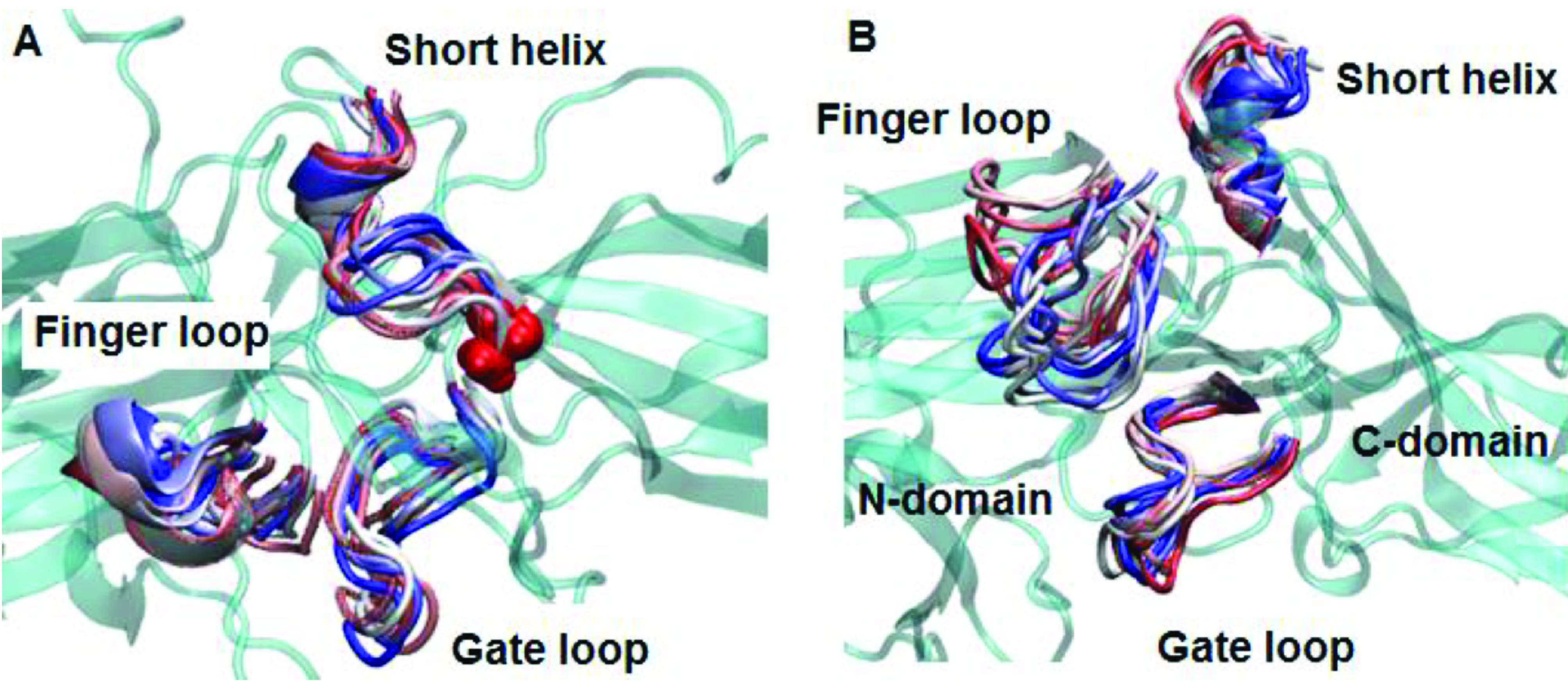
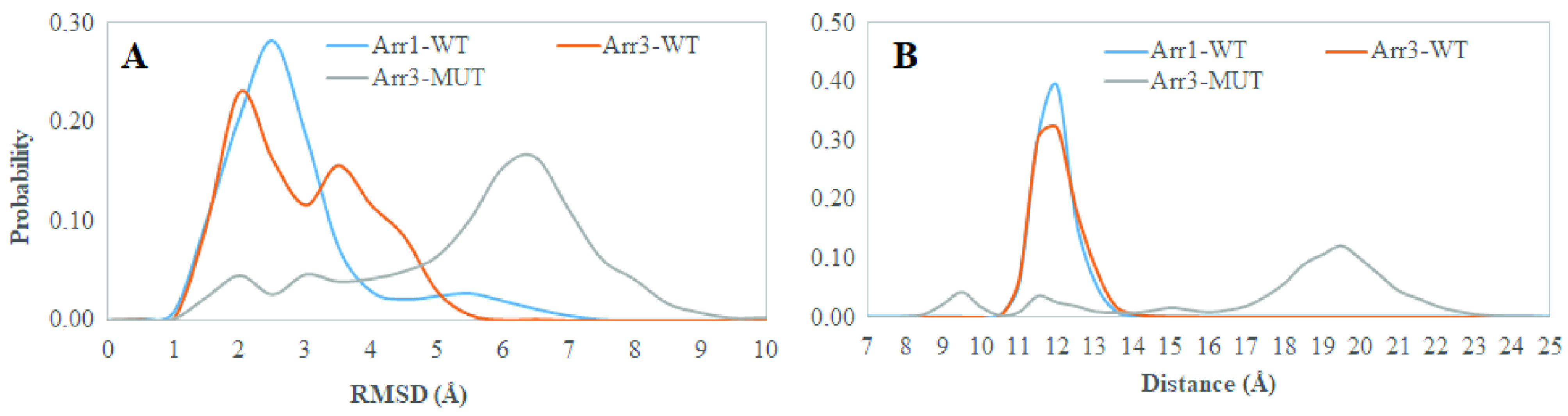
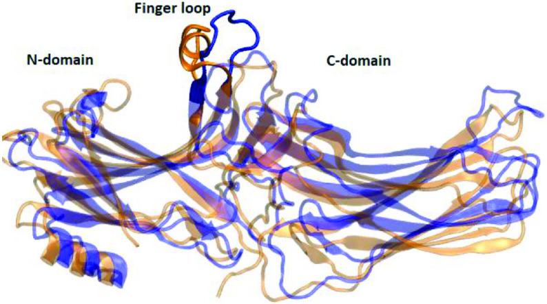
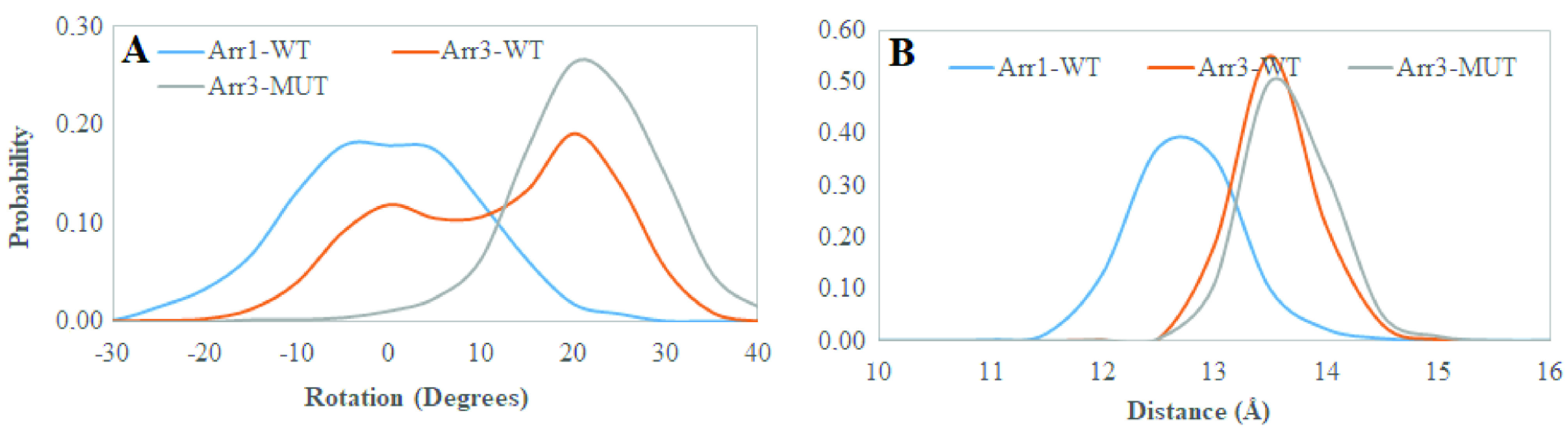
β -arrestins are responsible for termination of G protein-coupled receptor (GPCR)-mediated signaling. Association of single nucleotide variants with onset of crucial diseases has made this protein family hot targets in the field of GPCR-mediated pharmacology. However, impact of these mutations on function of these variants has remained elusive. In this study, structural and dynamical properties of one of β -arrestin2 (arrestin 3) variants, A248T, which has been identified in some cancer tissue samples, were investigated via molecular dynamics simulations. The results showed that the variant underwent structural rearrangements which are seen in crystal structures of active arrestin. Specifically, the "short helix" unravels and the "gate loop" swings forward as seen in crystal structures of receptor-bound and GPCR phosphopeptide-bound arrestin. Moreover, the "finger loop" samples upward position in the variant. Importantly, these regions harbor crucial residues that are involved in receptor binding interfaces. Cumulatively, these local structural rearrangements help the variant adopt active-like domain angle without perturbing the "polar core". Considering that phosphorylation of the receptor is required for activation of arrestin, A248T might serve as a model system to understand phosphorylation-independent activation mechanism, thus enabling modulation of function of arrestin variants which are activated independent of receptor phosphorylation as seen in cancer.

摘要

β -抑制蛋白负责终止G蛋白偶联受体(GPCR)介导的信号传导。单核苷酸变异与关键疾病发病的关联使这个蛋白家族成为GPCR介导药理学领域的热门靶点。然而,这些突变对这些变异体功能的影响仍不清楚。在本研究中,通过分子动力学模拟研究了在一些癌症组织样本中鉴定出的β -抑制蛋白2(抑制蛋白3)的一个变异体A248T的结构和动力学特性。结果表明,该变异体发生了在活性抑制蛋白晶体结构中可见的结构重排。具体而言,如在与受体结合和与GPCR磷酸肽结合的抑制蛋白晶体结构中所见,“短螺旋”解开,“门环”向前摆动。此外,“指环”在变异体中处于向上的位置。重要的是,这些区域含有参与受体结合界面的关键残基。总的来说,这些局部结构重排有助于变异体采用类似活性的结构域角度,而不会干扰“极性核心”。考虑到抑制蛋白的激活需要受体的磷酸化,A248T可能作为一个模型系统来理解磷酸化非依赖性激活机制,从而能够调节如在癌症中所见的独立于受体磷酸化而被激活的抑制蛋白变异体的功能。

https://cdn.ncbi.nlm.nih.gov/pmc/blobs/b7a4/7671214/2dd6f242dbd7/turkjchem-44-409-fig011.jpg
https://cdn.ncbi.nlm.nih.gov/pmc/blobs/b7a4/7671214/0bdf10c98ae4/turkjchem-44-409-fig001.jpg
https://cdn.ncbi.nlm.nih.gov/pmc/blobs/b7a4/7671214/73360132d994/turkjchem-44-409-fig002.jpg
https://cdn.ncbi.nlm.nih.gov/pmc/blobs/b7a4/7671214/055b4c3f3fdb/turkjchem-44-409-fig003.jpg
https://cdn.ncbi.nlm.nih.gov/pmc/blobs/b7a4/7671214/b4b223ca19f6/turkjchem-44-409-fig004.jpg
https://cdn.ncbi.nlm.nih.gov/pmc/blobs/b7a4/7671214/d38b3d61adb3/turkjchem-44-409-fig005.jpg
https://cdn.ncbi.nlm.nih.gov/pmc/blobs/b7a4/7671214/081f4d006a2f/turkjchem-44-409-fig006.jpg
https://cdn.ncbi.nlm.nih.gov/pmc/blobs/b7a4/7671214/5de81459e397/turkjchem-44-409-fig007.jpg
https://cdn.ncbi.nlm.nih.gov/pmc/blobs/b7a4/7671214/b7c2e0862840/turkjchem-44-409-fig008.jpg
https://cdn.ncbi.nlm.nih.gov/pmc/blobs/b7a4/7671214/c07bbd43c488/turkjchem-44-409-fig009.jpg
https://cdn.ncbi.nlm.nih.gov/pmc/blobs/b7a4/7671214/8946d178b1a0/turkjchem-44-409-fig010.jpg
https://cdn.ncbi.nlm.nih.gov/pmc/blobs/b7a4/7671214/2dd6f242dbd7/turkjchem-44-409-fig011.jpg

相似文献

1
The single nucleotide β -arrestin2 variant, A248T, resembles dynamical properties of activated arrestin.单核苷酸β-抑制蛋白2变体A248T类似于活化抑制蛋白的动力学特性。
Turk J Chem. 2020 Apr 1;44(2):409-420. doi: 10.3906/kim-1910-46. eCollection 2020.
2
Understanding the Differential Selectivity of Arrestins toward the Phosphorylation State of the Receptor.了解 arrestins 对受体磷酸化状态的差异选择性。
ACS Chem Neurosci. 2016 Sep 21;7(9):1212-24. doi: 10.1021/acschemneuro.6b00073. Epub 2016 Jul 22.
3
Beta-arrestin1 and beta-arrestin2 are differentially required for phosphorylation-dependent and -independent internalization of delta-opioid receptors.β-抑制蛋白1和β-抑制蛋白2对于δ-阿片受体磷酸化依赖性和非依赖性内化的需求不同。
J Neurochem. 2005 Oct;95(1):169-78. doi: 10.1111/j.1471-4159.2005.03352.x.
4
Termination of protease-activated receptor-1 signaling by beta-arrestins is independent of receptor phosphorylation.β-抑制蛋白介导的蛋白酶激活受体-1信号转导的终止不依赖于受体磷酸化。
J Biol Chem. 2004 Mar 12;279(11):10020-31. doi: 10.1074/jbc.M310590200. Epub 2003 Dec 29.
5
Mapping the arrestin-receptor interface. Structural elements responsible for receptor specificity of arrestin proteins.绘制抑制蛋白-受体界面图谱。负责抑制蛋白受体特异性的结构元件。
J Biol Chem. 2004 Jan 9;279(2):1262-8. doi: 10.1074/jbc.M308834200. Epub 2003 Oct 6.
6
A beta-arrestin binding determinant common to the second intracellular loops of rhodopsin family G protein-coupled receptors.视紫红质家族G蛋白偶联受体第二细胞内环共有的β-抑制蛋白结合决定簇。
J Biol Chem. 2006 Feb 3;281(5):2932-8. doi: 10.1074/jbc.M508074200. Epub 2005 Nov 30.
7
A Novel Polar Core and Weakly Fixed C-Tail in Squid Arrestin Provide New Insight into Interaction with Rhodopsin.鱿鱼 arrestin 中的新型极性核心和弱固定 C 端为其与视紫红质相互作用提供新的见解。
J Mol Biol. 2018 Oct 19;430(21):4102-4118. doi: 10.1016/j.jmb.2018.08.009. Epub 2018 Aug 16.
8
Heterologous phosphorylation-induced formation of a stability lock permits regulation of inactive receptors by β-arrestins.异源磷酸化诱导形成稳定锁,允许β-arrestins 调节无活性受体。
J Biol Chem. 2018 Jan 19;293(3):876-892. doi: 10.1074/jbc.M117.813139. Epub 2017 Nov 16.
9
beta-Arrestin scaffolding of the ERK cascade enhances cytosolic ERK activity but inhibits ERK-mediated transcription following angiotensin AT1a receptor stimulation.β-抑制蛋白对细胞外信号调节激酶(ERK)级联反应的支架作用增强了胞质ERK活性,但在血管紧张素AT1a受体刺激后抑制了ERK介导的转录。
J Biol Chem. 2002 Mar 15;277(11):9429-36. doi: 10.1074/jbc.M106457200. Epub 2002 Jan 2.
10
Phosphorylation of β-arrestin2 at Thr by MEK underlies β-arrestin-dependent activation of Erk1/2 by GPCRs.MEK介导的β-抑制蛋白2苏氨酸磷酸化是GPCRs通过β-抑制蛋白激活Erk1/2的基础。
Elife. 2017 Feb 7;6:e23777. doi: 10.7554/eLife.23777.

引用本文的文献

1
Structural rearrangement of Neisseria meningitidis transferrin binding protein A (TbpA) prior to human transferrin protein (hTf) binding.脑膜炎奈瑟菌转铁蛋白结合蛋白A(TbpA)在与人转铁蛋白(hTf)结合之前的结构重排。
Turk J Chem. 2021 Aug 27;45(4):1146-1154. doi: 10.3906/kim-2102-25. eCollection 2021.
2
Current Knowledge on the Background, Pathophysiology and Treatment of Levodopa-Induced Dyskinesia-Literature Review.左旋多巴诱发异动症的背景、病理生理学及治疗的当前知识——文献综述
J Clin Med. 2021 Sep 25;10(19):4377. doi: 10.3390/jcm10194377.

本文引用的文献

1
Critical role of the finger loop in arrestin binding to the receptors.指环在抑制蛋白与受体结合中的关键作用。
PLoS One. 2019 Mar 15;14(3):e0213792. doi: 10.1371/journal.pone.0213792. eCollection 2019.
2
Molecular mechanism of GPCR-mediated arrestin activation.GPCR 介导热激蛋白激活的分子机制。
Nature. 2018 May;557(7705):452-456. doi: 10.1038/s41586-018-0077-3. Epub 2018 May 2.
3
Identification of Phosphorylation Codes for Arrestin Recruitment by G Protein-Coupled Receptors.鉴定G蛋白偶联受体招募抑制蛋白的磷酸化编码
Cell. 2017 Jul 27;170(3):457-469.e13. doi: 10.1016/j.cell.2017.07.002.
4
Understanding the Differential Selectivity of Arrestins toward the Phosphorylation State of the Receptor.了解 arrestins 对受体磷酸化状态的差异选择性。
ACS Chem Neurosci. 2016 Sep 21;7(9):1212-24. doi: 10.1021/acschemneuro.6b00073. Epub 2016 Jul 22.
5
X-ray laser diffraction for structure determination of the rhodopsin-arrestin complex.X 射线激光衍射技术在视紫红质-抑制蛋白复合物结构测定中的应用。
Sci Data. 2016 Apr 12;3:160021. doi: 10.1038/sdata.2016.21.
6
GROMACS 4:  Algorithms for Highly Efficient, Load-Balanced, and Scalable Molecular Simulation.GROMACS 4:高效、负载均衡和可扩展的分子模拟算法。
J Chem Theory Comput. 2008 Mar;4(3):435-47. doi: 10.1021/ct700301q.
7
PROPKA3: Consistent Treatment of Internal and Surface Residues in Empirical pKa Predictions.PROPKA3:经验 pKa 预测中内部残基和表面残基的一致处理。
J Chem Theory Comput. 2011 Feb 8;7(2):525-37. doi: 10.1021/ct100578z. Epub 2011 Jan 6.
8
Structural evidence for the role of polar core residue Arg175 in arrestin activation.极性核心残基精氨酸175在抑制蛋白激活中作用的结构证据。
Sci Rep. 2015 Oct 29;5:15808. doi: 10.1038/srep15808.
9
Visualization of arrestin recruitment by a G-protein-coupled receptor.G 蛋白偶联受体介导的 arrestin 募集的可视化
Nature. 2014 Aug 14;512(7513):218-222. doi: 10.1038/nature13430. Epub 2014 Jun 22.
10
CHARMM36 all-atom additive protein force field: validation based on comparison to NMR data.CHARMM36 全原子加和蛋白力场:基于 NMR 数据比较的验证。
J Comput Chem. 2013 Sep 30;34(25):2135-45. doi: 10.1002/jcc.23354. Epub 2013 Jul 6.