Chair of Biomedical Physics, Department of Physics and Munich School of BioEngineering, Technical University of Munich, 85748, Garching, Germany.
Philips Research, 22335, Hamburg, Germany.
Eur Radiol Exp. 2021 Jan 26;5(1):6. doi: 10.1186/s41747-020-00201-1.
Grating-based x-ray dark-field and phase-contrast imaging allow extracting information about refraction and small-angle scatter, beyond conventional attenuation. A step towards clinical translation has recently been achieved, allowing further investigation on humans.
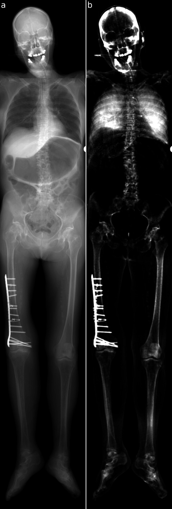
After the ethics committee approval, we scanned the full body of a human cadaver in anterior-posterior orientation. Six measurements were stitched together to form the whole-body image. All radiographs were taken at a three-grating large-object x-ray dark-field scanner, each lasting about 40 s. Signal intensities of different anatomical regions were assessed. The magnitude of visibility reduction caused by beam hardening instead of small-angle scatter was analysed using different phantom materials. Maximal effective dose was 0.3 mSv for the abdomen.
Combined attenuation and dark-field radiography are technically possible throughout a whole human body. High signal levels were found in several bony structures, foreign materials, and the lung. Signal levels were 0.25 ± 0.13 (mean ± standard deviation) for the lungs, 0.08 ± 0.06 for the bones, 0.023 ± 0.019 for soft tissue, and 0.30 ± 0.02 for an antibiotic bead chain. We found that phantom materials, which do not produce small-angle scatter, can generate a strong visibility reduction signal.
We acquired a whole-body x-ray dark-field radiograph of a human body in few minutes with an effective dose in a clinical acceptable range. Our findings suggest that the observed visibility reduction in the bone and metal is dominated by beam hardening and that the true dark-field signal in the lung is therefore much higher than that of the bone.
基于光栅的 X 射线暗场和相位对比成像是在传统衰减之外提取折射和小角度散射信息的方法。最近已经朝着临床转化迈出了一步,允许对人类进行进一步的研究。
在伦理委员会批准后,我们对一具人体尸体进行了前-后方向的全身扫描。将六次测量结果拼接在一起形成全身图像。所有射线照片均在一个三光栅大物体 X 射线暗场扫描仪上拍摄,每次拍摄持续约 40 秒。评估了不同解剖区域的信号强度。使用不同的模拟材料分析了由于束硬化而不是小角度散射引起的可见度降低的程度。腹部的最大有效剂量为 0.3 mSv。
整个人体的组合衰减和暗场射线照相技术是可行的。在几个骨结构、异物和肺部中发现了高信号水平。肺部的信号水平为 0.25±0.13(平均值±标准差),骨骼为 0.08±0.06,软组织为 0.023±0.019,抗生素珠链为 0.30±0.02。我们发现,不会产生小角度散射的模拟材料可以产生强烈的可见度降低信号。
我们在几分钟内获得了人体全身 X 射线暗场射线照片,有效剂量在临床可接受范围内。我们的研究结果表明,在骨和金属中观察到的可见度降低主要是由于束硬化引起的,因此肺部的真实暗场信号比骨骼高得多。