Zhang Liqin, Wu Calvin, Martel David T, West Michael, Sutton Michael A, Shore Susan E
Kresge Hearing Research Institute, Department of Otolaryngology, University of Michigan, Ann Arbor, Michigan, USA.
Molecular & Behavioral Neuroscience Institute, University of Michigan, Ann Arbor, Michigan, USA.
Neural Plast. 2021 Jan 14;2021:8833087. doi: 10.1155/2021/8833087. eCollection 2021.
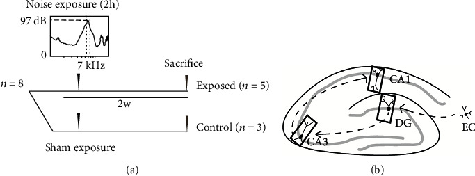
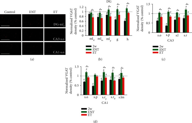
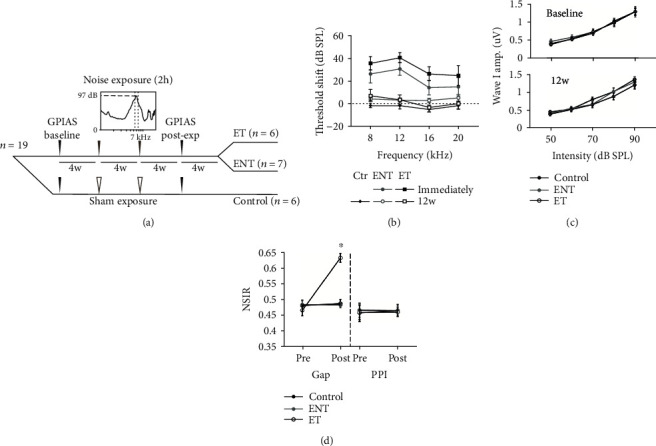
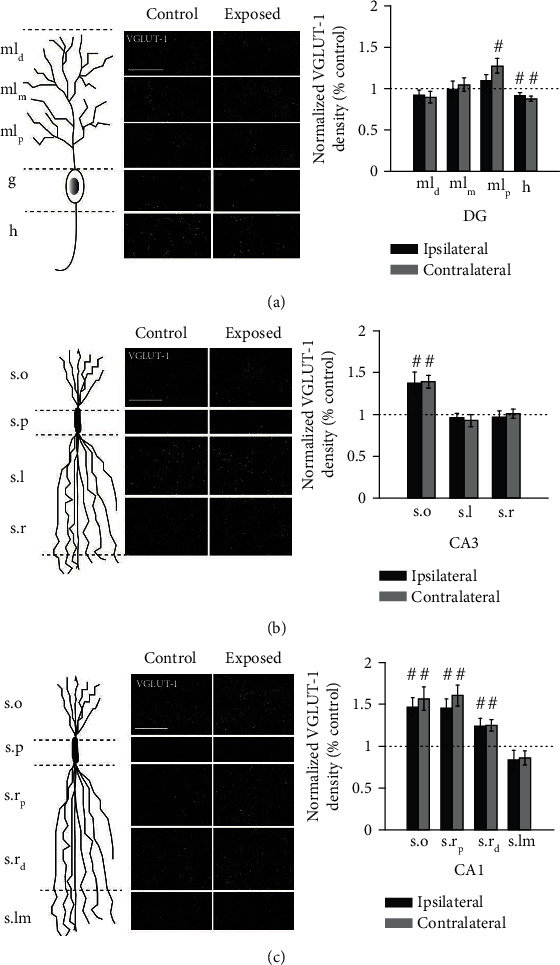
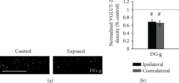
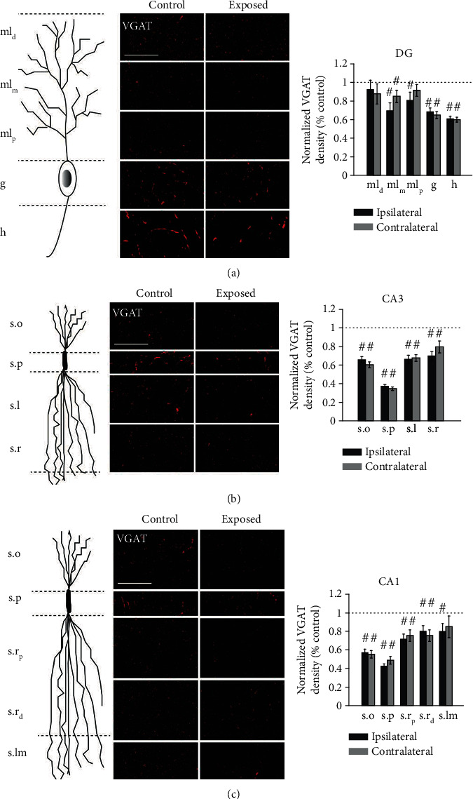
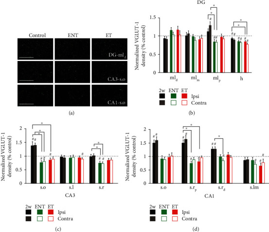
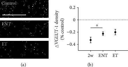
Accumulating evidence implicates a role for brain structures outside the ascending auditory pathway in tinnitus, the phantom perception of sound. In addition to other factors such as age-dependent hearing loss, high-level sound exposure is a prominent cause of tinnitus. Here, we examined how noise exposure altered the distribution of excitatory and inhibitory synaptic inputs in the guinea pig hippocampus and determined whether these changes were associated with tinnitus. In experiment one, guinea pigs were overexposed to unilateral narrow-band noise (98 dB SPL, 2 h). Two weeks later, the density of excitatory (VGLUT-1/2) and inhibitory (VGAT) synaptic terminals in CA1, CA3, and dentate gyrus hippocampal subregions was assessed by immunohistochemistry. Overall, VGLUT-1 density primarily increased, while VGAT density decreased significantly in many regions. Then, to assess whether the noise-induced alterations were persistent and related to tinnitus, experiment two utilized a noise-exposure paradigm shown to induce tinnitus and assessed tinnitus development which was assessed using gap-prepulse inhibition of the acoustic startle (GPIAS). Twelve weeks after sound overexposure, changes in excitatory synaptic terminal density had largely recovered regardless of tinnitus status, but the recovery of GABAergic terminal density was dramatically different in animals expressing tinnitus relative to animals resistant to tinnitus. In resistant animals, inhibitory synapse density recovered to preexposure levels, but in animals expressing tinnitus, inhibitory synapse density remained chronically diminished. Taken together, our results suggest that noise exposure induces striking changes in the balance of excitatory and inhibitory synaptic inputs throughout the hippocampus and reveal a potential role for rebounding inhibition in the hippocampus as a protective factor leading to tinnitus resilience.
越来越多的证据表明,在耳鸣(即声音的幻听)中,上升听觉通路之外的脑结构发挥了作用。除了诸如年龄相关性听力损失等其他因素外,高强度声音暴露是耳鸣的一个主要原因。在这里,我们研究了噪声暴露如何改变豚鼠海马体中兴奋性和抑制性突触输入的分布,并确定这些变化是否与耳鸣有关。在实验一中,豚鼠被单侧过度暴露于窄带噪声(98分贝声压级,2小时)。两周后,通过免疫组织化学评估海马体CA1、CA3和齿状回亚区中兴奋性(VGLUT - 1/2)和抑制性(VGAT)突触终末的密度。总体而言,VGLUT - 1密度主要增加,而在许多区域VGAT密度显著降低。然后,为了评估噪声诱导的改变是否持续以及与耳鸣相关,实验二采用了一种已被证明可诱导耳鸣的噪声暴露范式,并评估耳鸣的发展情况,这是通过对听觉惊吓的间隙前脉冲抑制(GPIAS)来评估的。声音过度暴露十二周后,无论耳鸣状态如何,兴奋性突触终末密度的变化在很大程度上已经恢复,但相对于抗耳鸣的动物,表达耳鸣的动物中GABA能终末密度的恢复情况有显著差异。在抗耳鸣的动物中,抑制性突触密度恢复到暴露前水平,但在表达耳鸣的动物中,抑制性突触密度长期保持降低。综上所述,我们的结果表明,噪声暴露会引起整个海马体中兴奋性和抑制性突触输入平衡的显著变化,并揭示出海马体中反弹抑制作为导致耳鸣恢复力的保护因素的潜在作用。