Department of Bioengineering, University of Pennsylvania, Philadelphia, PA, USA.
Nat Commun. 2021 Feb 2;12(1):753. doi: 10.1038/s41467-021-21029-2.
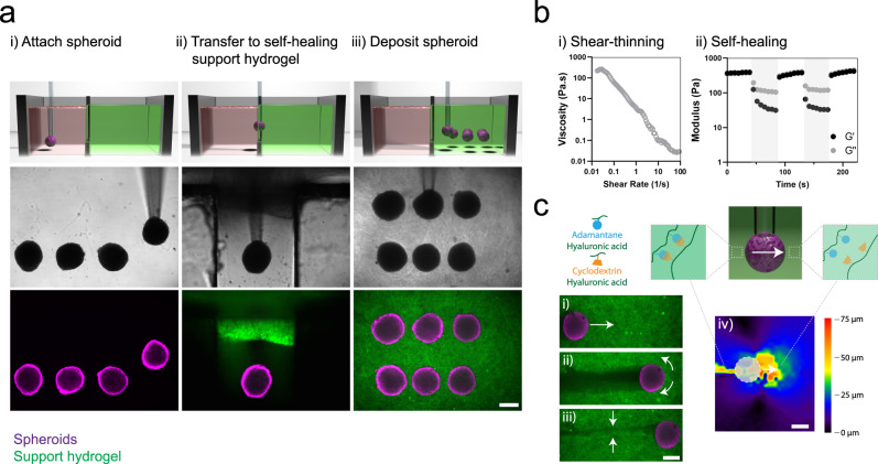
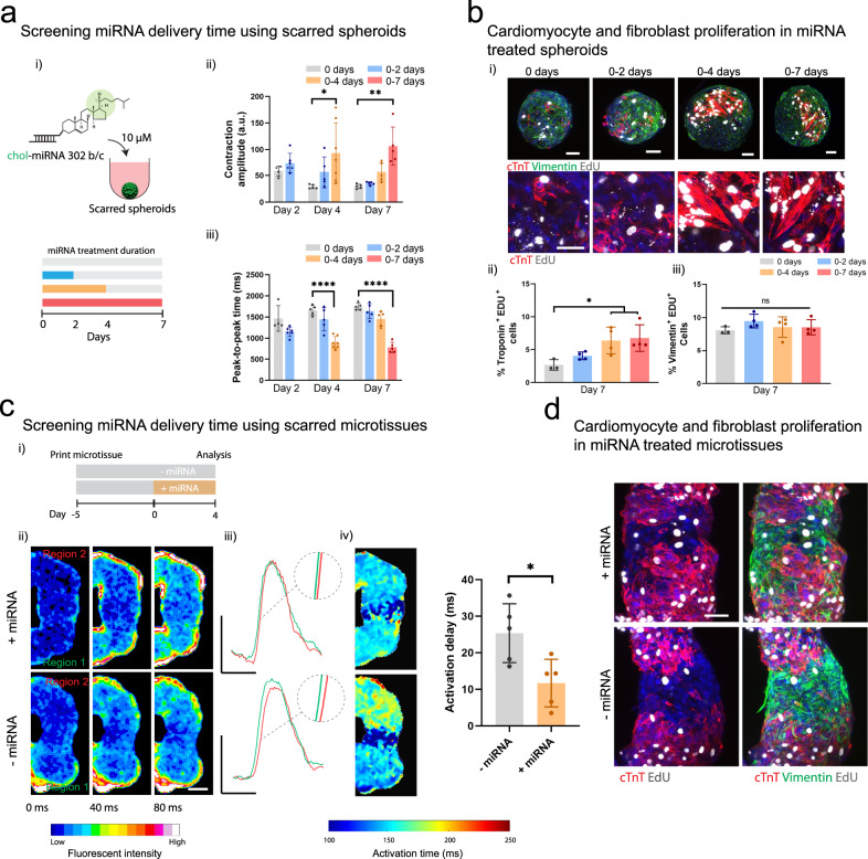
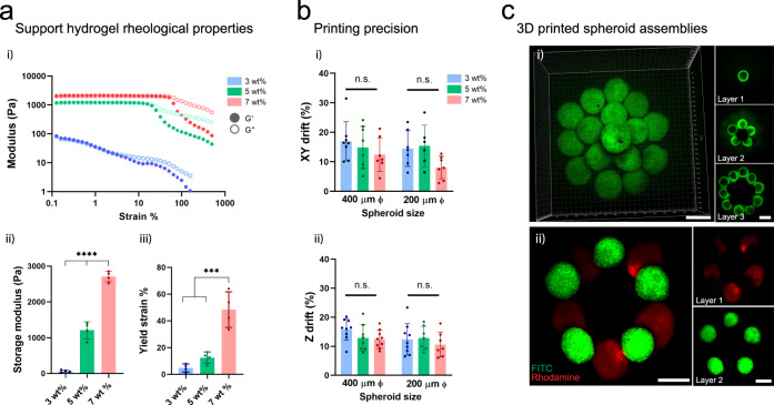
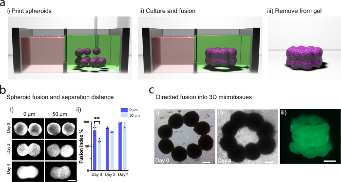
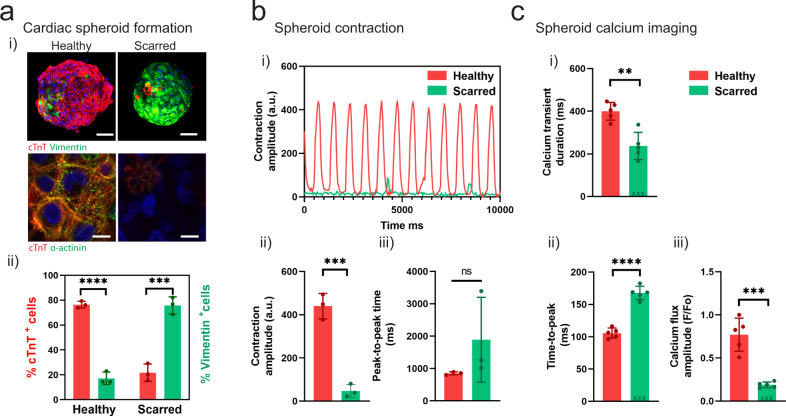
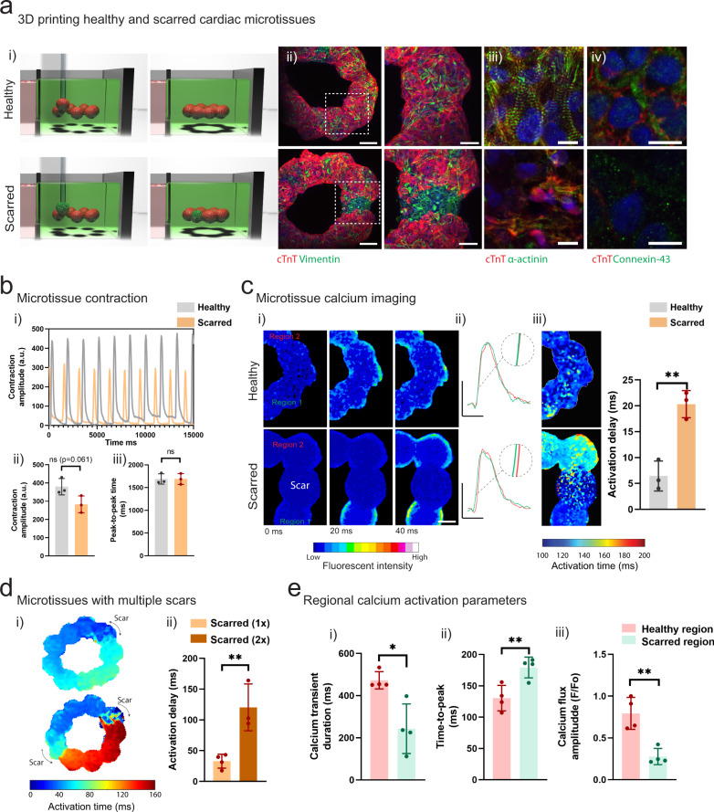
Cellular models are needed to study human development and disease in vitro, and to screen drugs for toxicity and efficacy. Current approaches are limited in the engineering of functional tissue models with requisite cell densities and heterogeneity to appropriately model cell and tissue behaviors. Here, we develop a bioprinting approach to transfer spheroids into self-healing support hydrogels at high resolution, which enables their patterning and fusion into high-cell density microtissues of prescribed spatial organization. As an example application, we bioprint induced pluripotent stem cell-derived cardiac microtissue models with spatially controlled cardiomyocyte and fibroblast cell ratios to replicate the structural and functional features of scarred cardiac tissue that arise following myocardial infarction, including reduced contractility and irregular electrical activity. The bioprinted in vitro model is combined with functional readouts to probe how various pro-regenerative microRNA treatment regimes influence tissue regeneration and recovery of function as a result of cardiomyocyte proliferation. This method is useful for a range of biomedical applications, including the development of precision models to mimic diseases and the screening of drugs, particularly where high cell densities and heterogeneity are important.
细胞模型对于在体外研究人类发育和疾病以及筛选毒性和疗效药物是必需的。目前的方法在构建具有必要细胞密度和异质性的功能性组织模型方面受到限制,无法很好地模拟细胞和组织行为。在这里,我们开发了一种生物打印方法,可将球体以高分辨率转移到自修复支撑水凝胶中,从而可以对其进行图案化和融合,形成具有规定空间组织的高细胞密度微组织。作为一个示例应用,我们生物打印了诱导多能干细胞衍生的心脏微组织模型,其中具有空间控制的心肌细胞和成纤维细胞比例,以复制心肌梗死后出现的瘢痕心脏组织的结构和功能特征,包括收缩力降低和不规则电活动。将生物打印的体外模型与功能读数相结合,以探究各种促再生 microRNA 治疗方案如何影响组织再生和功能恢复,这是由于心肌细胞增殖所致。该方法可用于一系列生物医学应用,包括开发模拟疾病的精确模型和药物筛选,特别是在需要高细胞密度和异质性的情况下。