Department of Computer Science and Engineering, Indian Institute of Technology Madras, Chennai, Tamil Nadu, India.
Department of Brain and Cognitive Sciences, Massachusetts Institute of Technology, Cambridge, Massachusetts, United States of America.
PLoS Comput Biol. 2021 Feb 4;17(2):e1008548. doi: 10.1371/journal.pcbi.1008548. eCollection 2021 Feb.
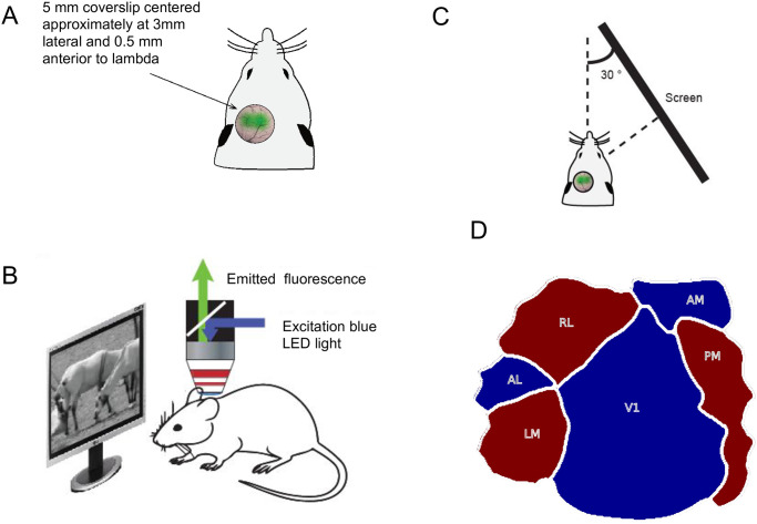
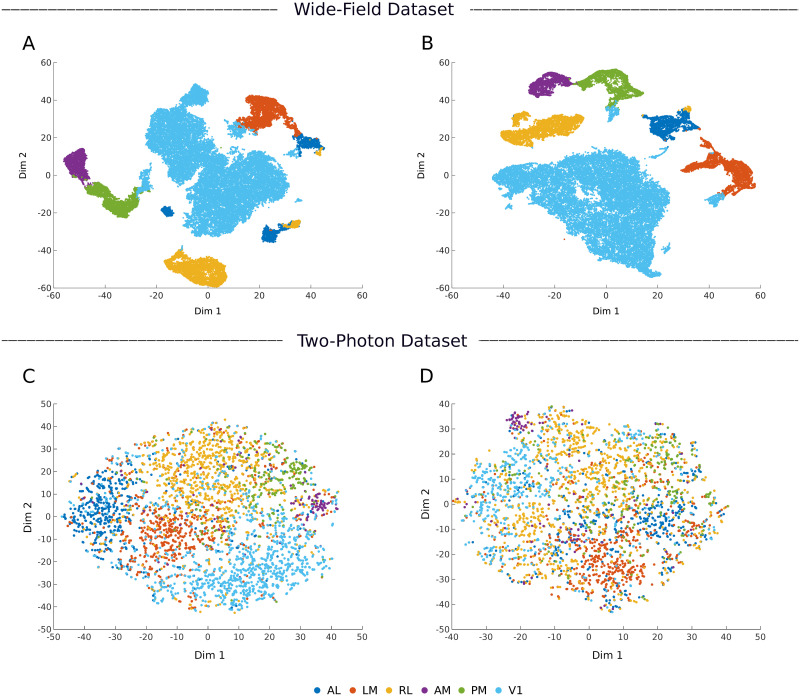
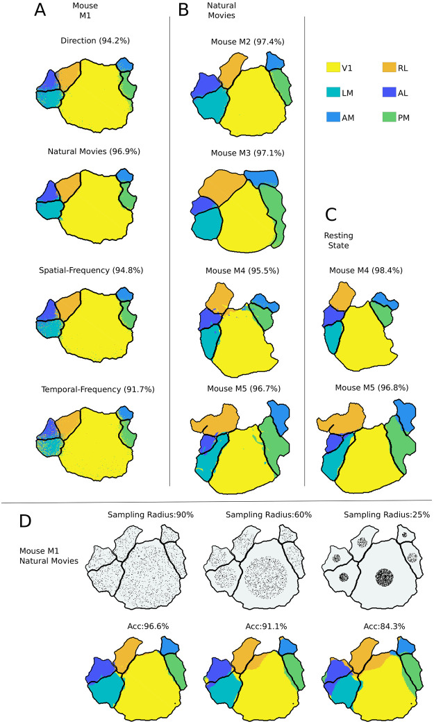
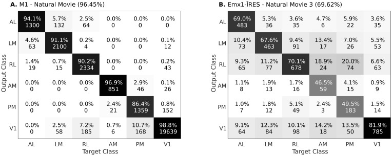
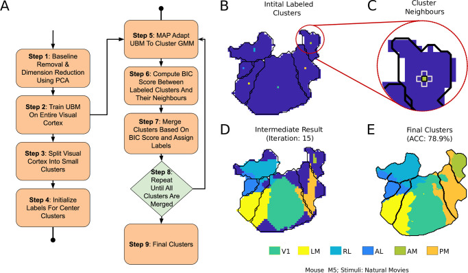
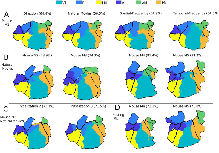
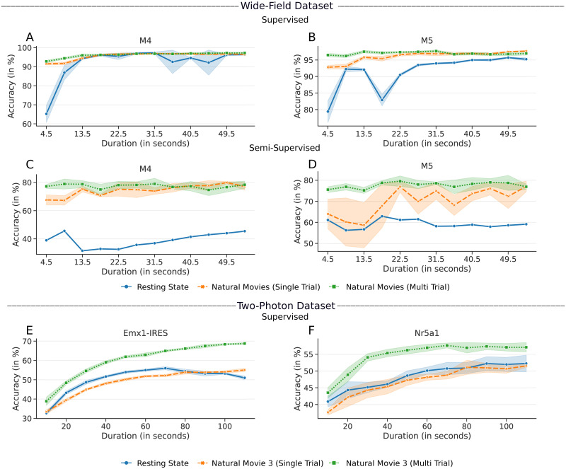
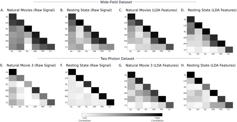
The visual cortex of the mouse brain can be divided into ten or more areas that each contain complete or partial retinotopic maps of the contralateral visual field. It is generally assumed that these areas represent discrete processing regions. In contrast to the conventional input-output characterizations of neuronal responses to standard visual stimuli, here we asked whether six of the core visual areas have responses that are functionally distinct from each other for a given visual stimulus set, by applying machine learning techniques to distinguish the areas based on their activity patterns. Visual areas defined by retinotopic mapping were examined using supervised classifiers applied to responses elicited by a range of stimuli. Using two distinct datasets obtained using wide-field and two-photon imaging, we show that the area labels predicted by the classifiers were highly consistent with the labels obtained using retinotopy. Furthermore, the classifiers were able to model the boundaries of visual areas using resting state cortical responses obtained without any overt stimulus, in both datasets. With the wide-field dataset, clustering neuronal responses using a constrained semi-supervised classifier showed graceful degradation of accuracy. The results suggest that responses from visual cortical areas can be classified effectively using data-driven models. These responses likely reflect unique circuits within each area that give rise to activity with stronger intra-areal than inter-areal correlations, and their responses to controlled visual stimuli across trials drive higher areal classification accuracy than resting state responses.
鼠脑的视皮层可分为十个或更多区域,每个区域都包含对侧视野的完整或部分视网膜映射。通常认为这些区域代表离散的处理区域。与传统的神经元对标准视觉刺激的输入-输出特征描述相反,我们通过应用机器学习技术来根据活动模式区分这些区域,从而询问核心视觉区域中的六个区域是否具有针对给定视觉刺激集的功能上不同的反应。通过使用视网膜映射定义的视觉区域,使用监督分类器来检查对一系列刺激引起的反应。使用广角和双光子成像获得的两个不同数据集,我们表明,分类器预测的区域标签与使用视网膜映射获得的标签高度一致。此外,分类器能够使用在两个数据集中都没有任何明显刺激的静息状态皮质反应来建模视觉区域的边界。使用广角数据集,使用受限的半监督分类器对神经元反应进行聚类显示出准确性的优雅降级。结果表明,可以使用基于数据的模型有效地对视觉皮质区域的反应进行分类。这些反应可能反映了每个区域内的独特回路,这些回路产生的活动具有比区域间更强的内在相关性,并且它们对跨试验的受控视觉刺激的反应比静息状态反应具有更高的区域分类准确性。