• 文献检索
  • 文档翻译
  • 深度研究
  • 学术资讯
  • Suppr Zotero 插件Zotero 插件
  • 邀请有礼
  • 套餐&价格
  • 历史记录
应用&插件
Suppr Zotero 插件Zotero 插件浏览器插件Mac 客户端Windows 客户端微信小程序
定价
高级版会员购买积分包购买API积分包
服务
文献检索文档翻译深度研究API 文档MCP 服务
关于我们
关于 Suppr公司介绍联系我们用户协议隐私条款
关注我们

Suppr 超能文献

核心技术专利:CN118964589B侵权必究
粤ICP备2023148730 号-1Suppr @ 2026

文献检索

告别复杂PubMed语法,用中文像聊天一样搜索,搜遍4000万医学文献。AI智能推荐,让科研检索更轻松。

立即免费搜索

文件翻译

保留排版,准确专业,支持PDF/Word/PPT等文件格式,支持 12+语言互译。

免费翻译文档

深度研究

AI帮你快速写综述,25分钟生成高质量综述,智能提取关键信息,辅助科研写作。

立即免费体验

IE-Map:人类内耳新型活体图谱和模板。

IE-Map: a novel in-vivo atlas and template of the human inner ear.

机构信息

German Center for Vertigo and Balance Disorders (DSGZ), Ludwig-Maximilians-University Munich, Fraunhoferstr. 20, 82152, Planegg, Germany.

Department of Informatics, Technical University of Munich, Boltzmannstr. 3, 85748, Garching, Germany.

出版信息

Sci Rep. 2021 Feb 8;11(1):3293. doi: 10.1038/s41598-021-82716-0.

DOI:10.1038/s41598-021-82716-0
PMID:33558581
原文链接:https://pmc.ncbi.nlm.nih.gov/articles/PMC7870663/
Abstract

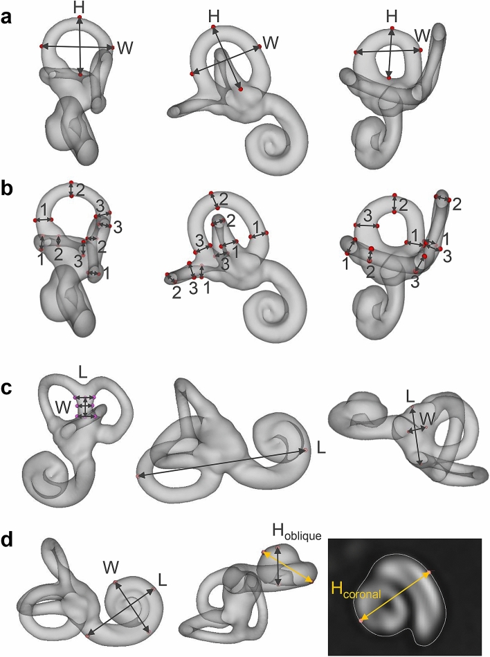
Brain atlases and templates are core tools in scientific research with increasing importance also in clinical applications. Advances in neuroimaging now allowed us to expand the atlas domain to the vestibular and auditory organ, the inner ear. In this study, we present IE-Map, an in-vivo template and atlas of the human labyrinth derived from multi-modal high-resolution magnetic resonance imaging (MRI) data, in a fully non-invasive manner without any contrast agent or radiation. We reconstructed a common template from 126 inner ears (63 normal subjects) and annotated it with 94 established landmarks and semi-automatic segmentations of all relevant macroscopic vestibular and auditory substructures. We validated the atlas by comparing MRI templates to a novel CT/micro-CT atlas, which we reconstructed from 21 publicly available post-mortem images of the bony labyrinth. Templates in MRI and micro-CT have a high overlap, and several key anatomical measures of the bony labyrinth in IE-Map are in line with micro-CT literature of the inner ear. A quantitative substructural analysis based on the new template, revealed a correlation of labyrinth parameters with total intracranial volume. No effects of gender or laterality were found. We provide the validated templates, atlas segmentations, surface meshes and landmark annotations as open-access material, to provide neuroscience researchers and clinicians in neurology, neurosurgery, and otorhinolaryngology with a widely applicable tool for computational neuro-otology.

摘要

脑图谱和模板是科学研究的核心工具,在临床应用中也越来越重要。神经影像学的进步现在使我们能够将图谱领域扩展到前庭和听觉器官,即内耳。在这项研究中,我们提出了 IE-Map,这是一种基于多模态高分辨率磁共振成像 (MRI) 数据的、完全非侵入性的、无需任何造影剂或辐射的人类迷路的模板和图谱。我们从 126 个内耳(63 个正常受试者)重建了一个通用模板,并使用 94 个已建立的标记和所有相关宏观前庭和听觉亚结构的半自动分割对其进行了注释。我们通过将 MRI 模板与我们从 21 个公开可用的骨骼迷路的死后 CT/微 CT 图谱重建进行比较来验证图谱。MRI 和微 CT 中的模板具有很高的重叠性,IE-Map 中骨骼迷路的几个关键解剖学测量值与内耳的微 CT 文献一致。基于新模板的定量亚结构分析显示,迷路参数与颅内总容积相关。未发现性别或侧性的影响。我们提供经过验证的模板、图谱分割、表面网格和标记注释作为开放访问材料,为神经科学研究人员和神经科、神经外科和耳鼻喉科的临床医生提供了一种广泛适用于计算神经耳科学的工具。

https://cdn.ncbi.nlm.nih.gov/pmc/blobs/7e27/7870663/040e64911659/41598_2021_82716_Fig5_HTML.jpg

相似文献

1
IE-Map: a novel in-vivo atlas and template of the human inner ear.IE-Map:人类内耳新型活体图谱和模板。
Sci Rep. 2021 Feb 8;11(1):3293. doi: 10.1038/s41598-021-82716-0.
2
A probabilistic atlas of the human inner ear's bony labyrinth enables reliable atlas-based segmentation of the total fluid space.人体内耳骨迷路概率图谱可实现总液腔基于图谱的可靠分割。
J Neurol. 2019 Sep;266(Suppl 1):52-61. doi: 10.1007/s00415-019-09488-6. Epub 2019 Aug 17.
3
Multi-atlas segmentation of the whole hippocampus and subfields using multiple automatically generated templates.使用多个自动生成的模板对整个海马体及其子区进行多图谱分割。
Neuroimage. 2014 Nov 1;101:494-512. doi: 10.1016/j.neuroimage.2014.04.054. Epub 2014 Apr 29.
4
Visualization of Reissner membrane and the spiral ganglion in human fetal cochlea by micro-computed tomography.通过微计算机断层扫描术对人类胎儿耳蜗中的Reissner膜和螺旋神经节进行可视化。
Am J Otolaryngol. 2009 Mar-Apr;30(2):112-20. doi: 10.1016/j.amjoto.2008.07.012.
5
Modulatory effects of magnetic vestibular stimulation on resting-state networks can be explained by subject-specific orientation of inner-ear anatomy in the MR static magnetic field.磁前庭刺激对静息态网络的调制作用可以用内耳解剖结构在磁共振静磁场中的个体特异性方位来解释。
J Neurol. 2020 Dec;267(Suppl 1):91-103. doi: 10.1007/s00415-020-09957-3. Epub 2020 Jun 11.
6
Automatic measurement of the labyrinth using image registration and a deformable inner ear atlas.使用图像配准和可变形内耳图谱自动测量迷路。
Acad Radiol. 2003 Sep;10(9):988-99. doi: 10.1016/s1076-6332(03)00121-1.
7
Comparing new templates and atlas-based segmentations in the volumetric analysis of brain magnetic resonance images for diagnosing Alzheimer's disease.比较新模板和基于图谱的脑磁共振图像容积分析在阿尔茨海默病诊断中的分割。
Alzheimers Dement. 2012 Sep;8(5):399-406. doi: 10.1016/j.jalz.2011.07.002.
8
Visual perception of the osseous labyrinth rendered from micro-CT scans of the petrous bone.从颞骨岩部的显微CT扫描图像重建出的骨迷路的视觉感知。
Folia Med Cracov. 2017;57(4):5-12.
9
Micro-CT analysis of the anatomical characteristics of the stapedial annular ligament.镫骨环形韧带解剖特征的显微CT分析
Anat Sci Int. 2017 Mar;92(2):262-266. doi: 10.1007/s12565-016-0331-4. Epub 2016 Feb 15.
10
A Bayesian approach to the creation of a study-customized neonatal brain atlas.一种用于创建针对特定研究的新生儿脑图谱的贝叶斯方法。
Neuroimage. 2014 Nov 1;101:256-67. doi: 10.1016/j.neuroimage.2014.07.001. Epub 2014 Jul 12.

引用本文的文献

1
Comparison of Inner Ear Volume Between Humans and Sheep Using MRI.使用磁共振成像(MRI)比较人与羊的内耳体积
J Assoc Res Otolaryngol. 2025 Jul 18. doi: 10.1007/s10162-025-01002-2.
2
Optimizing spatial normalization of multisubject inner ear MRI: comparison of different geometry-preserving co-registration approaches.优化多受试者内耳磁共振成像的空间归一化:不同几何保留配准方法的比较
Sci Rep. 2025 Feb 21;15(1):6414. doi: 10.1038/s41598-025-90842-2.
3
Applications of Artificial Intelligence in Temporal Bone Imaging: Advances and Future Challenges.

本文引用的文献

1
SciPy 1.0: fundamental algorithms for scientific computing in Python.SciPy 1.0:Python 中的科学计算基础算法。
Nat Methods. 2020 Mar;17(3):261-272. doi: 10.1038/s41592-019-0686-2. Epub 2020 Feb 3.
2
Human bony labyrinth dataset: Co-registered CT and micro-CT images, surface models and anatomical landmarks.人类骨迷路数据集:CT与显微CT图像配准、表面模型及解剖标志
Data Brief. 2019 Nov 9;27:104782. doi: 10.1016/j.dib.2019.104782. eCollection 2019 Dec.
3
A probabilistic atlas of the human inner ear's bony labyrinth enables reliable atlas-based segmentation of the total fluid space.
人工智能在颞骨成像中的应用:进展与未来挑战
Cureus. 2023 Sep 2;15(9):e44591. doi: 10.7759/cureus.44591. eCollection 2023 Sep.
4
IE-Vnet: Deep Learning-Based Segmentation of the Inner Ear's Total Fluid Space.IE-Vnet:基于深度学习的内耳全液腔分割
Front Neurol. 2022 May 11;13:663200. doi: 10.3389/fneur.2022.663200. eCollection 2022.
5
Persistent horizontal and vertical, MR-induced nystagmus in resting state Human Connectome Project data.静息态人类连接组计划数据中,MR 诱发的水平和垂直眼震持续存在。
Neuroimage. 2022 Jul 15;255:119170. doi: 10.1016/j.neuroimage.2022.119170. Epub 2022 Apr 1.
6
Intravenous Delayed Gadolinium-Enhanced MR Imaging of the Endolymphatic Space: A Methodological Comparative Study.内淋巴囊间隙的静脉注射延迟钆增强磁共振成像:一项方法学比较研究。
Front Neurol. 2021 Apr 22;12:647296. doi: 10.3389/fneur.2021.647296. eCollection 2021.
人体内耳骨迷路概率图谱可实现总液腔基于图谱的可靠分割。
J Neurol. 2019 Sep;266(Suppl 1):52-61. doi: 10.1007/s00415-019-09488-6. Epub 2019 Aug 17.
4
Human Inner-ear Malformation Types Captured in 3D.以3D形式呈现的人类内耳畸形类型
J Int Adv Otol. 2019 Apr;15(1):77-82. doi: 10.5152/iao.2019.6246.
5
Use of MRI to determine cochlear duct length in patients undergoing cochlear implantation.利用磁共振成像(MRI)确定接受人工耳蜗植入患者的蜗管长度。
Cochlear Implants Int. 2019 Mar;20(2):57-61. doi: 10.1080/14670100.2018.1549186. Epub 2018 Nov 22.
6
Anatomical basis of drug delivery to the inner ear.内耳药物递送的解剖学基础。
Hear Res. 2018 Oct;368:10-27. doi: 10.1016/j.heares.2018.06.017. Epub 2018 Jun 27.
7
Visualizing the 3D cytoarchitecture of the human cochlea in an intact temporal bone using synchrotron radiation phase contrast imaging.利用同步辐射相衬成像技术在完整颞骨中可视化人类耳蜗的三维细胞结构。
Biomed Opt Express. 2018 Jul 18;9(8):3757-3767. doi: 10.1364/BOE.9.003757. eCollection 2018 Aug 1.
8
Visualization of the Membranous Labyrinth and Nerve Fiber Pathways in Human and Animal Inner Ears Using MicroCT Imaging.使用显微计算机断层扫描成像技术可视化人类和动物内耳中的膜迷路和神经纤维通路。
Front Neurosci. 2018 Jul 31;12:501. doi: 10.3389/fnins.2018.00501. eCollection 2018.
9
Optimization of 3D-Visualization of Micro-Anatomical Structures of the Human Inner Ear in Osmium Tetroxide Contrast Enhanced Micro-CT Scans.锇酸增强显微CT扫描中人类内耳微观解剖结构的三维可视化优化
Front Neuroanat. 2018 May 22;12:41. doi: 10.3389/fnana.2018.00041. eCollection 2018.
10
Analysis of Vestibular Labyrinthine Geometry and Variation in the Human Temporal Bone.人类颞骨前庭迷路的几何形状及变异分析
Front Neurosci. 2018 Feb 26;12:107. doi: 10.3389/fnins.2018.00107. eCollection 2018.