Institut de Cancérologie de Lorraine, 6 Avenue de Bourgogne, 54519, Vandœuvre-lès-Nancy, France.
Sci Rep. 2021 Feb 8;11(1):3314. doi: 10.1038/s41598-021-82999-3.
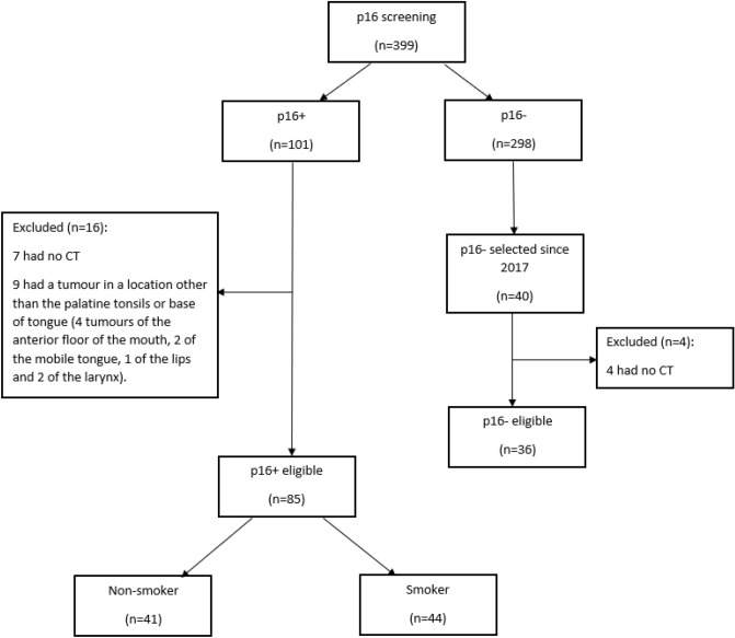
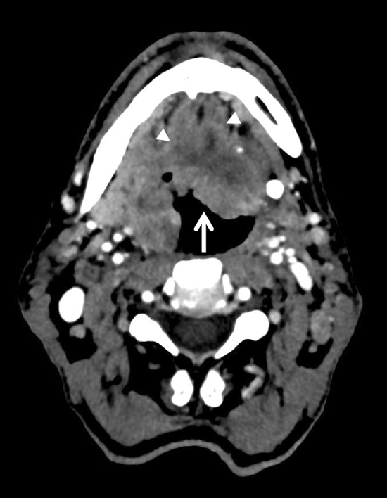
The eighth edition of the TNM classifies oropharyngeal squamous cell carcinomas (OSCCs) depending on p16 status. Some imaging features are reportedly associated more frequently with p16-positive (P16+) OSCC than p16-negative (p16-) OSCC. However, classical risk factors such as tobacco use were not specifically considered when assessing these imaging features. We aimed to evaluate whether P16+ OSCCs have different epidemiological, clinical, prognostic and imaging features depending on smoking status, and to compare P16+ and p16- groups. A retrospective study of data from 85 patients with P16+ OSCC (41 non-smokers, 44 smokers) and 36 with p16- OSCC from 2011 to 2020 was carried out, assessing epidemiological data, clinical aspects of the tumour and presence of adenopathy. Staging was assessed according to the seventh and eighth editions of the TNM. Compared with P16+ OSCC non-smokers, P16+ OSCC smokers had tumours that were less well-defined (36.6% vs 77.8%, p < 0.001), more ulcerated (85.4% vs 44.4%, p < 0.001) and more necrotic (53.7% vs 25%, p = 0.012). There was also less downstaging from N2 or N3 of the seventh edition of the TNM to N1 of the eighth edition for smokers than non-smokers (22.7% vs 43.9%, p = 0.042). Compared with p16- tumours, more P16+ tumours had well-defined contours (55.8% vs 22.2%, p = 0.001), were exophytic (89.6% vs 72.2%, p = 0.023), less necrotic (40.3% vs 80.6%, p < 0.001), less ulcerated (97.2% vs 66.2%, p = 0.006) and involved less muscle tissue (26.0% vs 47.2%, p = 0.027).P16+ OSCCs of smokers show clinical, imaging and prognostic differences with P16+ OSCCs of non-smokers.
第八版 TNM 分类根据 p16 状态将口咽鳞状细胞癌 (OSCC) 分类。据报道,一些影像学特征与 p16 阳性 (P16+) OSCC 比 p16 阴性 (p16-) OSCC 更频繁相关。然而,在评估这些影像学特征时,并没有专门考虑烟草使用等经典危险因素。我们旨在评估 P16+ OSCC 是否根据吸烟状况具有不同的流行病学、临床、预后和影像学特征,并比较 P16+ 和 p16- 组。对 2011 年至 2020 年间 85 例 P16+ OSCC 患者(41 例不吸烟者,44 例吸烟者)和 36 例 p16- OSCC 患者的数据进行了回顾性研究,评估了流行病学数据、肿瘤的临床特征和淋巴结肿大的存在。根据第七版和第八版 TNM 进行分期评估。与 P16+ OSCC 不吸烟者相比,P16+ OSCC 吸烟者的肿瘤边界更不清晰(36.6%对 77.8%,p<0.001),更溃疡(85.4%对 44.4%,p<0.001)和更坏死(53.7%对 25%,p=0.012)。与第七版 TNM 的 N2 或 N3 相比,吸烟者向第八版 TNM 的 N1 降期的比例也低于不吸烟者(22.7%对 43.9%,p=0.042)。与 p16- 肿瘤相比,更多的 P16+ 肿瘤具有清晰的轮廓(55.8%对 22.2%,p=0.001),更外生性(89.6%对 72.2%,p=0.023),更少坏死(40.3%对 80.6%,p<0.001),更少溃疡(97.2%对 66.2%,p=0.006)和更少肌肉组织受累(26.0%对 47.2%,p=0.027)。吸烟者的 P16+ OSCC 与不吸烟者的 P16+ OSCC 相比,具有不同的临床、影像学和预后特征。