Department of Orthopedics and Trauma Surgery, Medical University of Vienna, Spitalgasse 23, 1090, Vienna, Austria.
Department of Orthopedics and Traumatology, Hospital of the St. John of God Brothers Eisenstadt, Johannes von Gott-Platz 1, 7000, Eisenstadt, Austria.
Arch Orthop Trauma Surg. 2022 Jun;142(6):1075-1082. doi: 10.1007/s00402-021-03801-7. Epub 2021 Feb 8.
Distal radius fractures account for one-fifth of all fractures in the emergency department. Their classification based on standard radiographs is common practice although low inter-observer reliabilities and superiority of computer tomography (CT) scanning in evaluation of joint congruency have been reported.
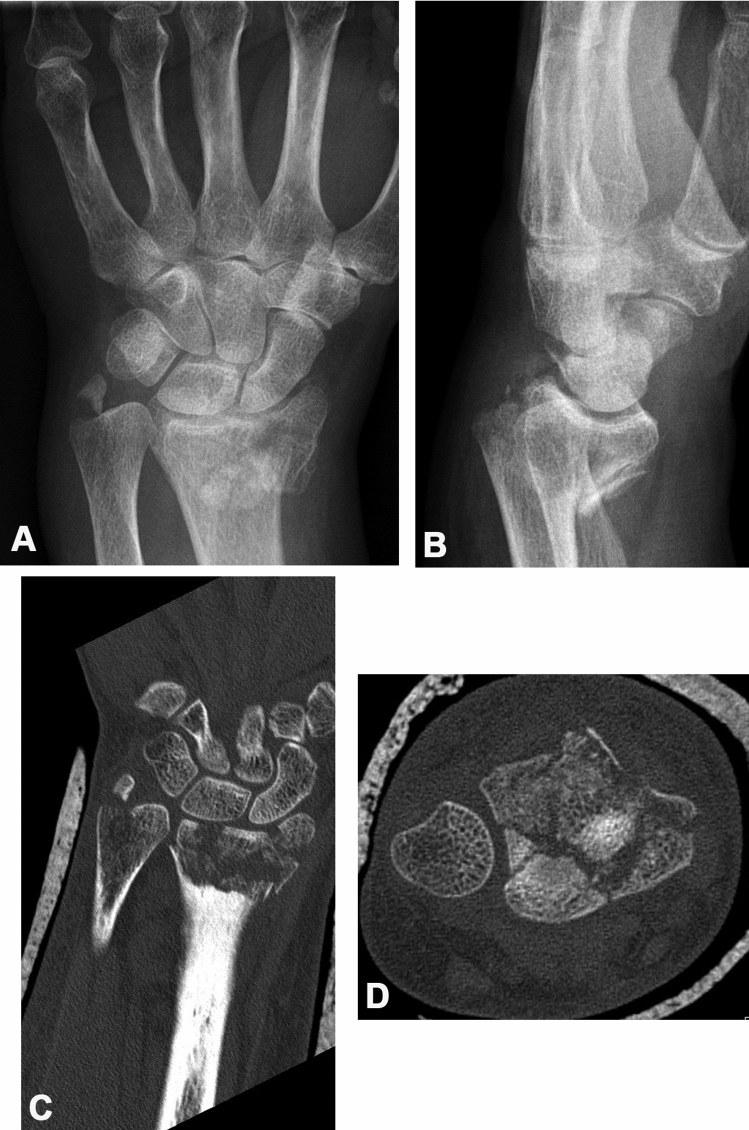
We retrospectively analyzed 96 displaced distal radius fractures scheduled for open reduction and internal fixation using standard radiographic assessment. The radiographs were classified with the Arbeitsgemeinschaft für Osteosynthesefragen/Orthopaedic Trauma Association (AO/OTA), Fernandez and Frykman classifications by three observers and inter-rater reliabilities were calculated. Additional CT scanning was performed in all cases and the following parameters were assessed: radiocarpal joint involvement, fracture extent into the radial sigmoid notch, i.e. the distal radio-ulnar joint, comminution of the metaphysis, and concomitant ulnar styloid fracture. The CT scans were used as a reference standard to determine sensitivity and accuracy of standard radiographic assessment in evaluation of distal radius fractures.
The inter-rater agreement for the AO classification was 35.4%, 68.8% for the Fernandez and 38.5% for the Frykman classification. Fracture extension into the radiocarpal joint was present in 81 cases (84.4%). Sigmoid notch involvement was found in 81 fractures (84.4%). Involvement of both joints was present in 72 cases (75%). The sensitivity of standard radiographs regarding radiocarpal joint involvement was 93.8%. Considering involvement of the distal radio-ulnar joint the false-negative rate using standard radiographs was 61.7% and the test's accuracy for sigmoid notch involvement was 45.8%.
This study demonstrates that involvement of the sigmoid notch is frequently missed in standard radiographs. The presented data support the frequent use of CT imaging to allow the holistic illustration of a fracture's complexion and to ensure optimal pre-operative planning.
桡骨远端骨折占急诊科所有骨折的五分之一。尽管已经报道了低观察者间可靠性和计算机断层扫描(CT)在评估关节吻合度方面的优势,但基于标准 X 线片的分类仍然是常见的做法。
我们回顾性分析了 96 例接受切开复位内固定术治疗的移位性桡骨远端骨折患者,这些患者均接受了标准的 X 线片评估。将 X 线片由 3 位观察者分别用 Arbeitsgemeinschaft für Osteosynthesefragen/Orthopaedic Trauma Association(AO/OTA)、Fernandez 和 Frykman 分类进行分类,并计算了观察者间的可靠性。所有病例均进行了额外的 CT 扫描,并评估了以下参数:桡腕关节受累、桡骨切迹延伸至桡尺远侧关节的程度、干骺端粉碎程度和尺骨茎突骨折。将 CT 扫描作为参考标准,以确定标准 X 线片评估在桡骨远端骨折中的敏感性和准确性。
AO 分类的观察者间一致性为 35.4%,Fernandez 分类为 68.8%,Frykman 分类为 38.5%。81 例(84.4%)骨折延伸至桡腕关节。81 例(84.4%)骨折累及桡骨切迹。72 例(75%)存在两个关节受累。标准 X 线片对桡腕关节受累的敏感性为 93.8%。考虑到桡尺远侧关节受累,标准 X 线片的假阴性率为 61.7%,用于评估桡骨切迹受累的测试准确性为 45.8%。
本研究表明,标准 X 线片常漏诊桡骨切迹受累。所提供的数据支持经常使用 CT 成像来全面显示骨折的复杂性,并确保术前的最佳规划。