Barrera-Alcocer Jorge, García-Benavides Leonel, Muñoz-Valle José F, de la Cruz-Mosso Ulises, González Ramón A, Luquín Sonia, Alarcón-Romero Luz Del C, Marino-Ortega Linda A, Matia-Garcia Ines, Parra-Rojas Isela
Facultad de Ciencias Químico Biológicas, Universidad Autónoma de Guerrero, Chilpancingo, Guerrero, México.
Instituto de Terapéutica Experimental y Clínica, Centro Universitario de Ciencias de la Salud, Universidad de Guadalajara, Guadalajara, Jalisco, México.
Diabetes Metab Syndr Obes. 2021 Feb 3;14:477-486. doi: 10.2147/DMSO.S285341. eCollection 2021.
Human adenovirus 36 (HAd36) infection has been associated with obesity. Experiments using 3T3-L1 adipocyte cultured cells and human adipose stem cells (hASCc) have shown that HAd36 stimulates the expression of genes implicated in cell differentiation and increased lipid accumulation. The presence of HAd36 in adipose tissue of overweight and obese women has also been confirmed. This study aims to analyze the presence of HAd36 DNA in the adipose tissue of women undergoing surgery for weight reduction and its relationship with obesity through changes in adipocyte morphology as well as the expression of C/EBPβ and HIF-1α.
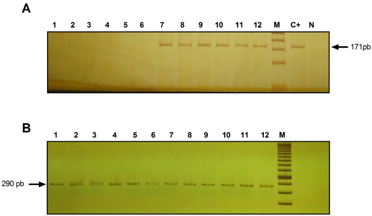
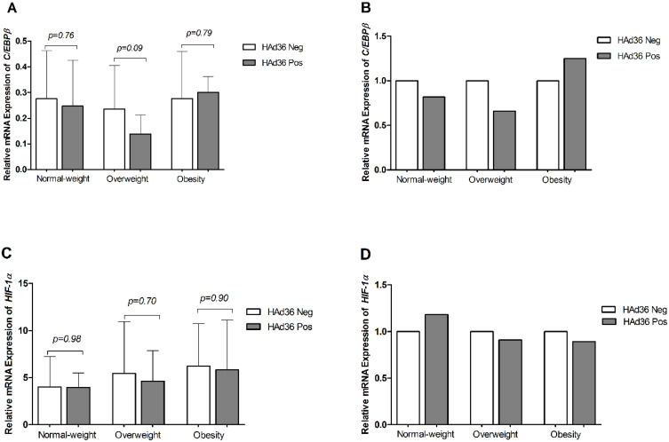
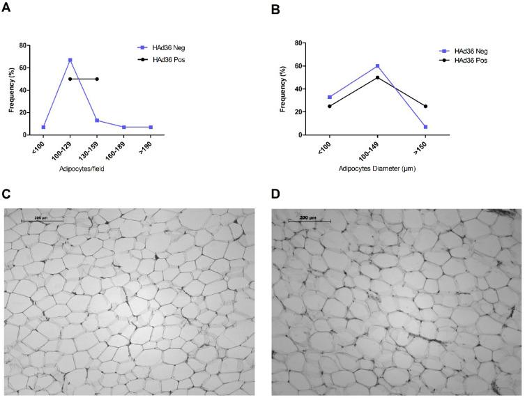
Fifty-two subcutaneous adipose tissue biopsies were collected. The anthropometric parameters measured were weight, height, skin folds, body circumferences, and body fat percentage. Biochemical measures were performed for glucose, cholesterol, triglycerides, cholesterol HDL-c, and LDL-c. The presence of HAd36 DNA was performed by conventional PCR. Adipocyte morphology was analyzed in H&E-stained sections using ImageJ/Fiji software. The expression of genes C/EBPβ, HIF-1α and β-actin was determined using TaqMan probes.
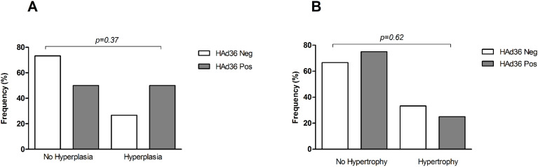
HAd36 DNA was detected in 31% of adipose tissue samples. The presence of viral DNA was not significantly associated with anthropometric, clinical, or metabolic measurements, or with changes in adipose tissue morphology. The levels of mRNA expression for C/EBPβ and HIF-1α did not show significant differences between positive and negative samples for HAd36 (p>0.05).
The presence of HAd36 DNA in adipose tissue was identified, but it was not related to morphological changes of adipocytes, or the expression of C/EBPβ and HIF-1α. Further studies are needed to confirm these findings.
人腺病毒36型(HAd36)感染与肥胖有关。使用3T3-L1脂肪细胞培养细胞和人脂肪干细胞(hASCc)进行的实验表明,HAd36刺激参与细胞分化的基因表达并增加脂质积累。超重和肥胖女性的脂肪组织中也已证实存在HAd36。本研究旨在分析接受减肥手术的女性脂肪组织中HAd36 DNA的存在情况,并通过脂肪细胞形态变化以及C/EBPβ和HIF-1α的表达来分析其与肥胖的关系。
收集了52份皮下脂肪组织活检样本。测量的人体测量参数包括体重、身高、皮褶厚度、体围和体脂百分比。对葡萄糖、胆固醇、甘油三酯、高密度脂蛋白胆固醇(HDL-c)和低密度脂蛋白胆固醇(LDL-c)进行生化检测。通过常规PCR检测HAd36 DNA的存在情况。使用ImageJ/Fiji软件在苏木精-伊红(H&E)染色切片中分析脂肪细胞形态。使用TaqMan探针测定C/EBPβ、HIF-1α和β-肌动蛋白基因的表达。
在31%的脂肪组织样本中检测到HAd36 DNA。病毒DNA的存在与人体测量、临床或代谢指标,或与脂肪组织形态变化均无显著相关性。HAd36阳性和阴性样本之间C/EBPβ和HIF-1α的mRNA表达水平无显著差异(p>0.05)。
已确定脂肪组织中存在HAd36 DNA,但它与脂肪细胞的形态变化、C/EBPβ和HIF-1α的表达均无关。需要进一步研究来证实这些发现。