Medical Imaging, Anatomy, Radboud University Medical Center, Donders Institute for Brain, Cognition and Behaviour, Nijmegen, the Netherlands.
Department of Neurology, Donders Institute for Brain Cognition and Behaviour, Radboud University Medical Center, Nijmegen, the Netherlands.
Neuropathology. 2021 Feb;41(1):3-20. doi: 10.1111/neup.12721.
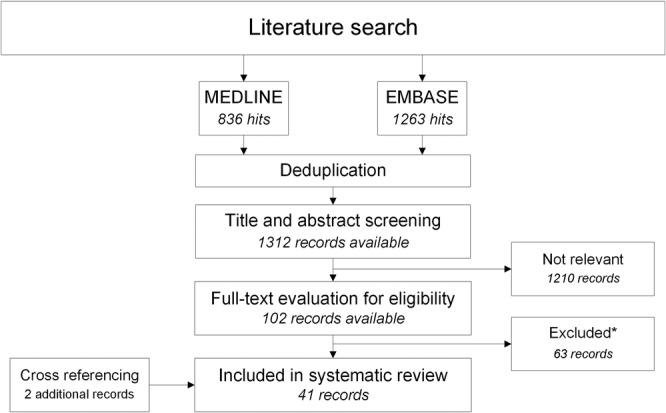
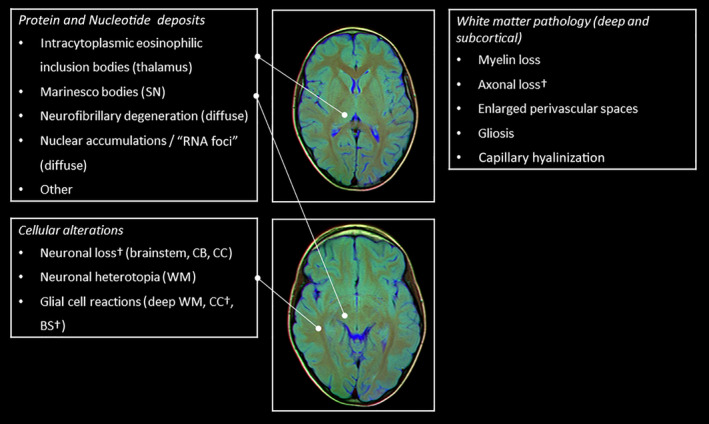
Brain involvement in myotonic dystrophy type 1 (DM1) is characterized by heterogeneous cognitive, behavioral, and affective symptoms and imaging alterations indicative of widespread grey and white matter involvement. The aim of the present study was to systematically review the literature on brain pathology in DM1. We conducted a structured search in EMBASE (index period 1974-2017) and MEDLINE (index period 1887-2017) on December 11, 2017, using free text and index search terms related to myotonic dystrophy type 1 and brain structures or regions. Eligible studies were full-text studies reporting on microscopic brain pathology of DM1 patients without potentially interfering comorbidity. We discussed the findings based on the anatomical region and the nature of the anomaly. Neuropathological findings in DM1 can be classified as follows: (1) protein and nucleotide deposits; (2) changes in neurons and glial cells; and (3) white matter alterations. Most findings are unspecific to DM1 and may occur with physiological aging, albeit to a lesser degree. There are similarities and contrasts with Alzheimer's disease; both show the appearance of neurofibrillary tangles in the limbic system without plaque occurrence. Likewise, there is myelin loss and gliosis, and there are dilated perivascular spaces in the white matter resemblant of cerebral small vessel disease. However, we did not find evidence of lacunar infarction or microbleeding. The various neuropathological findings in DM1 are reflective of the heterogeneous clinical and neuroimaging features of the disease. The strength of conclusions from this study's findings is bounded by limited numbers of participants in studies, methodological constraints, and lack of assessed associations between histopathology and clinical or neuroimaging findings.
脑肌病 1 型(DM1)的脑受累表现为异质性认知、行为和情感症状,影像学改变提示广泛的灰质和白质受累。本研究旨在系统回顾 DM1 脑病理学的文献。我们于 2017 年 12 月 11 日在 EMBASE(索引期 1974-2017 年)和 MEDLINE(索引期 1887-2017 年)中进行了结构化检索,使用了与肌强直性营养不良 1 型和脑结构或区域相关的自由文本和索引搜索词。符合条件的研究为报告无潜在混杂疾病的 DM1 患者的脑微观病理学的全文研究。我们根据解剖区域和异常性质对发现进行了讨论。DM1 的神经病理学发现可分为以下几类:(1)蛋白和核苷酸沉积;(2)神经元和神经胶质细胞变化;(3)白质改变。大多数发现对 DM1 不具有特异性,可能会随着生理性衰老而出现,但程度较轻。与阿尔茨海默病有相似之处和对比;两者均在边缘系统出现神经纤维缠结,而无斑块发生。同样,有髓鞘丢失和神经胶质增生,白质中有扩张的血管周围间隙,类似于脑小血管病。然而,我们未发现腔隙性梗死或微出血的证据。DM1 的各种神经病理学发现反映了该病异质性的临床和神经影像学特征。本研究结果的结论强度受到研究中参与者数量有限、方法学限制以及组织病理学与临床或神经影像学发现之间缺乏评估关联的限制。