• 文献检索
  • 文档翻译
  • 深度研究
  • 学术资讯
  • Suppr Zotero 插件Zotero 插件
  • 邀请有礼
  • 套餐&价格
  • 历史记录
应用&插件
Suppr Zotero 插件Zotero 插件浏览器插件Mac 客户端Windows 客户端微信小程序
定价
高级版会员购买积分包购买API积分包
服务
文献检索文档翻译深度研究API 文档MCP 服务
关于我们
关于 Suppr公司介绍联系我们用户协议隐私条款
关注我们

Suppr 超能文献

核心技术专利:CN118964589B侵权必究
粤ICP备2023148730 号-1Suppr @ 2026

文献检索

告别复杂PubMed语法,用中文像聊天一样搜索,搜遍4000万医学文献。AI智能推荐,让科研检索更轻松。

立即免费搜索

文件翻译

保留排版,准确专业,支持PDF/Word/PPT等文件格式,支持 12+语言互译。

免费翻译文档

深度研究

AI帮你快速写综述,25分钟生成高质量综述,智能提取关键信息,辅助科研写作。

立即免费体验

基于胸部 X 射线图像的 COVID-19 与常见病毒性肺炎鉴别诊断的混合集成模型。

Hybrid ensemble model for differential diagnosis between COVID-19 and common viral pneumonia by chest X-ray radiograph.

机构信息

School of Medicine, Shanghai Jiao Tong University, 200025, Shanghai, PR China.

School of Traffic and Transportation Engineering, Central South University, 410075, Hunan, PR China.

出版信息

Comput Biol Med. 2021 Apr;131:104252. doi: 10.1016/j.compbiomed.2021.104252. Epub 2021 Feb 2.

DOI:10.1016/j.compbiomed.2021.104252
PMID:33610001
原文链接:https://pmc.ncbi.nlm.nih.gov/articles/PMC7966819/
Abstract

BACKGROUND

Chest X-ray radiography (CXR) has been widely considered as an accessible, feasible, and convenient method to evaluate suspected patients' lung involvement during the COVID-19 pandemic. However, with the escalating number of suspected cases, traditional diagnosis via CXR fails to deliver results within a short period of time. Therefore, it is crucial to employ artificial intelligence (AI) to enhance CXRs for obtaining quick and accurate diagnoses. Previous studies have reported the feasibility of utilizing deep learning methods to screen for COVID-19 using CXR and CT results. However, these models only use a single deep learning network for chest radiograph detection; the accuracy of this approach required further improvement.

METHODS

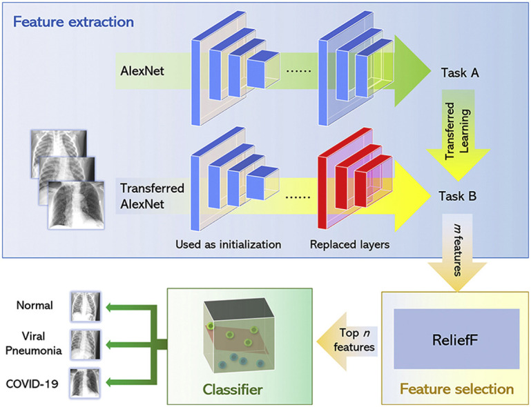
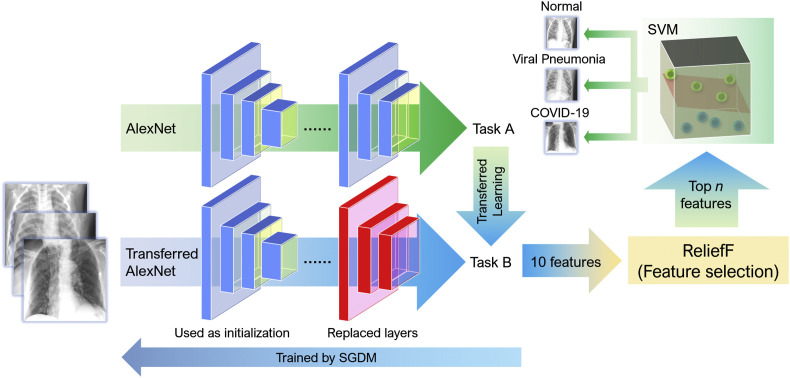
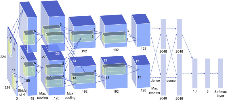
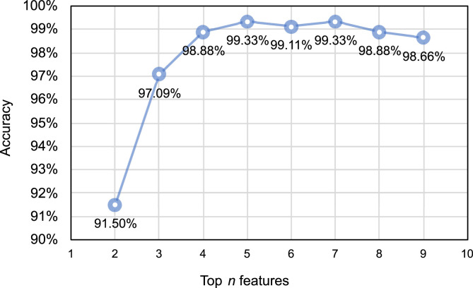
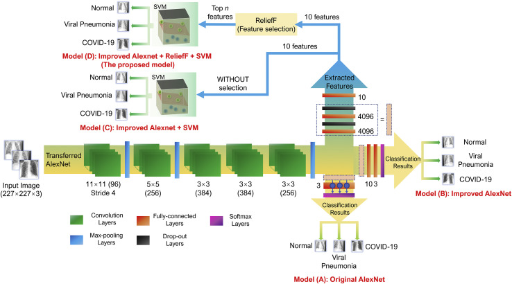
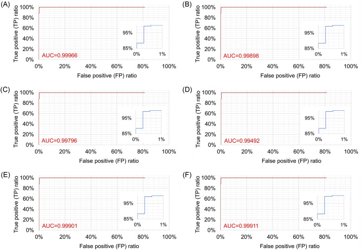
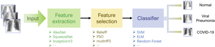
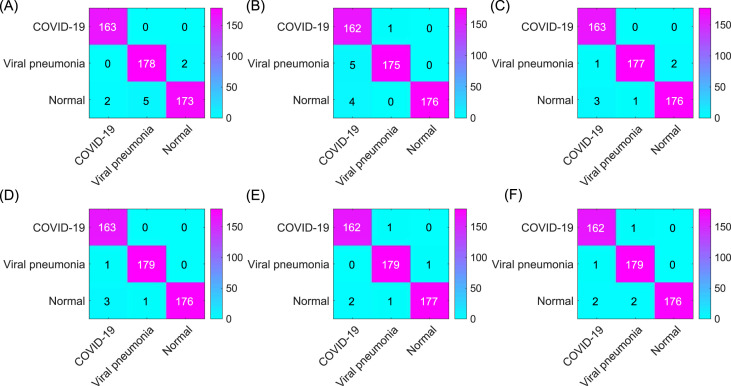
In this study, we propose a three-step hybrid ensemble model, including a feature extractor, a feature selector, and a classifier. First, a pre-trained AlexNet with an improved structure extracts the original image features. Then, the ReliefF algorithm is adopted to sort the extracted features, and a trial-and-error approach is used to select the n most important features to reduce the feature dimension. Finally, an SVM classifier provides classification results based on the n selected features.

RESULTS

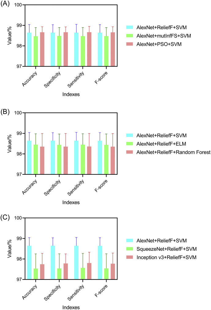
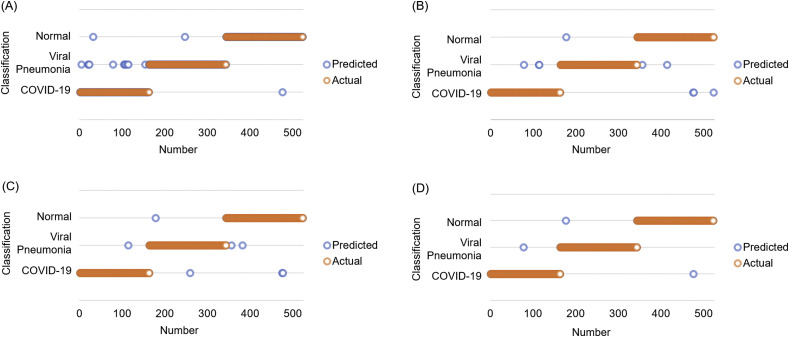
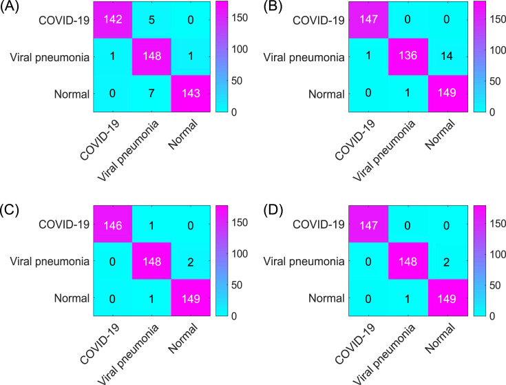
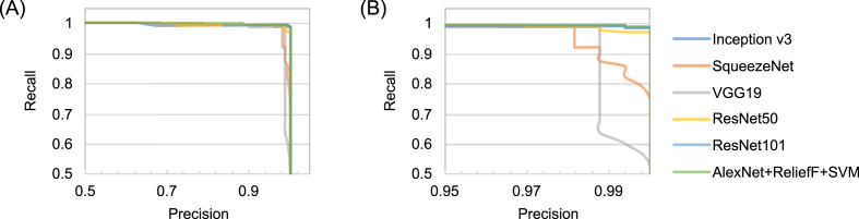
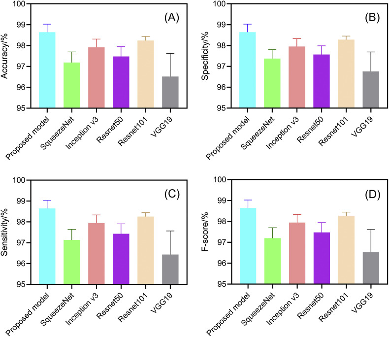
Compared to five existing models (InceptionV3: 97.916 ± 0.408%; SqueezeNet: 97.189 ± 0.526%; VGG19: 96.520 ± 1.220%; ResNet50: 97.476 ± 0.513%; ResNet101: 98.241 ± 0.209%), the proposed model demonstrated the best performance in terms of overall accuracy rate (98.642 ± 0.398%). Additionally, compared to the existing models, the proposed model demonstrates a considerable improvement in classification time efficiency (SqueezeNet: 6.602 ± 0.001s; InceptionV3: 12.376 ± 0.002s; ResNet50: 10.952 ± 0.001s; ResNet101: 18.040 ± 0.002s; VGG19: 16.632 ± 0.002s; proposed model: 5.917 ± 0.001s).

CONCLUSION

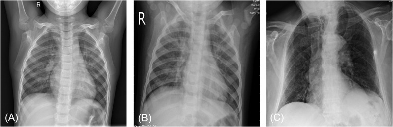
The model proposed in this article is practical and effective, and can provide high-precision COVID-19 CXR detection. We demonstrated its suitability to aid medical professionals in distinguishing normal CXRs, viral pneumonia CXRs and COVID-19 CXRs efficiently on small sample sizes.

摘要

背景

在 COVID-19 大流行期间,胸部 X 射线摄影(CXR)被广泛认为是一种易于获得、可行且方便的方法,可用于评估疑似患者的肺部受累情况。然而,随着疑似病例数量的不断增加,传统的 CXR 诊断方法无法在短时间内得出结果。因此,利用人工智能(AI)增强 CXR 以快速准确诊断至关重要。先前的研究报告了使用 CXR 和 CT 结果利用深度学习方法筛查 COVID-19 的可行性。然而,这些模型仅使用单个深度学习网络进行胸部 X 射线检测;该方法的准确性需要进一步提高。

方法

在这项研究中,我们提出了一种三步骤混合集成模型,包括特征提取器、特征选择器和分类器。首先,使用经过改进结构的预训练 AlexNet 提取原始图像特征。然后,采用 ReliefF 算法对提取的特征进行排序,并采用试错法选择最重要的 n 个特征来降低特征维度。最后,SVM 分类器根据 n 个选择的特征提供分类结果。

结果

与五个现有模型(InceptionV3:97.916 ± 0.408%;SqueezeNet:97.189 ± 0.526%;VGG19:96.520 ± 1.220%;ResNet50:97.476 ± 0.513%;ResNet101:98.241 ± 0.209%)相比,所提出的模型在总准确率(98.642 ± 0.398%)方面表现最佳。此外,与现有模型相比,所提出的模型在分类时间效率方面有了相当大的提高(SqueezeNet:6.602 ± 0.001s;InceptionV3:12.376 ± 0.002s;ResNet50:10.952 ± 0.001s;ResNet101:18.040 ± 0.002s;VGG19:16.632 ± 0.002s;所提出的模型:5.917 ± 0.001s)。

结论

本文提出的模型实用有效,可提供高精度的 COVID-19 CXR 检测。我们证明了它适用于帮助医疗专业人员在小样本量上有效区分正常 CXR、病毒性肺炎 CXR 和 COVID-19 CXR。

https://cdn.ncbi.nlm.nih.gov/pmc/blobs/44c9/7966819/391289fb6bf9/gr15_lrg.jpg
https://cdn.ncbi.nlm.nih.gov/pmc/blobs/44c9/7966819/87a8187ddf75/fx1_lrg.jpg
https://cdn.ncbi.nlm.nih.gov/pmc/blobs/44c9/7966819/264a6077d984/gr1_lrg.jpg
https://cdn.ncbi.nlm.nih.gov/pmc/blobs/44c9/7966819/d546f238ecf8/gr2_lrg.jpg
https://cdn.ncbi.nlm.nih.gov/pmc/blobs/44c9/7966819/9d48cc0c982c/gr3_lrg.jpg
https://cdn.ncbi.nlm.nih.gov/pmc/blobs/44c9/7966819/ed7745de6625/gr4_lrg.jpg
https://cdn.ncbi.nlm.nih.gov/pmc/blobs/44c9/7966819/b9bf3efa9dff/gr5_lrg.jpg
https://cdn.ncbi.nlm.nih.gov/pmc/blobs/44c9/7966819/d3f34867f256/gr6_lrg.jpg
https://cdn.ncbi.nlm.nih.gov/pmc/blobs/44c9/7966819/70c5c9b6052e/gr7_lrg.jpg
https://cdn.ncbi.nlm.nih.gov/pmc/blobs/44c9/7966819/242b6ba9c746/gr8_lrg.jpg
https://cdn.ncbi.nlm.nih.gov/pmc/blobs/44c9/7966819/a906118f5f2e/gr9_lrg.jpg
https://cdn.ncbi.nlm.nih.gov/pmc/blobs/44c9/7966819/f74e2119e23e/gr10_lrg.jpg
https://cdn.ncbi.nlm.nih.gov/pmc/blobs/44c9/7966819/b3ec0d7ef6dc/gr11_lrg.jpg
https://cdn.ncbi.nlm.nih.gov/pmc/blobs/44c9/7966819/f2ab505141ed/gr12_lrg.jpg
https://cdn.ncbi.nlm.nih.gov/pmc/blobs/44c9/7966819/659194c21bef/gr13_lrg.jpg
https://cdn.ncbi.nlm.nih.gov/pmc/blobs/44c9/7966819/74cb9e9c75ca/gr14_lrg.jpg
https://cdn.ncbi.nlm.nih.gov/pmc/blobs/44c9/7966819/391289fb6bf9/gr15_lrg.jpg

相似文献

1
Hybrid ensemble model for differential diagnosis between COVID-19 and common viral pneumonia by chest X-ray radiograph.基于胸部 X 射线图像的 COVID-19 与常见病毒性肺炎鉴别诊断的混合集成模型。
Comput Biol Med. 2021 Apr;131:104252. doi: 10.1016/j.compbiomed.2021.104252. Epub 2021 Feb 2.
2
Computer aid screening of COVID-19 using X-ray and CT scan images: An inner comparison.基于 X 射线和 CT 扫描图像的 COVID-19 计算机辅助筛查:内部比较。
J Xray Sci Technol. 2021;29(2):197-210. doi: 10.3233/XST-200784.
3
MCSC-Net: COVID-19 detection using deep-Q-neural network classification with RFNN-based hybrid whale optimization.MCSC-Net:基于深度 Q 神经网络分类和基于 RFNN 的混合鲸鱼优化算法的 COVID-19 检测。
J Xray Sci Technol. 2023;31(3):483-509. doi: 10.3233/XST-221360.
4
A Deep Learning Model for Diagnosing COVID-19 and Pneumonia through X-ray.一种通过X射线诊断新冠肺炎和肺炎的深度学习模型。
Curr Med Imaging. 2023;19(4):333-346. doi: 10.2174/1573405618666220610093740.
5
Thorax computed tomography (CTX) guided ground truth annotation of CHEST radiographs (CXR) for improved classification and detection of COVID-19.胸部 CT 引导的 CHEST 射线照片(CXR)标注以提高 COVID-19 的分类和检测。
Int J Numer Method Biomed Eng. 2024 Jun;40(6):e3823. doi: 10.1002/cnm.3823. Epub 2024 Apr 8.
6
Deep Learning Algorithm for COVID-19 Classification Using Chest X-Ray Images.基于胸部 X 光图像的 COVID-19 分类深度学习算法。
Comput Math Methods Med. 2021 Nov 9;2021:9269173. doi: 10.1155/2021/9269173. eCollection 2021.
7
COVID-DSNet: A novel deep convolutional neural network for detection of coronavirus (SARS-CoV-2) cases from CT and Chest X-Ray images.COVID-DSNet:一种新型深度卷积神经网络,用于从 CT 和胸部 X 光图像中检测冠状病毒(SARS-CoV-2)病例。
Artif Intell Med. 2022 Dec;134:102427. doi: 10.1016/j.artmed.2022.102427. Epub 2022 Oct 17.
8
COVID-19 detection in CT and CXR images using deep learning models.使用深度学习模型进行 CT 和 CXR 图像中的 COVID-19 检测。
Biogerontology. 2022 Feb;23(1):65-84. doi: 10.1007/s10522-021-09946-7. Epub 2022 Jan 22.
9
Chest X-ray image phase features for improved diagnosis of COVID-19 using convolutional neural network.基于卷积神经网络的胸部 X 射线图像相位特征提高 COVID-19 诊断性能
Int J Comput Assist Radiol Surg. 2021 Feb;16(2):197-206. doi: 10.1007/s11548-020-02305-w. Epub 2021 Jan 9.
10
COVID-19 classification of X-ray images using deep neural networks.基于深度神经网络的 X 射线图像 COVID-19 分类。
Eur Radiol. 2021 Dec;31(12):9654-9663. doi: 10.1007/s00330-021-08050-1. Epub 2021 May 29.

引用本文的文献

1
A systematic literature review: exploring the challenges of ensemble model for medical imaging.一项系统的文献综述:探索医学成像集成模型的挑战。
BMC Med Imaging. 2025 Apr 18;25(1):128. doi: 10.1186/s12880-025-01667-4.
2
Deep Learning for Pneumonia Detection in Chest X-ray Images: A Comprehensive Survey.胸部X光图像中肺炎检测的深度学习:全面综述。
J Imaging. 2024 Jul 23;10(8):176. doi: 10.3390/jimaging10080176.
3
A brief review and scientometric analysis on ensemble learning methods for handling COVID-19.关于处理新冠肺炎的集成学习方法的简要综述与科学计量分析

本文引用的文献

1
Classification of COVID-19 in chest X-ray images using DeTraC deep convolutional neural network.使用DeTraC深度卷积神经网络对胸部X光图像中的新冠肺炎进行分类。
Appl Intell (Dordr). 2021;51(2):854-864. doi: 10.1007/s10489-020-01829-7. Epub 2020 Sep 5.
2
RANDGAN: Randomized generative adversarial network for detection of COVID-19 in chest X-ray.RANDGAN:用于胸部 X 光 COVID-19 检测的随机生成对抗网络。
Sci Rep. 2021 Apr 21;11(1):8602. doi: 10.1038/s41598-021-87994-2.
3
Application of deep learning techniques for detection of COVID-19 cases using chest X-ray images: A comprehensive study.
Heliyon. 2024 Feb 20;10(4):e26694. doi: 10.1016/j.heliyon.2024.e26694. eCollection 2024 Feb 29.
4
Ensemble learning for multi-class COVID-19 detection from big data.基于大数据的多类别 COVID-19 检测的集成学习。
PLoS One. 2023 Oct 11;18(10):e0292587. doi: 10.1371/journal.pone.0292587. eCollection 2023.
5
The role of machine learning in health policies during the COVID-19 pandemic and in long COVID management.机器学习在 COVID-19 大流行期间和长新冠管理中的卫生政策中的作用。
Front Public Health. 2023 Apr 11;11:1140353. doi: 10.3389/fpubh.2023.1140353. eCollection 2023.
6
AI-based radiodiagnosis using chest X-rays: A review.基于人工智能的胸部X光放射诊断:综述
Front Big Data. 2023 Apr 6;6:1120989. doi: 10.3389/fdata.2023.1120989. eCollection 2023.
7
Boosting automatic COVID-19 detection performance with self-supervised learning and batch knowledge ensembling.基于自监督学习和批量知识集成提升 COVID-19 自动检测性能
Comput Biol Med. 2023 May;158:106877. doi: 10.1016/j.compbiomed.2023.106877. Epub 2023 Mar 31.
8
Feature selection of pre-trained shallow CNN using the QLESCA optimizer: COVID-19 detection as a case study.使用QLESCA优化器对预训练浅层卷积神经网络进行特征选择:以新冠病毒疾病检测为例
Appl Intell (Dordr). 2023 Feb 6:1-23. doi: 10.1007/s10489-022-04446-8.
9
Towards smart diagnostic methods for COVID-19: Review of deep learning for medical imaging.迈向COVID-19的智能诊断方法:医学成像深度学习综述
IPEM Transl. 2022 Nov-Dec;3:100008. doi: 10.1016/j.ipemt.2022.100008. Epub 2022 Oct 26.
10
Explainable artificial intelligence-based edge fuzzy images for COVID-19 detection and identification.基于可解释人工智能的边缘模糊图像用于新冠病毒病的检测与识别
Appl Soft Comput. 2022 Jul;123:108966. doi: 10.1016/j.asoc.2022.108966. Epub 2022 May 13.
利用胸部X光图像的深度学习技术检测新冠肺炎病例的应用:一项综合研究。
Biomed Signal Process Control. 2021 Feb;64:102365. doi: 10.1016/j.bspc.2020.102365. Epub 2020 Nov 19.
4
COVID-Net: a tailored deep convolutional neural network design for detection of COVID-19 cases from chest X-ray images.COVID-Net:一种针对胸部 X 光图像中 COVID-19 病例检测的定制化深度卷积神经网络设计。
Sci Rep. 2020 Nov 11;10(1):19549. doi: 10.1038/s41598-020-76550-z.
5
Evaluation of Scalability and Degree of Fine-Tuning of Deep Convolutional Neural Networks for COVID-19 Screening on Chest X-ray Images Using Explainable Deep-Learning Algorithm.使用可解释深度学习算法对基于胸部X光图像的COVID-19筛查的深度卷积神经网络的可扩展性和微调程度进行评估。
J Pers Med. 2020 Nov 7;10(4):213. doi: 10.3390/jpm10040213.
6
A five-layer deep convolutional neural network with stochastic pooling for chest CT-based COVID-19 diagnosis.一种用于基于胸部CT的COVID-19诊断的带有随机池化的五层深度卷积神经网络。
Mach Vis Appl. 2021;32(1):14. doi: 10.1007/s00138-020-01128-8. Epub 2020 Nov 3.
7
CGNet: A graph-knowledge embedded convolutional neural network for detection of pneumonia.CGNet:一种用于肺炎检测的图知识嵌入卷积神经网络。
Inf Process Manag. 2021 Jan;58(1):102411. doi: 10.1016/j.ipm.2020.102411. Epub 2020 Oct 19.
8
A light CNN for detecting COVID-19 from CT scans of the chest.一种用于从胸部CT扫描中检测新冠肺炎的轻量级卷积神经网络。
Pattern Recognit Lett. 2020 Dec;140:95-100. doi: 10.1016/j.patrec.2020.10.001. Epub 2020 Oct 3.
9
A new approach for classifying coronavirus COVID-19 based on its manifestation on chest X-rays using texture features and neural networks.一种基于胸部X光片表现,利用纹理特征和神经网络对新型冠状病毒COVID-19进行分类的新方法。
Inf Sci (N Y). 2021 Feb 4;545:403-414. doi: 10.1016/j.ins.2020.09.041. Epub 2020 Sep 24.
10
CVDNet: A novel deep learning architecture for detection of coronavirus (Covid-19) from chest x-ray images.CVDNet:一种用于从胸部X光图像中检测冠状病毒(COVID-19)的新型深度学习架构。
Chaos Solitons Fractals. 2020 Nov;140:110245. doi: 10.1016/j.chaos.2020.110245. Epub 2020 Sep 4.