• 文献检索
  • 文档翻译
  • 深度研究
  • 学术资讯
  • Suppr Zotero 插件Zotero 插件
  • 邀请有礼
  • 套餐&价格
  • 历史记录
应用&插件
Suppr Zotero 插件Zotero 插件浏览器插件Mac 客户端Windows 客户端微信小程序
定价
高级版会员购买积分包购买API积分包
服务
文献检索文档翻译深度研究API 文档MCP 服务
关于我们
关于 Suppr公司介绍联系我们用户协议隐私条款
关注我们

Suppr 超能文献

核心技术专利:CN118964589B侵权必究
粤ICP备2023148730 号-1Suppr @ 2026

文献检索

告别复杂PubMed语法,用中文像聊天一样搜索,搜遍4000万医学文献。AI智能推荐,让科研检索更轻松。

立即免费搜索

文件翻译

保留排版,准确专业,支持PDF/Word/PPT等文件格式,支持 12+语言互译。

免费翻译文档

深度研究

AI帮你快速写综述,25分钟生成高质量综述,智能提取关键信息,辅助科研写作。

立即免费体验

管状上皮细胞来源的含 microRNA-21 的微囊泡促进心肌细胞肥大。

MicroRNA-21-containing microvesicles from tubular epithelial cells promote cardiomyocyte hypertrophy.

机构信息

Department of Nephrology, The First People's Hospital of Changzhou, Changzhou, China.

Department of Pathology, The Second People's Hospital of Changzhou, Changzhou, China.

出版信息

Ren Fail. 2021 Dec;43(1):391-400. doi: 10.1080/0886022X.2021.1891098.

DOI:10.1080/0886022X.2021.1891098
PMID:33632070
原文链接:https://pmc.ncbi.nlm.nih.gov/articles/PMC7919913/
Abstract

BACKGROUND

Cardiomyocyte hypertrophy has been reported as one of the important mechanisms for cardiovascular disease (CVD) in patients with chronic kidney disease (CKD). MiroRNA-21(miR-21) was determined to play an important role in myocardial hypertrophy. However, the role of microvesicles (MVs) containing miR-21 in CKD-related cardiomyocyte hypertrophy remains largely unexplored.

METHODS

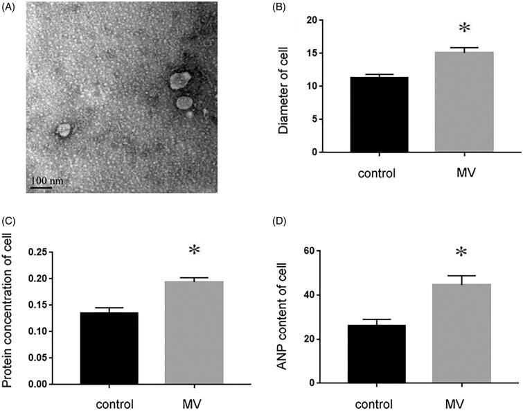
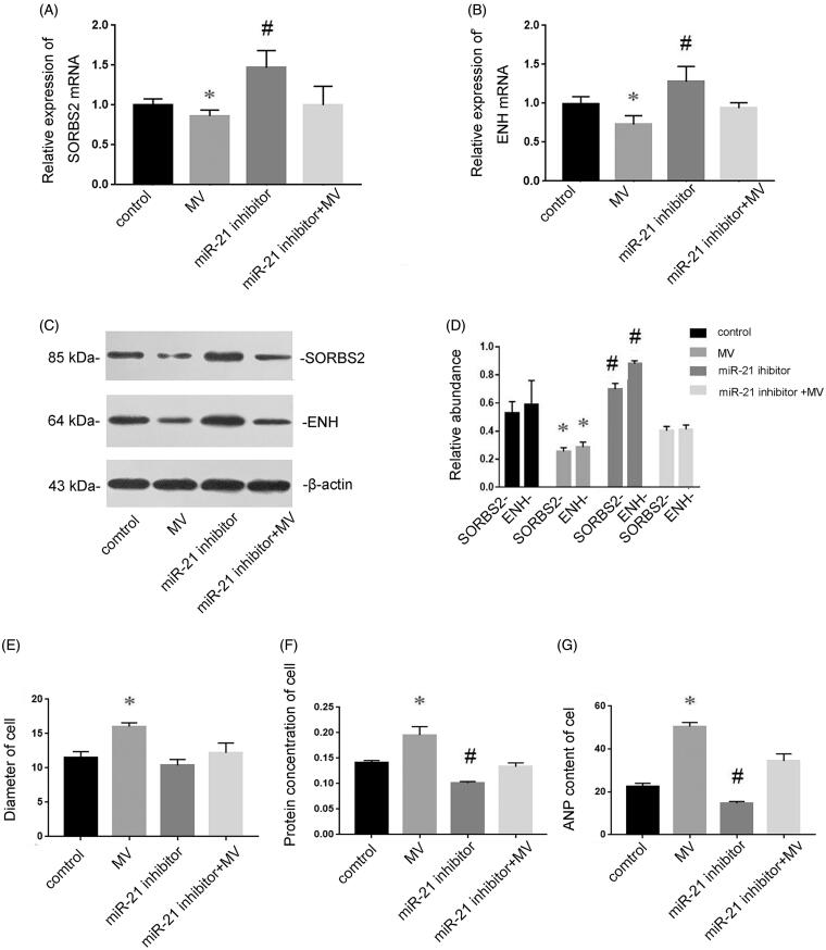
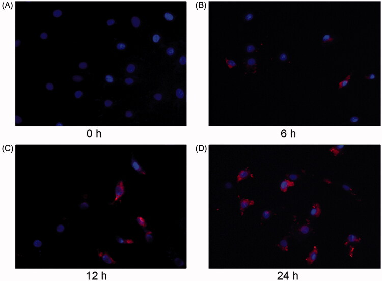
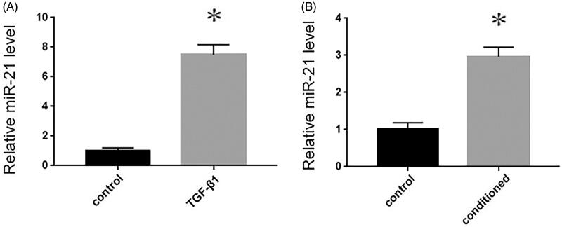
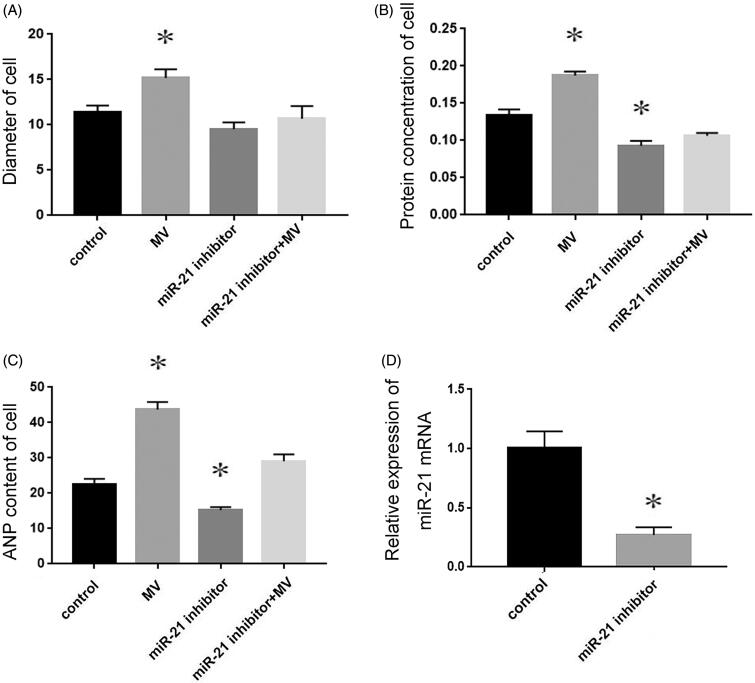
Renal tubular epithelial cells were stimulated by transforming growth factor (TGF-β1), and the conditioned medium was extracted by differential centrifugation. Renal tubular epithelial cells were labeled with Dil-C18 dye and the recipient cardiomyocytes were observed by fluorescence microscope. MiR-21 level in MVs was detected by qRT-PCR, and the length and diameter of cardiomyocytes were measured by microscope. BCA protein kit and ANP kit were used to detect the content of cell protein and the level of ANP. MiR-21 inhibitor was transfected into cardiomyocytes to observe the effect of miR-21 on myocardial hypertrophy.

RESULTS

TGF-β1 could induce donor renal tubular epithelial cells to produce MVs and delivered into cardiomyocytes, followed by the diameter, protein concentration and ANP content of cardiomyocytes significantly increased. Meanwhile, MiR-21 levels were markedly increased in MVs isolated from donor renal tubular epithelial cells and recipient cardiomyocytes. Pre-transfection of miR-21 inhibitors could inhibit MV-induced cardiomyocyte hypertrophy.

CONCLUSION

Tubular cells could secrete miR-21 by MVs and deliver it into recipient cardiomyocytes to induce cardiomyocyte hypertrophy. It might shed a new light on the mechanism and treatment of CKD-related cardiac dysfunction.

摘要

背景

心肌细胞肥大已被报道为慢性肾脏病(CKD)患者心血管疾病(CVD)的重要机制之一。miRNA-21(miR-21)被确定在心肌肥大中发挥重要作用。然而,含 miR-21 的微小泡(MVs)在 CKD 相关心肌细胞肥大中的作用在很大程度上仍未得到探索。

方法

用转化生长因子(TGF-β1)刺激肾小管上皮细胞,通过差速离心提取条件培养基。用 Dil-C18 染料标记肾小管上皮细胞,荧光显微镜观察受体心肌细胞。用 qRT-PCR 检测 MVs 中的 miR-21 水平,用显微镜测量心肌细胞的长度和直径。用 BCA 蛋白试剂盒和 ANP 试剂盒检测细胞蛋白含量和 ANP 水平。将 miR-21 抑制剂转染到心肌细胞中,观察 miR-21 对心肌肥大的影响。

结果

TGF-β1 可诱导供体肾小管上皮细胞产生 MVs 并传递到心肌细胞,随后心肌细胞的直径、蛋白浓度和 ANP 含量明显增加。同时,供体肾小管上皮细胞和受体心肌细胞中分离的 MVs 中 miR-21 水平明显升高。转染前 miR-21 抑制剂可抑制 MV 诱导的心肌细胞肥大。

结论

肾小管细胞可通过 MVs 分泌 miR-21 并将其传递到受体心肌细胞,从而诱导心肌细胞肥大。这可能为 CKD 相关心功能障碍的机制和治疗提供新的思路。

https://cdn.ncbi.nlm.nih.gov/pmc/blobs/4b69/7919913/398662a53096/IRNF_A_1891098_F0006_B.jpg
https://cdn.ncbi.nlm.nih.gov/pmc/blobs/4b69/7919913/2af329f85d13/IRNF_A_1891098_F0001_B.jpg
https://cdn.ncbi.nlm.nih.gov/pmc/blobs/4b69/7919913/7a59d10dc487/IRNF_A_1891098_F0002_C.jpg
https://cdn.ncbi.nlm.nih.gov/pmc/blobs/4b69/7919913/bb6be6726b2e/IRNF_A_1891098_F0003_B.jpg
https://cdn.ncbi.nlm.nih.gov/pmc/blobs/4b69/7919913/c70bd25b7684/IRNF_A_1891098_F0004_B.jpg
https://cdn.ncbi.nlm.nih.gov/pmc/blobs/4b69/7919913/5c60f2bfe587/IRNF_A_1891098_F0005_B.jpg
https://cdn.ncbi.nlm.nih.gov/pmc/blobs/4b69/7919913/398662a53096/IRNF_A_1891098_F0006_B.jpg

相似文献

1
MicroRNA-21-containing microvesicles from tubular epithelial cells promote cardiomyocyte hypertrophy.管状上皮细胞来源的含 microRNA-21 的微囊泡促进心肌细胞肥大。
Ren Fail. 2021 Dec;43(1):391-400. doi: 10.1080/0886022X.2021.1891098.
2
Micro-vesicles from mesenchymal stem cells over-expressing miR-34a inhibit transforming growth factor-β1-induced epithelial-mesenchymal transition in renal tubular epithelial cells in vitro.间充质干细胞来源的微小囊泡过表达 miR-34a 抑制转化生长因子-β1 诱导的肾小管上皮细胞上皮-间充质转化。
Chin Med J (Engl). 2020 Apr 5;133(7):800-807. doi: 10.1097/CM9.0000000000000720.
3
[MicroRNA-133a attenuates isoproterenol-induced neonatal rat cardiomyocyte hypertrophy by downregulating L-type calcium channel α1C subunit gene expression].[微小RNA-133a通过下调L型钙通道α1C亚基基因表达减轻异丙肾上腺素诱导的新生大鼠心肌细胞肥大]
Zhonghua Xin Xue Guan Bing Za Zhi. 2013 Jun;41(6):507-13.
4
miR-21-containing microvesicles from injured tubular epithelial cells promote tubular phenotype transition by targeting PTEN protein.损伤的肾小管上皮细胞来源的含 miR-21 的微囊泡通过靶向 PTEN 蛋白促进肾小管表型转化。
Am J Pathol. 2013 Oct;183(4):1183-1196. doi: 10.1016/j.ajpath.2013.06.032. Epub 2013 Aug 23.
5
Mechanical stretch via transforming growth factor-β1 activates microRNA-208a to regulate hypertrophy in cultured rat cardiac myocytes.机械拉伸通过转化生长因子-β1 激活 microRNA-208a 调节培养的大鼠心肌细胞肥大。
J Formos Med Assoc. 2013 Oct;112(10):635-43. doi: 10.1016/j.jfma.2013.01.002. Epub 2013 Feb 4.
6
A microRNA-30e/mitochondrial uncoupling protein 2 axis mediates TGF-β1-induced tubular epithelial cell extracellular matrix production and kidney fibrosis.微小 RNA-30e/解偶联蛋白 2 轴介导 TGF-β1 诱导的肾小管上皮细胞细胞外基质产生和肾脏纤维化。
Kidney Int. 2013 Aug;84(2):285-96. doi: 10.1038/ki.2013.80. Epub 2013 Mar 20.
7
Microvesicles containing microRNA-216a secreted by tubular epithelial cells participate in renal interstitial fibrosis through activating PTEN/AKT pathway.管状上皮细胞分泌的含有 microRNA-216a 的微囊泡通过激活 PTEN/AKT 通路参与肾间质纤维化。
Eur Rev Med Pharmacol Sci. 2019 Aug;23(15):6629-6636. doi: 10.26355/eurrev_201908_18552.
8
Deficiency of cardiomyocyte-specific microRNA-378 contributes to the development of cardiac fibrosis involving a transforming growth factor β (TGFβ1)-dependent paracrine mechanism.心肌细胞特异性微小RNA - 378的缺乏通过一种依赖转化生长因子β1(TGFβ1)的旁分泌机制,促进心脏纤维化的发展。
J Biol Chem. 2014 Sep 26;289(39):27199-27215. doi: 10.1074/jbc.M114.580977. Epub 2014 Aug 7.
9
Klotho inhibits angiotensin II-induced cardiac hypertrophy, fibrosis, and dysfunction in mice through suppression of transforming growth factor-β1 signaling pathway.Klotho 通过抑制转化生长因子-β1 信号通路抑制血管紧张素 II 诱导的小鼠心肌肥厚、纤维化和功能障碍。
Eur J Pharmacol. 2019 Sep 15;859:172549. doi: 10.1016/j.ejphar.2019.172549. Epub 2019 Jul 17.
10
Sp1 mediates microRNA-29c-regulated type I collagen production in renal tubular epithelial cells.Sp1 介导微小 RNA-29c 调控的肾小管上皮细胞 I 型胶原产生。
Exp Cell Res. 2013 Aug 15;319(14):2254-65. doi: 10.1016/j.yexcr.2013.06.007. Epub 2013 Jun 24.

引用本文的文献

1
Extracellular vesicular microRNAs and cardiac hypertrophy.细胞外囊泡微小RNA与心肌肥大
Front Endocrinol (Lausanne). 2025 Jan 9;15:1444940. doi: 10.3389/fendo.2024.1444940. eCollection 2024.
2
Progress and Challenges of Understanding Cardiorenal Syndrome Type 3.理解3型心肾综合征的进展与挑战
Rev Cardiovasc Med. 2023 Jan 4;24(1):8. doi: 10.31083/j.rcm2401008. eCollection 2023 Jan.
3
Basic Pathogenic Mechanisms and Epigenetic Players Promoted by Extracellular Vesicles in Vascular Damage.细胞外囊泡促进血管损伤的基本发病机制和表观遗传调控因子。

本文引用的文献

1
Prevention of heart failure with preserved ejection fraction (HFpEF): reexamining microRNA-21 inhibition in the era of oligonucleotide-based therapeutics.预防射血分数保留型心力衰竭(HFpEF):在寡核苷酸治疗时代重新审视 microRNA-21 抑制。
Cardiovasc Pathol. 2020 Nov-Dec;49:107243. doi: 10.1016/j.carpath.2020.107243. Epub 2020 May 19.
2
miR-21, Mediator, and Potential Therapeutic Target in the Cardiorenal Syndrome.微小RNA-21、中介体与心肾综合征中的潜在治疗靶点
Front Pharmacol. 2020 May 15;11:726. doi: 10.3389/fphar.2020.00726. eCollection 2020.
3
Circulating miRNA-21 as a diagnostic biomarker in elderly patients with type 2 cardiorenal syndrome.
Int J Mol Sci. 2023 Apr 19;24(8):7509. doi: 10.3390/ijms24087509.
4
New Insight in Cardiorenal Syndrome: From Biomarkers to Therapy.心肾综合征的新认识:从生物标志物到治疗。
Int J Mol Sci. 2023 Mar 7;24(6):5089. doi: 10.3390/ijms24065089.
5
Beyond the Cardiorenal Syndrome: Pathophysiological Approaches and Biomarkers for Renal and Cardiac Crosstalk.超越心肾综合征:肾脏与心脏相互作用的病理生理学方法及生物标志物
Diagnostics (Basel). 2022 Mar 22;12(4):773. doi: 10.3390/diagnostics12040773.
循环 miRNA-21 作为老年 2 型心肾综合征患者的诊断生物标志物。
Sci Rep. 2020 Mar 17;10(1):4894. doi: 10.1038/s41598-020-61836-z.
4
Circulating microRNAs as possible biomarkers for coronary artery disease: a narrative review.循环微小RNA作为冠状动脉疾病潜在生物标志物的叙述性综述
EJIFCC. 2019 Jun 24;30(2):179-194. eCollection 2019 Jun.
5
New insights into extracellular vesicle biogenesis and function.对细胞外囊泡的生物发生和功能的新认识。
J Cell Sci. 2019 Jul 1;132(13):jcs222406. doi: 10.1242/jcs.222406.
6
Mir-21 Promotes Cardiac Fibrosis After Myocardial Infarction Via Targeting Smad7.微小RNA-21通过靶向Smad7促进心肌梗死后的心脏纤维化。
Cell Physiol Biochem. 2017;42(6):2207-2219. doi: 10.1159/000479995. Epub 2017 Aug 16.
7
MicroRNA-21 regulates peroxisome proliferator-activated receptor alpha, a molecular mechanism of cardiac pathology in Cardiorenal Syndrome Type 4.miRNA-21 调节过氧化物酶体增殖物激活受体 α,这是 4 型心肾综合征中心脏病理的分子机制。
Kidney Int. 2018 Feb;93(2):375-389. doi: 10.1016/j.kint.2017.05.014. Epub 2017 Jul 29.
8
Cardiovascular events and death in Japanese patients with chronic kidney disease.日本慢性肾脏病患者的心血管事件和死亡。
Kidney Int. 2017 Jan;91(1):227-234. doi: 10.1016/j.kint.2016.09.015. Epub 2016 Nov 22.
9
MicroRNAs in Valvular Heart Diseases: Potential Role as Markers and Actors of Valvular and Cardiac Remodeling.瓣膜性心脏病中的微小RNA:作为瓣膜和心脏重塑的标志物及参与者的潜在作用
Int J Mol Sci. 2016 Jul 13;17(7):1120. doi: 10.3390/ijms17071120.
10
Advanced research on the microRNA mechanism in heart failure.心力衰竭中微小RNA机制的前沿研究。
Int J Cardiol. 2016 Oct 1;220:61-4. doi: 10.1016/j.ijcard.2016.06.185. Epub 2016 Jun 26.