• 文献检索
  • 文档翻译
  • 深度研究
  • 学术资讯
  • Suppr Zotero 插件Zotero 插件
  • 邀请有礼
  • 套餐&价格
  • 历史记录
应用&插件
Suppr Zotero 插件Zotero 插件浏览器插件Mac 客户端Windows 客户端微信小程序
定价
高级版会员购买积分包购买API积分包
服务
文献检索文档翻译深度研究API 文档MCP 服务
关于我们
关于 Suppr公司介绍联系我们用户协议隐私条款
关注我们

Suppr 超能文献

核心技术专利:CN118964589B侵权必究
粤ICP备2023148730 号-1Suppr @ 2026

文献检索

告别复杂PubMed语法,用中文像聊天一样搜索,搜遍4000万医学文献。AI智能推荐,让科研检索更轻松。

立即免费搜索

文件翻译

保留排版,准确专业,支持PDF/Word/PPT等文件格式,支持 12+语言互译。

免费翻译文档

深度研究

AI帮你快速写综述,25分钟生成高质量综述,智能提取关键信息,辅助科研写作。

立即免费体验

粒径对无定形二氧化硅细胞毒性的影响:体外毒理学研究。

The Effect of Particle Size on the Cytotoxicity of Amorphous Silicon Dioxide: An in Vitro Toxicological Study.

机构信息

Student Research Committee, School of Public Health and Safety, Shahid Beheshti University of Medical Sciences, Tehran, Iran.

School of Public Health and Safety, Shahid Beheshti University of Medical Sciences, Tehran, Iran.

出版信息

Asian Pac J Cancer Prev. 2021 Feb 1;22(2):325-332. doi: 10.31557/APJCP.2021.22.2.325.

DOI:10.31557/APJCP.2021.22.2.325
PMID:33639644
原文链接:https://pmc.ncbi.nlm.nih.gov/articles/PMC8190354/
Abstract

INTRODUCTION

Amorphous silicon dioxide (A-SiO2) is abundant in the Earth's crust, the A-SiO2 nano and microparticles are released into the air through industrial and manufacturing activities. Due to the limited available toxicological information, the objective of the present study was to evaluate the toxicity of different sizes of A-SiO2 particles on the A549 cell-lines in an in vitro study.

MATERIALS AND METHODS

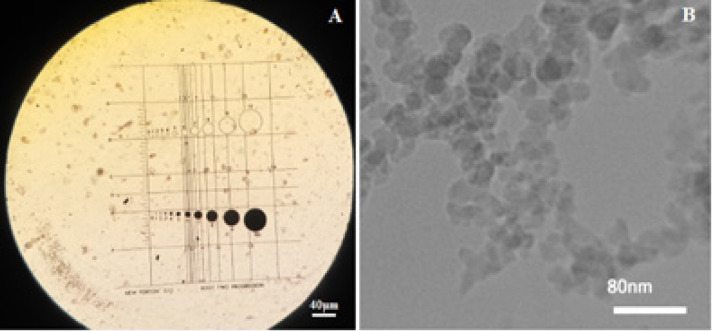
The A-SiO2 particles in two categories of nano (10-100 nm) and micro (< 5um) were used in this study. The human lung A549 cell-line was exposed to either nano- or micro-sized A-SiO2 particles at 10, 50, 100, and 250 μg/ml, and the effects were investigated.

RESULTS

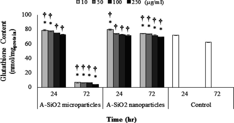
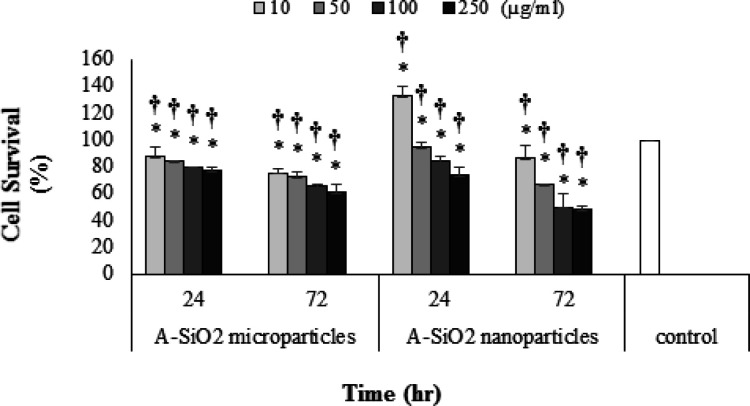
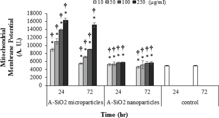
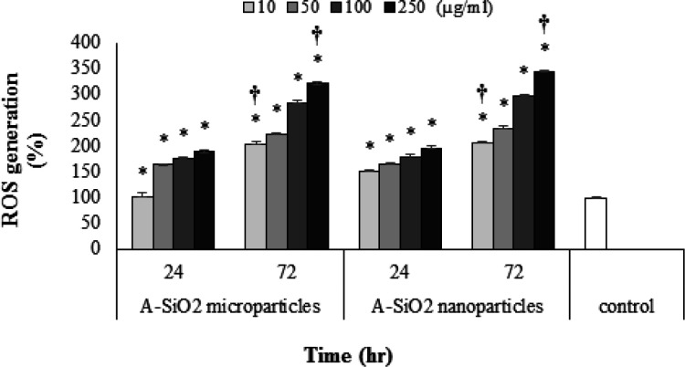
The cytotoxicity of A-SiO2 nano and microparticles in both 24- and 72-hour exposure times resulted in decreased cell survival, mitochondrial membrane potential, and increased ROS generation which was concentration-time dependent (P <0.05) but glutathione content was not affected in a time-dependent manner. Cytotoxicity of nanoparticles, contrary to the previous study, was not higher than microparticles in the comparable dose and exposure times.

CONCLUSION

The rate of ROS generation in the A549 cell-line exposed to A-SiO2 nanoparticles was higher than microparticles. And at the same time, cell survival for exposed cells to A-SiO2 nano and microparticles were higher for nanoparticles in shorter exposure periods and was inversely concentration- and time-dependent. Further studies on exploring the effect of size and its possible toxic mechanism are recommended to achieve a more credible risk assessment.

摘要

简介

非晶态二氧化硅(A-SiO2)在地壳中含量丰富,A-SiO2纳米和微粒子通过工业和制造活动释放到空气中。由于毒理学信息有限,本研究的目的是评估不同尺寸的 A-SiO2 颗粒对体外 A549 细胞系的毒性。

材料和方法

本研究使用了两类 A-SiO2 纳米颗粒(10-100nm)和微颗粒(<5μm)。将人肺 A549 细胞系暴露于纳米或微尺寸的 A-SiO2 颗粒中,浓度分别为 10、50、100 和 250μg/ml,并观察其效果。

结果

A-SiO2 纳米和微颗粒在 24 小时和 72 小时暴露时间内的细胞毒性导致细胞存活率降低,线粒体膜电位降低,ROS 生成增加,呈浓度和时间依赖性(P<0.05),但谷胱甘肽含量无时间依赖性变化。与之前的研究相反,纳米颗粒的细胞毒性在可比剂量和暴露时间内并不高于微颗粒。

结论

暴露于 A-SiO2 纳米颗粒的 A549 细胞系中 ROS 生成速率高于微颗粒。同时,暴露于 A-SiO2 纳米和微颗粒的细胞的细胞存活率在较短的暴露时间内对纳米颗粒更高,呈浓度和时间依赖性反比。建议进一步研究尺寸的影响及其可能的毒性机制,以实现更可信的风险评估。

https://cdn.ncbi.nlm.nih.gov/pmc/blobs/bd1a/8190354/bad031b036fa/APJCP-22-325-g006.jpg
https://cdn.ncbi.nlm.nih.gov/pmc/blobs/bd1a/8190354/a73f1d3b82a6/APJCP-22-325-g001.jpg
https://cdn.ncbi.nlm.nih.gov/pmc/blobs/bd1a/8190354/c873ddc7a9d4/APJCP-22-325-g002.jpg
https://cdn.ncbi.nlm.nih.gov/pmc/blobs/bd1a/8190354/e2b82688bc7c/APJCP-22-325-g003.jpg
https://cdn.ncbi.nlm.nih.gov/pmc/blobs/bd1a/8190354/4206880823a6/APJCP-22-325-g004.jpg
https://cdn.ncbi.nlm.nih.gov/pmc/blobs/bd1a/8190354/7129e031066b/APJCP-22-325-g005.jpg
https://cdn.ncbi.nlm.nih.gov/pmc/blobs/bd1a/8190354/bad031b036fa/APJCP-22-325-g006.jpg

相似文献

1
The Effect of Particle Size on the Cytotoxicity of Amorphous Silicon Dioxide: An in Vitro Toxicological Study.粒径对无定形二氧化硅细胞毒性的影响:体外毒理学研究。
Asian Pac J Cancer Prev. 2021 Feb 1;22(2):325-332. doi: 10.31557/APJCP.2021.22.2.325.
2
Cytotoxic effects of crystalline silica in form of micro and nanoparticles on the human lung cell line A549.微米和纳米级晶体二氧化硅对人肺细胞系A549的细胞毒性作用。
Toxicol Ind Health. 2023 Jan;39(1):23-35. doi: 10.1177/07482337221140644. Epub 2022 Nov 26.
3
Combined exposure to nano-silica and lead induced potentiation of oxidative stress and DNA damage in human lung epithelial cells.纳米二氧化硅和铅联合暴露致肺上皮细胞氧化应激和 DNA 损伤增强。
Ecotoxicol Environ Saf. 2015 Dec;122:537-44. doi: 10.1016/j.ecoenv.2015.09.030. Epub 2015 Sep 29.
4
Investigation of the effect of magnetite iron oxide particles size on cytotoxicity in A cell line.磁铁矿氧化铁颗粒大小对A细胞系细胞毒性影响的研究。
Toxicol Ind Health. 2019 Nov-Dec;35(11-12):703-713. doi: 10.1177/0748233719888077.
5
The effect of single and combined exposures to magnetite and polymorphous silicon dioxide nanoparticles on the human A cell line: in vitro study.单一和联合暴露于磁铁矿和多晶态二氧化硅纳米颗粒对人 A 细胞系的影响:体外研究。
Environ Sci Pollut Res Int. 2019 Nov;26(31):31752-31762. doi: 10.1007/s11356-019-06229-0. Epub 2019 Sep 4.
6
SiO2 nanoparticles induce cytotoxicity and protein expression alteration in HaCaT cells.二氧化硅纳米颗粒诱导 HaCaT 细胞产生细胞毒性和蛋白表达改变。
Part Fibre Toxicol. 2010 Jan 19;7:1. doi: 10.1186/1743-8977-7-1.
7
Assessment of temporal dose-toxicity relationship of fumed silica nanoparticle in human lung A549 cells by conventional cytotoxicity and ¹H-NMR-based extracellular metabonomic assays.通过常规细胞毒性测定和基于¹H-NMR 的细胞外代谢组学检测评估人肺 A549 细胞中烟炱二氧化硅纳米颗粒的时间剂量-毒性关系。
Toxicol Sci. 2014 Apr;138(2):354-64. doi: 10.1093/toxsci/kfu009. Epub 2014 Jan 21.
8
Combined toxicity of amorphous silica nanoparticles and methylmercury to human lung epithelial cells.无定形二氧化硅纳米颗粒与甲基汞对人肺上皮细胞的联合毒性。
Ecotoxicol Environ Saf. 2015 Feb;112:144-52. doi: 10.1016/j.ecoenv.2014.10.026. Epub 2014 Nov 15.
9
Amphipathic silica nanoparticles induce cytotoxicity through oxidative stress mediated and p53 dependent apoptosis pathway in human liver cell line HL-7702 and rat liver cell line BRL-3A.两亲性二氧化硅纳米颗粒通过氧化应激介导的和p53依赖性凋亡途径在人肝细胞系HL-7702和大鼠肝细胞系BRL-3A中诱导细胞毒性。
Colloids Surf B Biointerfaces. 2016 Sep 1;145:232-240. doi: 10.1016/j.colsurfb.2016.05.006. Epub 2016 May 5.
10
Interactive toxicity effect of combined exposure to hematite and amorphous silicon dioxide nanoparticles in human A cell line.人 A 细胞系中赤铁矿和无定形二氧化硅纳米颗粒联合暴露的交互毒性效应。
Toxicol Ind Health. 2021 May;37(5):289-302. doi: 10.1177/07482337211002373.

引用本文的文献

1
Benchmark dose determining airborne crystalline silica particles based on A549 lung-cell line survival in an in vitro study.基于体外 A549 肺细胞系生存试验的空气中结晶二氧化硅颗粒基准剂量的确定。
Sci Rep. 2024 Sep 13;14(1):21458. doi: 10.1038/s41598-024-72607-5.
2
Nanoparticles and Airway Epithelial Cells: Exploring the Impacts and Methodologies in Toxicity Assessment.纳米颗粒与气道上皮细胞:毒性评估中的影响与方法探索。
Int J Mol Sci. 2024 Jul 18;25(14):7885. doi: 10.3390/ijms25147885.
3
Silica nanoparticles-induced cytotoxicity and genotoxicity in A549 cell lines.

本文引用的文献

1
The toxicity of silica nanoparticles to the immune system.硅纳米颗粒对免疫系统的毒性。
Nanomedicine (Lond). 2018 Aug 1;13(15):1939-1962. doi: 10.2217/nnm-2018-0076. Epub 2018 Aug 28.
2
Subacute inhalation toxicity study of synthetic amorphous silica nanoparticles in Sprague-Dawley rats.合成无定形二氧化硅纳米颗粒对Sprague-Dawley大鼠的亚急性吸入毒性研究。
Inhal Toxicol. 2017 Oct-Dec;29(12-14):567-576. doi: 10.1080/08958378.2018.1426661. Epub 2018 Feb 5.
3
Estimates and 25-year trends of the global burden of disease attributable to ambient air pollution: an analysis of data from the Global Burden of Diseases Study 2015.
硅纳米颗粒诱导 A549 细胞系的细胞毒性和遗传毒性。
Sci Rep. 2024 Jun 24;14(1):14484. doi: 10.1038/s41598-024-65333-5.
4
Safety Considerations for Lyophilized Human Amniotic Membrane Impregnated with Colistin and Silver Nanoparticles.载有黏菌素和银纳米粒子的冻干人羊膜的安全性考虑。
Appl Biochem Biotechnol. 2024 Mar;196(3):1419-1434. doi: 10.1007/s12010-023-04618-3. Epub 2023 Jul 7.
5
Airborne LTA Nanozeolites Characterization during the Manufacturing Process and External Sources Interaction with the Workplace Background.制造过程中及与工作场所背景的外部源相互作用期间的空气传播LTA纳米沸石特性
Nanomaterials (Basel). 2022 Apr 24;12(9):1448. doi: 10.3390/nano12091448.
6
Mechanistic study of silica nanoparticles on the size-dependent retinal toxicity in vitro and in vivo.关于体外和体内二氧化硅纳米颗粒的大小依赖性视网膜毒性的机制研究。
J Nanobiotechnology. 2022 Mar 19;20(1):146. doi: 10.1186/s12951-022-01326-8.
归因于环境空气污染的全球疾病负担估计数和 25 年趋势:2015 年全球疾病负担研究数据分析。
Lancet. 2017 May 13;389(10082):1907-1918. doi: 10.1016/S0140-6736(17)30505-6. Epub 2017 Apr 10.
4
Nanopurification of silicon from 84% to 99.999% purity with a simple and scalable process.通过简单且可扩展的工艺将硅的纯度从84%提高到99.999%。
Proc Natl Acad Sci U S A. 2015 Nov 3;112(44):13473-7. doi: 10.1073/pnas.1513012112. Epub 2015 Oct 19.
5
Mesoporous silica nanoparticles in drug delivery and biomedical applications.介孔硅纳米粒子在药物传递和生物医学应用中的作用。
Nanomedicine. 2015 Feb;11(2):313-27. doi: 10.1016/j.nano.2014.09.014. Epub 2014 Nov 13.
6
Silica nanoparticle uptake induces survival mechanism in A549 cells by the activation of autophagy but not apoptosis.硅纳米颗粒摄取通过自噬而不是细胞凋亡激活 A549 细胞的存活机制。
Toxicol Lett. 2014 Jan 3;224(1):84-92. doi: 10.1016/j.toxlet.2013.10.003. Epub 2013 Oct 16.
7
Diterpenylhydroquinones from natural ent-labdanes induce apoptosis through decreased mitochondrial membrane potential.天然 ent-劳丹烷的二萜基对苯二酚通过降低线粒体膜电位诱导细胞凋亡。
Molecules. 2013 May 10;18(5):5348-59. doi: 10.3390/molecules18055348.
8
Silica nanoparticles-induced cytotoxicity, oxidative stress and apoptosis in cultured A431 and A549 cells.二氧化硅纳米颗粒诱导培养的 A431 和 A549 细胞的细胞毒性、氧化应激和凋亡。
Hum Exp Toxicol. 2013 Feb;32(2):186-95. doi: 10.1177/0960327112459206.
9
Silicosis.矽肺。
Lancet. 2012 May 26;379(9830):2008-18. doi: 10.1016/S0140-6736(12)60235-9. Epub 2012 Apr 24.
10
Cytotoxicity and genotoxicity of nanosized and microsized titanium dioxide and iron oxide particles in Syrian hamster embryo cells.纳米级和微米级二氧化钛及氧化铁颗粒对叙利亚仓鼠胚胎细胞的细胞毒性和遗传毒性
Ann Occup Hyg. 2012 Jul;56(5):631-44. doi: 10.1093/annhyg/mes006. Epub 2012 Mar 26.