• 文献检索
  • 文档翻译
  • 深度研究
  • 学术资讯
  • Suppr Zotero 插件Zotero 插件
  • 邀请有礼
  • 套餐&价格
  • 历史记录
应用&插件
Suppr Zotero 插件Zotero 插件浏览器插件Mac 客户端Windows 客户端微信小程序
定价
高级版会员购买积分包购买API积分包
服务
文献检索文档翻译深度研究API 文档MCP 服务
关于我们
关于 Suppr公司介绍联系我们用户协议隐私条款
关注我们

Suppr 超能文献

核心技术专利:CN118964589B侵权必究
粤ICP备2023148730 号-1Suppr @ 2026

文献检索

告别复杂PubMed语法,用中文像聊天一样搜索,搜遍4000万医学文献。AI智能推荐,让科研检索更轻松。

立即免费搜索

文件翻译

保留排版,准确专业,支持PDF/Word/PPT等文件格式,支持 12+语言互译。

免费翻译文档

深度研究

AI帮你快速写综述,25分钟生成高质量综述,智能提取关键信息,辅助科研写作。

立即免费体验

使用条件生成对抗神经网络对睑板腺功能障碍进行基于图像的分析。

Image based analysis of meibomian gland dysfunction using conditional generative adversarial neural network.

作者信息

Khan Zakir Khan, Umar Arif Iqbal, Shirazi Syed Hamad, Rasheed Asad, Qadir Abdul, Gul Sarah

机构信息

Information Technology, Hazara University, Mansehra, Pakistan.

Biological Sciences, International Islamic University, Islamabad, Pakistan.

出版信息

BMJ Open Ophthalmol. 2021 Feb 12;6(1):e000436. doi: 10.1136/bmjophth-2020-000436. eCollection 2021.

DOI:10.1136/bmjophth-2020-000436
PMID:33644402
原文链接:https://pmc.ncbi.nlm.nih.gov/articles/PMC7883862/
Abstract

OBJECTIVE

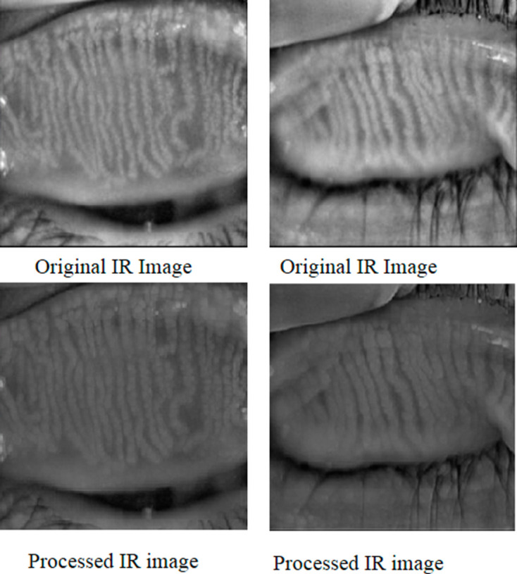
Meibomian gland dysfunction (MGD) is a primary cause of dry eye disease. Analysis of MGD, its severity, shapes and variation in the acini of the meibomian glands (MGs) is receiving much attention in ophthalmology clinics. Existing methods for diagnosing, detection and analysing meibomianitis are not capable to quantify the irregularities to IR (infrared) images of MG area such as light reflection, interglands and intraglands boundaries, the improper focus of the light and positioning, and eyelid eversion.

METHODS AND ANALYSIS

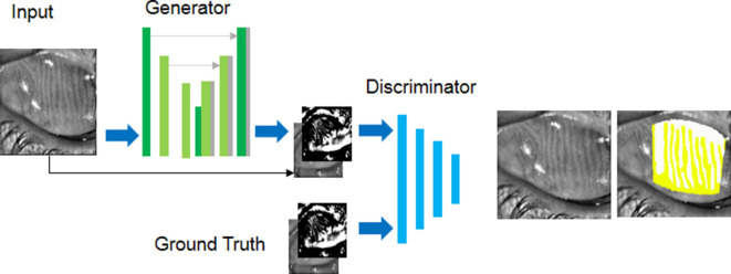
We proposed a model that is based on adversarial learning that is, conditional generative adversarial network that can overcome these blatant challenges. The generator of the model learns the mapping from the IR images of the MG to a confidence map specifying the probabilities of being a pixel of MG. The discriminative part of the model is responsible to penalise the mismatch between the IR images of the MG and confidence map. Furthermore, the adversarial learning assists the generator to produce a qualitative confidence map which is transformed into binary images with the help of fixed thresholding to fulfil the segmentation of MG. We identified MGs and interglands boundaries from IR images.

RESULTS

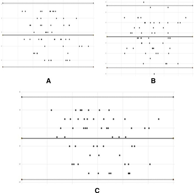
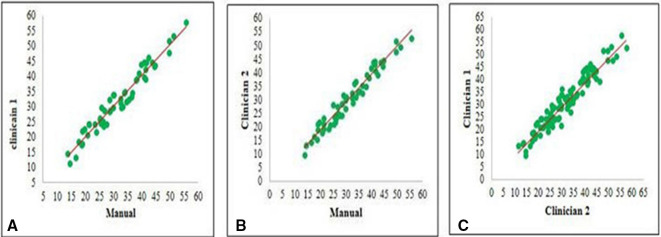
This method is evaluated by meiboscoring, grading, Pearson correlation and Bland-Altman analysis. We also judged the quality of our method through average Pompeiu-Hausdorff distance, and Aggregated Jaccard Index.

CONCLUSIONS

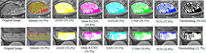
This technique provides a significant improvement in the quantification of the irregularities to IR. This technique has outperformed the state-of-art results for the detection and analysis of the dropout area of MGD.

摘要

目的

睑板腺功能障碍(MGD)是干眼疾病的主要病因。睑板腺(MG)腺泡中MGD及其严重程度、形态和变化的分析在眼科诊所备受关注。现有的睑板腺炎诊断、检测和分析方法无法对MG区域红外(IR)图像的不规则性进行量化,如光反射、腺间和腺内边界、光聚焦和定位不当以及眼睑外翻。

方法与分析

我们提出了一种基于对抗学习的模型,即条件生成对抗网络,该模型可以克服这些明显的挑战。该模型的生成器学习从MG的IR图像到指定为MG像素概率的置信图的映射。模型的判别部分负责惩罚MG的IR图像与置信图之间的不匹配。此外,对抗学习有助于生成器生成定性置信图,该置信图在固定阈值的帮助下转换为二值图像,以完成MG的分割。我们从IR图像中识别出MG和腺间边界。

结果

该方法通过睑板腺评分、分级、Pearson相关性和Bland-Altman分析进行评估。我们还通过平均Pompeiu-Hausdorff距离和聚合Jaccard指数来判断我们方法的质量。

结论

该技术在对IR图像不规则性的量化方面有显著改进。该技术在MGD缺失区域的检测和分析方面优于现有技术成果。

https://cdn.ncbi.nlm.nih.gov/pmc/blobs/d828/7883862/0f64f82765db/bmjophth-2020-000436f05.jpg
https://cdn.ncbi.nlm.nih.gov/pmc/blobs/d828/7883862/f8bda849d347/bmjophth-2020-000436f01.jpg
https://cdn.ncbi.nlm.nih.gov/pmc/blobs/d828/7883862/9f06968c41f0/bmjophth-2020-000436f02.jpg
https://cdn.ncbi.nlm.nih.gov/pmc/blobs/d828/7883862/8fda9c8b3e66/bmjophth-2020-000436f03.jpg
https://cdn.ncbi.nlm.nih.gov/pmc/blobs/d828/7883862/062bca87276a/bmjophth-2020-000436f04.jpg
https://cdn.ncbi.nlm.nih.gov/pmc/blobs/d828/7883862/0f64f82765db/bmjophth-2020-000436f05.jpg

相似文献

1
Image based analysis of meibomian gland dysfunction using conditional generative adversarial neural network.使用条件生成对抗神经网络对睑板腺功能障碍进行基于图像的分析。
BMJ Open Ophthalmol. 2021 Feb 12;6(1):e000436. doi: 10.1136/bmjophth-2020-000436. eCollection 2021.
2
Examination of Gland Dropout Detected on Infrared Meibography by Using Optical Coherence Tomography Meibography.应用光学相干断层扫描眼表仪检查红外照相仪检测到的睑板腺缺失。
Ocul Surf. 2017 Jan;15(1):130-138.e1. doi: 10.1016/j.jtos.2016.10.001. Epub 2016 Nov 2.
3
Image-based quantitative analysis of tear film lipid layer thickness for meibomian gland evaluation.基于图像的泪膜脂质层厚度定量分析用于评估睑板腺。
Biomed Eng Online. 2017 Nov 23;16(1):135. doi: 10.1186/s12938-017-0426-8.
4
Conditional generative adversarial network for 3D rigid-body motion correction in MRI.条件生成对抗网络在 MRI 中用于 3D 刚体运动校正。
Magn Reson Med. 2019 Sep;82(3):901-910. doi: 10.1002/mrm.27772. Epub 2019 Apr 22.
5
[A new classification for meibomian gland diseases with in vivo confocal microscopy].[一种用于睑板腺疾病的新型体内共聚焦显微镜分类法]
J Fr Ophtalmol. 2016 Mar;39(3):239-47. doi: 10.1016/j.jfo.2015.07.015. Epub 2016 Feb 16.
6
Learning from adversarial medical images for X-ray breast mass segmentation.从对抗性医学图像中学习 X 射线乳房肿块分割。
Comput Methods Programs Biomed. 2019 Oct;180:105012. doi: 10.1016/j.cmpb.2019.105012. Epub 2019 Aug 5.
7
A Deep Learning Approach for Meibomian Gland Appearance Evaluation.一种用于睑板腺外观评估的深度学习方法。
Ophthalmol Sci. 2023 May 22;3(4):100334. doi: 10.1016/j.xops.2023.100334. eCollection 2023 Dec.
8
Automatic Meniscus Segmentation Using Adversarial Learning-Based Segmentation Network with Object-Aware Map in Knee MR Images.基于对抗学习的分割网络与目标感知映射在膝关节磁共振成像中自动分割半月板
Diagnostics (Basel). 2021 Sep 3;11(9):1612. doi: 10.3390/diagnostics11091612.
9
Meibomian Gland Morphology Is a Sensitive Early Indicator of Meibomian Gland Dysfunction.睑板腺形态是睑板腺功能障碍的敏感早期指标。
Am J Ophthalmol. 2019 Apr;200:16-25. doi: 10.1016/j.ajo.2018.12.006. Epub 2018 Dec 20.
10
Deep learning-based automatic meibomian gland segmentation and morphology assessment in infrared meibography.基于深度学习的红外睑板腺图像中睑板腺自动分割与形态评估。
Sci Rep. 2021 Apr 7;11(1):7649. doi: 10.1038/s41598-021-87314-8.

引用本文的文献

1
A new digital biomarker of blepharitis: energy curve of the meibomian edge.睑缘炎的一种新型数字生物标志物:睑板边缘能量曲线。
Front Cell Dev Biol. 2025 Aug 5;13:1627327. doi: 10.3389/fcell.2025.1627327. eCollection 2025.
2
Advances in AI-assisted quantification of dry eye indicators.人工智能辅助干眼指标量化的进展。
Front Med (Lausanne). 2025 Jul 18;12:1628311. doi: 10.3389/fmed.2025.1628311. eCollection 2025.
3
Development and multicenter validation of an AI driven model for quantitative meibomian gland evaluation.一种用于睑板腺定量评估的人工智能驱动模型的开发与多中心验证

本文引用的文献

1
Deep Neural Network-Based Method for Detecting Obstructive Meibomian Gland Dysfunction With in Vivo Laser Confocal Microscopy.基于深度神经网络的活体共焦激光显微镜下阻塞性睑板腺功能障碍检测方法。
Cornea. 2020 Jun;39(6):720-725. doi: 10.1097/ICO.0000000000002279.
2
A Deep Learning Approach for Meibomian Gland Atrophy Evaluation in Meibography Images.一种用于睑板腺造影图像中睑板腺萎缩评估的深度学习方法。
Transl Vis Sci Technol. 2019 Dec 18;8(6):37. doi: 10.1167/tvst.8.6.37. eCollection 2019 Nov.
3
DeepLab: Semantic Image Segmentation with Deep Convolutional Nets, Atrous Convolution, and Fully Connected CRFs.
NPJ Digit Med. 2025 Jul 4;8(1):403. doi: 10.1038/s41746-025-01753-5.
4
Ophthalmic Image Synthesis and Analysis with Generative Adversarial Network Artificial Intelligence.基于生成对抗网络人工智能的眼科图像合成与分析
J Imaging Inform Med. 2025 May 20. doi: 10.1007/s10278-025-01519-1.
5
A Future Picture: A Review of Current Generative Adversarial Neural Networks in Vitreoretinal Pathologies and Their Future Potentials.未来图景:当前生成对抗神经网络在玻璃体视网膜病变中的应用及其未来潜力综述
Biomedicines. 2025 Jan 24;13(2):284. doi: 10.3390/biomedicines13020284.
6
Artificial Intelligence Applications in Ophthalmology.人工智能在眼科中的应用。
JMA J. 2025 Jan 15;8(1):66-75. doi: 10.31662/jmaj.2024-0139. Epub 2024 Sep 13.
7
Internal validation of a convolutional neural network pipeline for assessing meibomian gland structure from meibography.用于从睑板腺造影评估睑板腺结构的卷积神经网络流程的内部验证
Optom Vis Sci. 2025 Jan 1;102(1):28-36. doi: 10.1097/OPX.0000000000002208. Epub 2025 Jan 13.
8
Artificial intelligence in corneal diseases: A narrative review.人工智能在角膜疾病中的应用:综述。
Cont Lens Anterior Eye. 2024 Dec;47(6):102284. doi: 10.1016/j.clae.2024.102284. Epub 2024 Aug 27.
9
Meibomian glands segmentation in infrared images with limited annotation.基于有限标注的红外图像中睑板腺分割
Int J Ophthalmol. 2024 Mar 18;17(3):401-407. doi: 10.18240/ijo.2024.03.01. eCollection 2024.
10
A Deep Learning Approach for Meibomian Gland Appearance Evaluation.一种用于睑板腺外观评估的深度学习方法。
Ophthalmol Sci. 2023 May 22;3(4):100334. doi: 10.1016/j.xops.2023.100334. eCollection 2023 Dec.
DeepLab:基于深度卷积网络、空洞卷积和全连接条件随机场的语义图像分割。
IEEE Trans Pattern Anal Mach Intell. 2018 Apr;40(4):834-848. doi: 10.1109/TPAMI.2017.2699184. Epub 2017 Apr 27.
4
A Dataset and a Technique for Generalized Nuclear Segmentation for Computational Pathology.用于计算病理学中通用核分割的数据集和技术。
IEEE Trans Med Imaging. 2017 Jul;36(7):1550-1560. doi: 10.1109/TMI.2017.2677499. Epub 2017 Mar 6.
5
Interexaminer Reliability of Meibography: Upper or Lower Eyelid?睑板腺照相术的检查者间可靠性:上眼睑还是下眼睑?
Eye Contact Lens. 2018 Mar;44(2):113-117. doi: 10.1097/ICL.0000000000000307.
6
Utility of meibography in the evaluation of meibomian glands morphology in normal and diseased eyelids.睑板腺造影术在评估正常及患病眼睑睑板腺形态中的应用价值。
Saudi J Ophthalmol. 2011 Jan;25(1):61-6. doi: 10.1016/j.sjopt.2010.10.005. Epub 2010 Oct 13.
7
Statistics corner: A guide to appropriate use of correlation coefficient in medical research.统计专栏:医学研究中相关系数合理应用指南
Malawi Med J. 2012 Sep;24(3):69-71.
8
Comparison of subjective grading and objective assessment in meibography.主观分级与客观评估在眼表相干光断层扫描中的比较。
Cont Lens Anterior Eye. 2013 Feb;36(1):22-7. doi: 10.1016/j.clae.2012.10.074. Epub 2012 Oct 27.
9
Interrater reliability: the kappa statistic.组内一致性:kappa 统计量。
Biochem Med (Zagreb). 2012;22(3):276-82.
10
A newly developed noninvasive and mobile pen-shaped meibography system.一种新开发的非侵入式、可移动的笔形睑板腺照相系统。
Cornea. 2013 Mar;32(3):242-7. doi: 10.1097/ICO.0b013e31825425ef.