• 文献检索
  • 文档翻译
  • 深度研究
  • 学术资讯
  • Suppr Zotero 插件Zotero 插件
  • 邀请有礼
  • 套餐&价格
  • 历史记录
应用&插件
Suppr Zotero 插件Zotero 插件浏览器插件Mac 客户端Windows 客户端微信小程序
定价
高级版会员购买积分包购买API积分包
服务
文献检索文档翻译深度研究API 文档MCP 服务
关于我们
关于 Suppr公司介绍联系我们用户协议隐私条款
关注我们

Suppr 超能文献

核心技术专利:CN118964589B侵权必究
粤ICP备2023148730 号-1Suppr @ 2026

文献检索

告别复杂PubMed语法,用中文像聊天一样搜索,搜遍4000万医学文献。AI智能推荐,让科研检索更轻松。

立即免费搜索

文件翻译

保留排版,准确专业,支持PDF/Word/PPT等文件格式,支持 12+语言互译。

免费翻译文档

深度研究

AI帮你快速写综述,25分钟生成高质量综述,智能提取关键信息,辅助科研写作。

立即免费体验

利用微晶体电子衍射进行大分子晶体学研究。

Macromolecular crystallography using microcrystal electron diffraction.

机构信息

Department of Materials and Environmental Chemistry, Stockholm University, 106 91 Stockholm, Sweden.

出版信息

Acta Crystallogr D Struct Biol. 2021 Mar 1;77(Pt 3):313-324. doi: 10.1107/S2059798320016368. Epub 2021 Feb 17.

DOI:10.1107/S2059798320016368
PMID:33645535
原文链接:https://pmc.ncbi.nlm.nih.gov/articles/PMC7919406/
Abstract

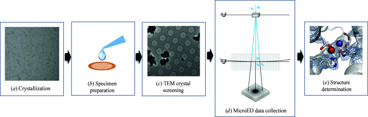
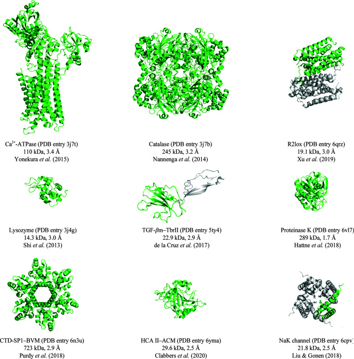
Microcrystal electron diffraction (MicroED) has recently emerged as a promising method for macromolecular structure determination in structural biology. Since the first protein structure was determined in 2013, the method has been evolving rapidly. Several protein structures have been determined and various studies indicate that MicroED is capable of (i) revealing atomic structures with charges, (ii) solving new protein structures by molecular replacement, (iii) visualizing ligand-binding interactions and (iv) determining membrane-protein structures from microcrystals embedded in lipidic mesophases. However, further development and optimization is required to make MicroED experiments more accurate and more accessible to the structural biology community. Here, we provide an overview of the current status of the field, and highlight the ongoing development, to provide an indication of where the field may be going in the coming years. We anticipate that MicroED will become a robust method for macromolecular structure determination, complementing existing methods in structural biology.

摘要

微晶电子衍射(MicroED)最近作为一种有前途的方法在结构生物学中用于确定大分子结构而出现。自 2013 年首次确定蛋白质结构以来,该方法一直在迅速发展。已经确定了几个蛋白质结构,并且各种研究表明 MicroED 能够 (i) 揭示带电荷的原子结构,(ii) 通过分子置换解决新的蛋白质结构,(iii) 可视化配体结合相互作用,以及 (iv) 从嵌入脂质中间相的微晶中确定膜蛋白结构。然而,需要进一步的开发和优化,以使 MicroED 实验更准确,并更容易被结构生物学界所接受。在这里,我们提供了该领域的现状概述,并强调了正在进行的发展,以表明该领域在未来几年可能的发展方向。我们预计 MicroED 将成为一种用于确定大分子结构的强大方法,补充结构生物学中现有的方法。

https://cdn.ncbi.nlm.nih.gov/pmc/blobs/1c35/7919406/57b9dfbe884a/d-77-00313-fig4.jpg
https://cdn.ncbi.nlm.nih.gov/pmc/blobs/1c35/7919406/0666f360aebe/d-77-00313-fig1.jpg
https://cdn.ncbi.nlm.nih.gov/pmc/blobs/1c35/7919406/54bba9d8fdcf/d-77-00313-fig2.jpg
https://cdn.ncbi.nlm.nih.gov/pmc/blobs/1c35/7919406/597d0eb21439/d-77-00313-fig3.jpg
https://cdn.ncbi.nlm.nih.gov/pmc/blobs/1c35/7919406/57b9dfbe884a/d-77-00313-fig4.jpg

相似文献

1
Macromolecular crystallography using microcrystal electron diffraction.利用微晶体电子衍射进行大分子晶体学研究。
Acta Crystallogr D Struct Biol. 2021 Mar 1;77(Pt 3):313-324. doi: 10.1107/S2059798320016368. Epub 2021 Feb 17.
2
Advances and applications of microcrystal electron diffraction (MicroED).微晶体电子衍射(MicroED)的进展与应用。
Curr Opin Struct Biol. 2024 Feb;84:102741. doi: 10.1016/j.sbi.2023.102741. Epub 2023 Dec 11.
3
Microcrystal electron diffraction in macromolecular and pharmaceutical structure determination.微晶电子衍射在大分子和药物结构测定中的应用。
Drug Discov Today Technol. 2020 Dec;37:93-105. doi: 10.1016/j.ddtec.2020.12.002. Epub 2020 Dec 31.
4
Comparing serial X-ray crystallography and microcrystal electron diffraction (MicroED) as methods for routine structure determination from small macromolecular crystals.比较串行X射线晶体学和微晶电子衍射(MicroED)作为从小分子晶体进行常规结构测定的方法。
IUCrJ. 2020 Feb 26;7(Pt 2):306-323. doi: 10.1107/S205225252000072X. eCollection 2020 Mar 1.
5
Visualizing drug binding interactions using microcrystal electron diffraction.利用微晶体电子衍射可视化药物结合相互作用。
Commun Biol. 2020 Jul 31;3(1):417. doi: 10.1038/s42003-020-01155-1.
6
An Overview of Microcrystal Electron Diffraction (MicroED).微晶体电子衍射(MicroED)概述。
Annu Rev Biochem. 2021 Jun 20;90:431-450. doi: 10.1146/annurev-biochem-081720-020121.
7
Studying membrane proteins with MicroED.用微晶电子衍射技术研究膜蛋白。
Biochem Soc Trans. 2022 Feb 28;50(1):231-239. doi: 10.1042/BST20210911.
8
Protocol for the use of focused ion-beam milling to prepare crystalline lamellae for microcrystal electron diffraction (MicroED).使用聚焦离子束研磨制备用于微晶电子衍射(MicroED)的晶体薄片的协议。
STAR Protoc. 2021 Jul 28;2(3):100686. doi: 10.1016/j.xpro.2021.100686. eCollection 2021 Sep 17.
9
The complementarity of serial femtosecond crystallography and MicroED for structure determination from microcrystals.串行飞秒晶体学和微束电子衍射在微晶体结构测定中的互补性。
Curr Opin Struct Biol. 2019 Oct;58:286-293. doi: 10.1016/j.sbi.2019.06.004. Epub 2019 Jul 22.
10
High-Resolution Macromolecular Structure Determination by MicroED, a cryo-EM Method.通过MicroED(一种冷冻电镜方法)进行高分辨率大分子结构测定。
Methods Enzymol. 2016;579:369-92. doi: 10.1016/bs.mie.2016.04.017. Epub 2016 Jun 16.

引用本文的文献

1
Small but mighty: the power of microcrystals in structural biology.小而强大:微晶在结构生物学中的力量。
IUCrJ. 2025 May 1;12(Pt 3):262-279. doi: 10.1107/S2052252525001484.
2
Comprehensive microcrystal electron diffraction sample preparation for cryo-EM.用于冷冻电子显微镜的综合微晶电子衍射样品制备
Nat Protoc. 2025 May;20(5):1275-1309. doi: 10.1038/s41596-024-01088-7. Epub 2024 Dec 20.
3
How to use DIALS to process chemical crystallography 3D ED rotation data from pixel array detectors.如何使用DIALS处理来自像素阵列探测器的化学晶体学3D ED旋转数据。

本文引用的文献

1
Statistically correcting dynamical electron scattering improves the refinement of protein nanocrystals, including charge refinement of coordinated metals.统计校正动力学电子散射提高了蛋白质纳米晶体的精修,包括配位金属的电荷精修。
Acta Crystallogr D Struct Biol. 2021 Jan 1;77(Pt 1):75-85. doi: 10.1107/S2059798320014540.
2
Demonstration of electron diffraction from membrane protein crystals grown in a lipidic mesophase after lamella preparation by focused ion beam milling at cryogenic temperatures.在低温下通过聚焦离子束铣削制备薄片后,对脂质中间相生长的膜蛋白晶体进行电子衍射演示。
J Appl Crystallogr. 2020 Oct 13;53(Pt 6):1416-1424. doi: 10.1107/S1600576720013096. eCollection 2020 Dec 1.
3
Acta Crystallogr C Struct Chem. 2025 Jan 1;81(Pt 1):1-13. doi: 10.1107/S2053229624011148.
4
Microcrystal electron diffraction structure of Toll-like receptor 2 TIR-domain-nucleated MyD88 TIR-domain higher-order assembly.Toll 样受体 2 TIR 结构域引发的 MyD88 TIR 结构域高阶组装的微晶电子衍射结构。
Acta Crystallogr D Struct Biol. 2024 Sep 1;80(Pt 9):699-712. doi: 10.1107/S2059798324008210. Epub 2024 Sep 4.
5
On the structure refinement of metal complexes against 3D electron diffraction data using multipolar scattering factors.关于使用多极散射因子对金属配合物进行三维电子衍射数据结构精修的研究
IUCrJ. 2024 Sep 1;11(Pt 5):878-890. doi: 10.1107/S2052252524006730.
6
Structure determination of a DNA crystal by MicroED.通过微晶电子衍射法测定DNA晶体的结构。
Structure. 2023 Dec 7;31(12):1499-1503.e2. doi: 10.1016/j.str.2023.07.005. Epub 2023 Aug 3.
7
Protein Lipidation Types: Current Strategies for Enrichment and Characterization.蛋白质脂化类型:富集与表征的当前策略
Int J Mol Sci. 2022 Feb 21;23(4):2365. doi: 10.3390/ijms23042365.
8
DIALS as a toolkit.DIALS 工具包。
Protein Sci. 2022 Jan;31(1):232-250. doi: 10.1002/pro.4224. Epub 2021 Nov 24.
9
Metalloprotein catalysis: structural and mechanistic insights into oxidoreductases from neutron protein crystallography.金属蛋白酶催化:从中子蛋白晶体学角度对氧化还原酶的结构和机制见解。
Acta Crystallogr D Struct Biol. 2021 Oct 1;77(Pt 10):1251-1269. doi: 10.1107/S2059798321009025. Epub 2021 Sep 27.
MicroED structure of lipid-embedded mammalian mitochondrial voltage-dependent anion channel.
脂质嵌入的哺乳动物线粒体电压依赖性阴离子通道的 MicroED 结构。
Proc Natl Acad Sci U S A. 2020 Dec 22;117(51):32380-32385. doi: 10.1073/pnas.2020010117. Epub 2020 Dec 8.
4
Through-grid wicking enables high-speed cryoEM specimen preparation.通过网格吸液法可实现高速冷冻电镜样品制备。
Acta Crystallogr D Struct Biol. 2020 Nov 1;76(Pt 11):1092-1103. doi: 10.1107/S2059798320012474. Epub 2020 Oct 13.
5
Single-particle cryo-EM at atomic resolution.单颗粒 cryo-EM 在原子分辨率下。
Nature. 2020 Nov;587(7832):152-156. doi: 10.1038/s41586-020-2829-0. Epub 2020 Oct 21.
6
Atomic-resolution protein structure determination by cryo-EM.利用冷冻电镜技术进行原子分辨率的蛋白质结构测定。
Nature. 2020 Nov;587(7832):157-161. doi: 10.1038/s41586-020-2833-4. Epub 2020 Oct 21.
7
A Workflow for Protein Structure Determination From Thin Crystal Lamella by Micro-Electron Diffraction.一种通过微电子衍射从薄晶体薄片确定蛋白质结构的工作流程。
Front Mol Biosci. 2020 Aug 4;7:179. doi: 10.3389/fmolb.2020.00179. eCollection 2020.
8
Inelastic scattering and solvent scattering reduce dynamical diffraction in biological crystals.非弹性散射和溶剂散射会降低生物晶体中的动态衍射。
Acta Crystallogr B Struct Sci Cryst Eng Mater. 2019 Aug 1;75(Pt 4):523-531. doi: 10.1107/S2052520619009661.
9
Fragment-based determination of a proteinase K structure from MicroED data using ARCIMBOLDO_SHREDDER.基于碎片的 MicroED 数据的蛋白酶 K 结构解析,使用 ARCIMBOLDO_SHREDDER。
Acta Crystallogr D Struct Biol. 2020 Aug 1;76(Pt 8):703-712. doi: 10.1107/S2059798320008049. Epub 2020 Jul 27.
10
Visualizing drug binding interactions using microcrystal electron diffraction.利用微晶体电子衍射可视化药物结合相互作用。
Commun Biol. 2020 Jul 31;3(1):417. doi: 10.1038/s42003-020-01155-1.