Department of Biochemistry, University of Illinois, Urbana, Illinois 61801, USA.
Cancer Center@Illinois, University of Illinois, Urbana, Illinois 61801, USA.
Genome Res. 2021 Apr;31(4):576-591. doi: 10.1101/gr.267013.120. Epub 2021 Mar 1.
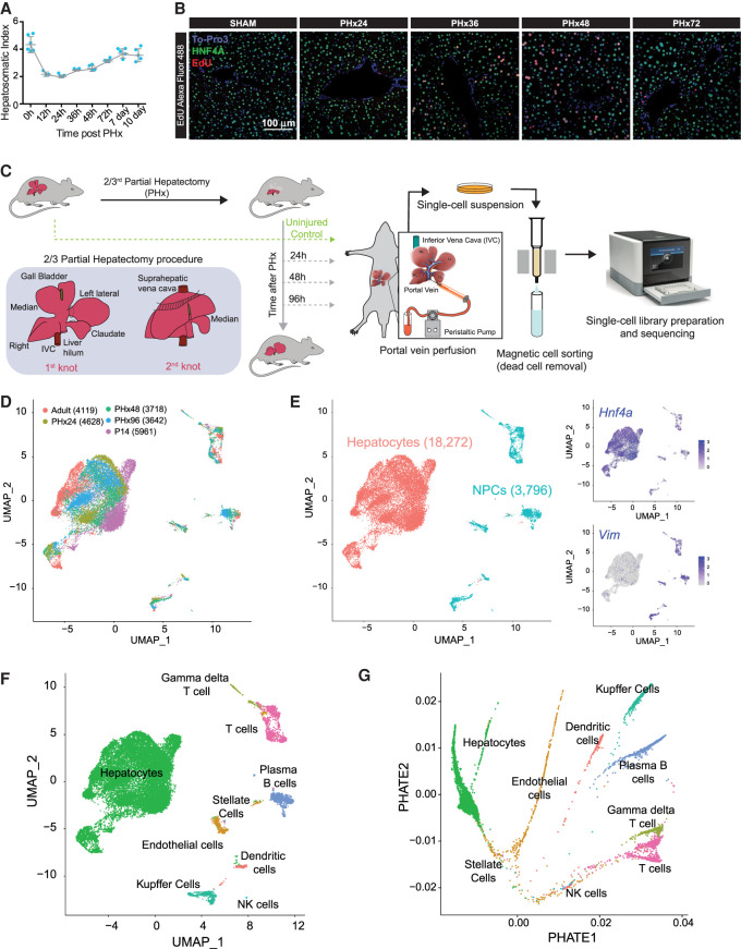
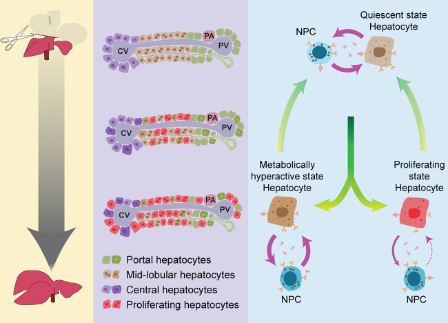
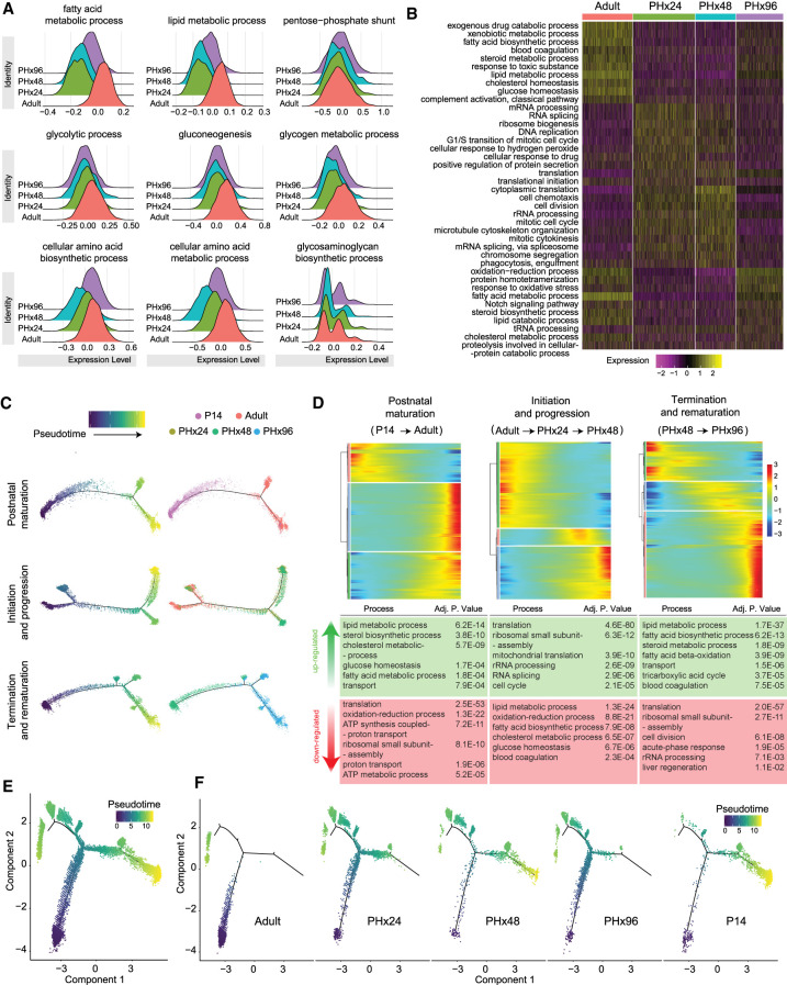
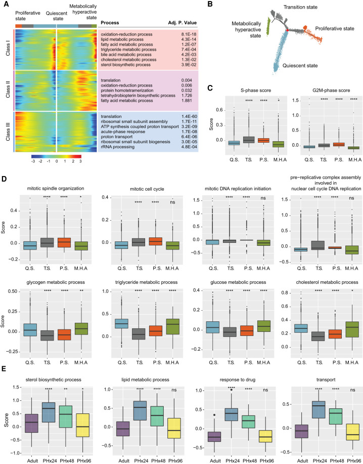
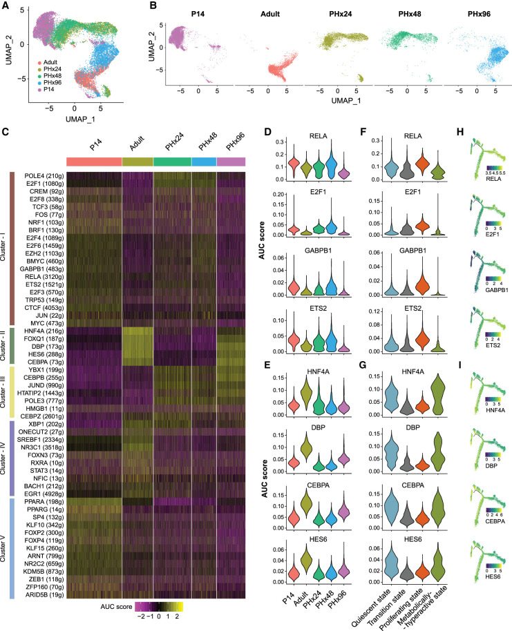
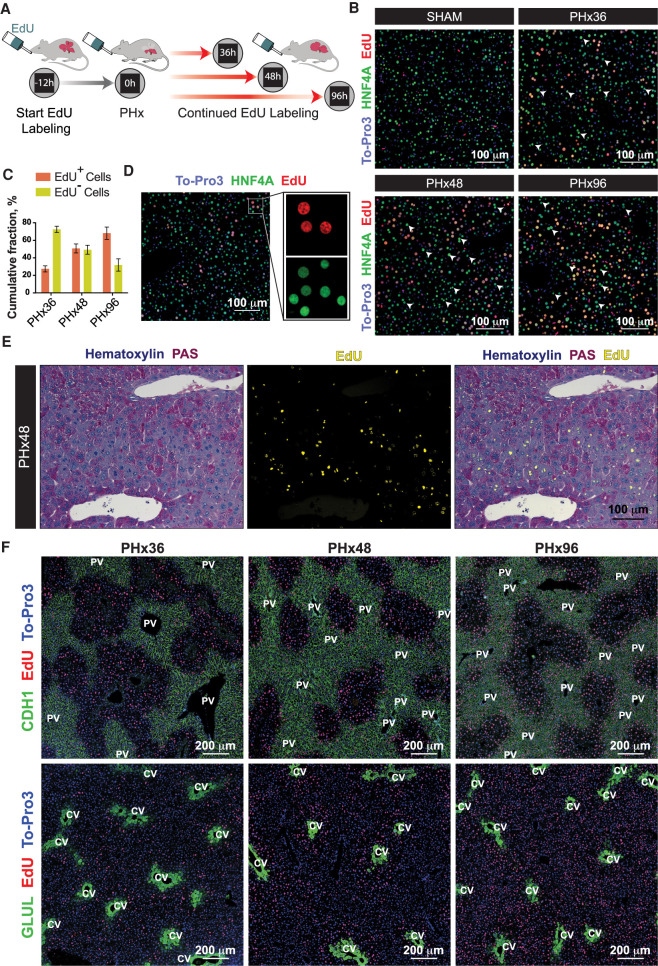
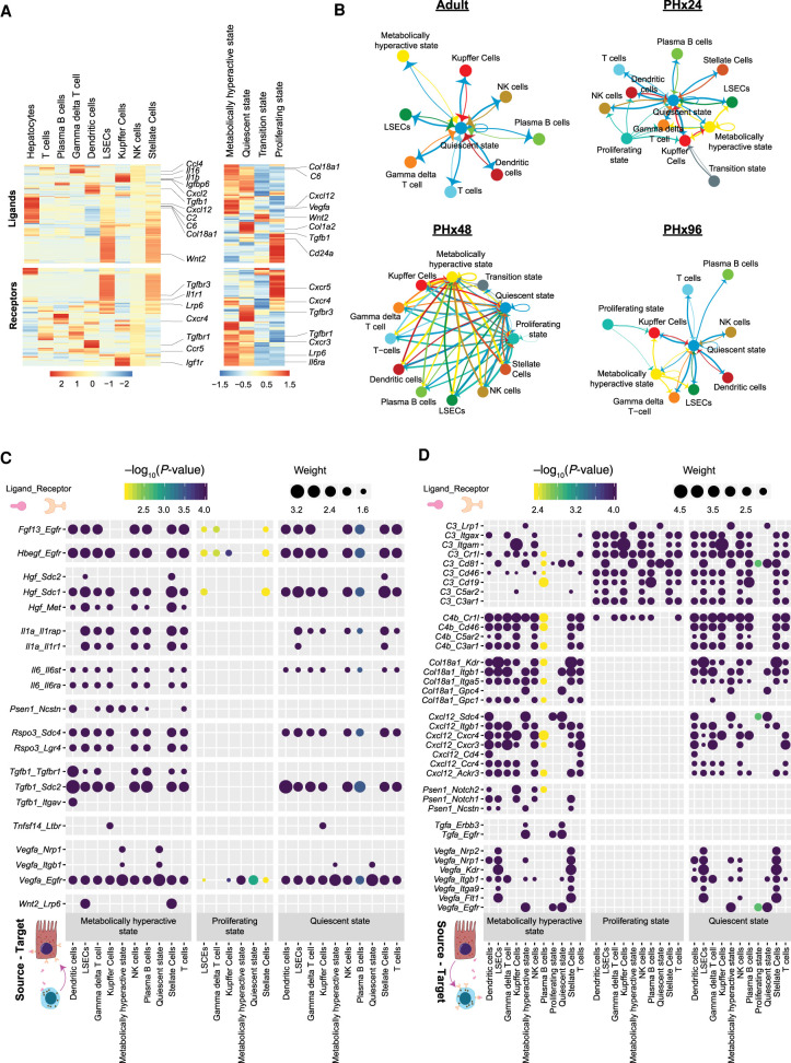
The adult liver has an exceptional ability to regenerate, but how it maintains its specialized functions during regeneration is unclear. Here, we used partial hepatectomy (PHx) in tandem with single-cell transcriptomics to track cellular transitions and heterogeneities of ∼22,000 liver cells through the initiation, progression, and termination phases of mouse liver regeneration. Our results uncovered that, following PHx, a subset of hepatocytes transiently reactivates an early-postnatal-like gene expression program to proliferate, while a distinct population of metabolically hyperactive cells appears to compensate for any temporary deficits in liver function. Cumulative EdU labeling and immunostaining of metabolic, portal, and central vein-specific markers revealed that hepatocyte proliferation after PHx initiates in the midlobular region before proceeding toward the periportal and pericentral areas. We further demonstrate that portal and central vein proximal hepatocytes retain their metabolically active state to preserve essential liver functions while midlobular cells proliferate nearby. Through combined analysis of gene regulatory networks and cell-cell interaction maps, we found that regenerating hepatocytes redeploy key developmental regulons, which are guided by extensive ligand-receptor-mediated signaling events between hepatocytes and nonparenchymal cells. Altogether, our study offers a detailed blueprint of the intercellular crosstalk and cellular reprogramming that balances the metabolic and proliferative requirements of a regenerating liver.
成年肝脏具有非凡的再生能力,但它如何在再生过程中维持其特化功能尚不清楚。在这里,我们使用部分肝切除术 (PHx) 与单细胞转录组学相结合,通过跟踪约 22000 个肝细胞在小鼠肝再生的起始、进展和终止阶段的细胞转变和异质性。我们的结果揭示了,在 PHx 之后,一小部分肝细胞短暂地重新激活类似于出生后早期的基因表达程序来增殖,而一群代谢活性较高的细胞似乎可以弥补任何暂时的肝功能缺陷。累积 EdU 标记和代谢、门脉和中央静脉特异性标志物的免疫染色显示,PHx 后肝细胞的增殖首先从中小叶区域开始,然后向门周和中央区域推进。我们进一步证明,门脉和中央静脉近端肝细胞保持其代谢活跃状态,以维持重要的肝功能,而中小叶细胞在附近增殖。通过基因调控网络和细胞-细胞相互作用图谱的综合分析,我们发现再生的肝细胞重新利用关键的发育调控网络,这是由肝细胞和非实质细胞之间广泛的配体-受体介导的信号事件指导的。总的来说,我们的研究提供了一个详细的细胞间通讯和细胞重编程蓝图,平衡了再生肝脏的代谢和增殖需求。