Yao Mengyu, Cheng Shi, Zhong Guoqing, Zhou Jielong, Shao Hongwei, Ma Limin, Du Chang, Peng Feng, Zhang Yu
Department of Biomedical Engineering, School of Materials Science and Engineering, South China University of Technology, Guangzhou, 510641, China.
Department of Orthopedics, Guangdong Provincial People's Hospital, Guangdong Academy of Medical Sciences, Guangzhou, Guangdong, 510080, China.
Bioact Mater. 2021 Feb 13;6(9):2729-2741. doi: 10.1016/j.bioactmat.2021.02.003. eCollection 2021 Sep.
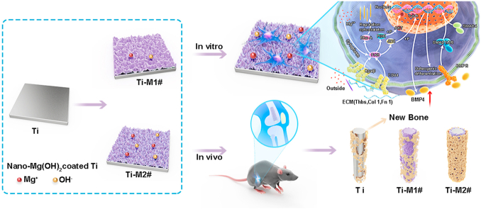
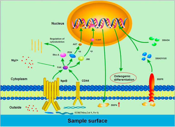
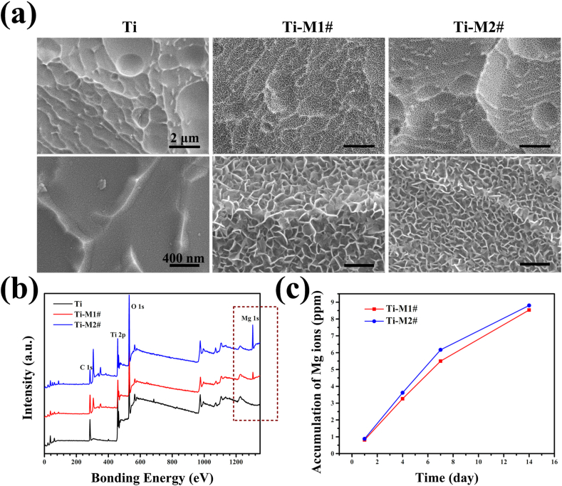
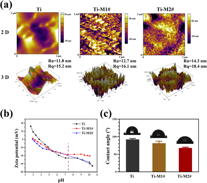
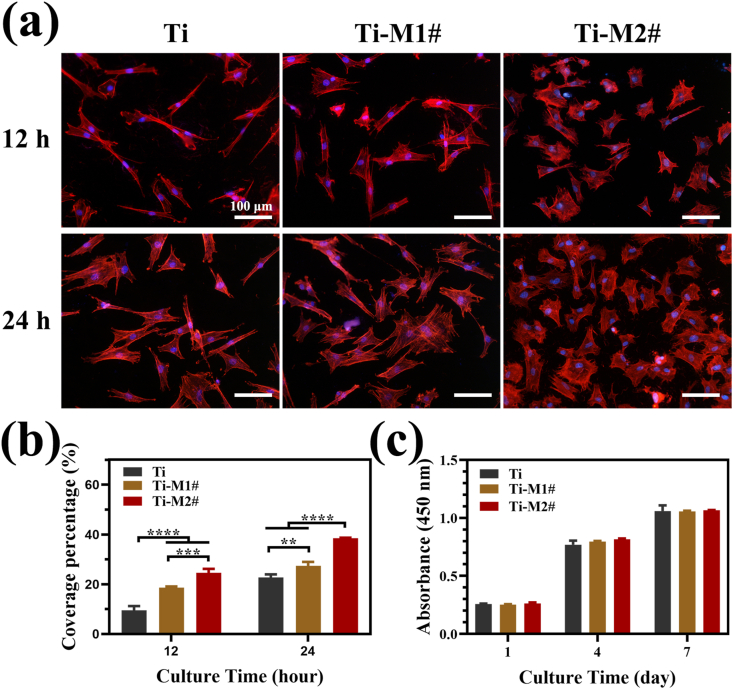
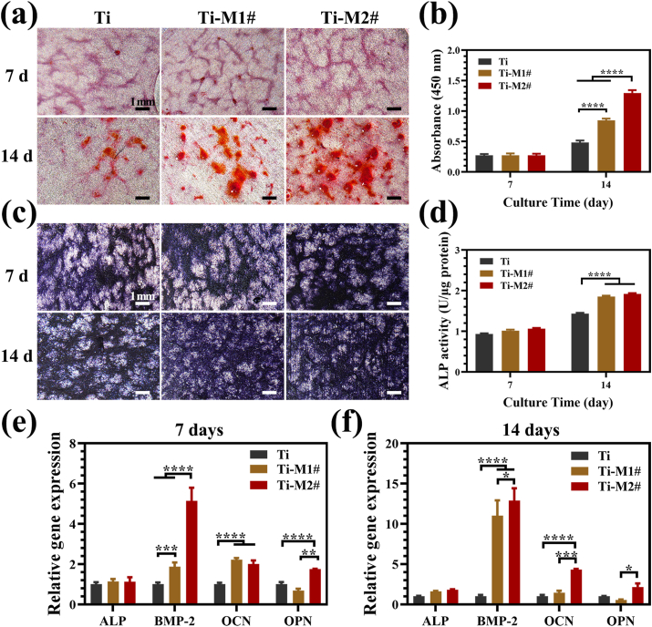
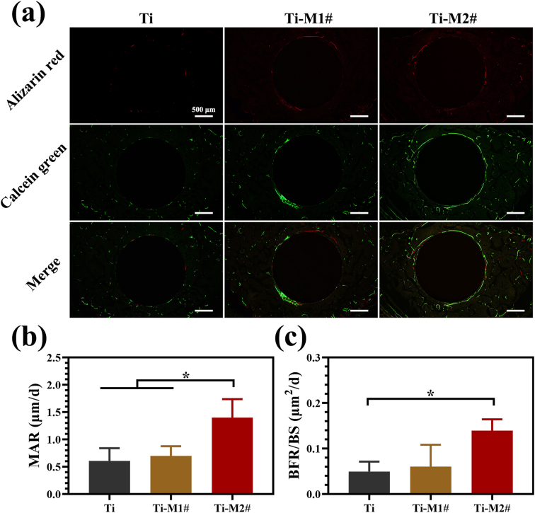
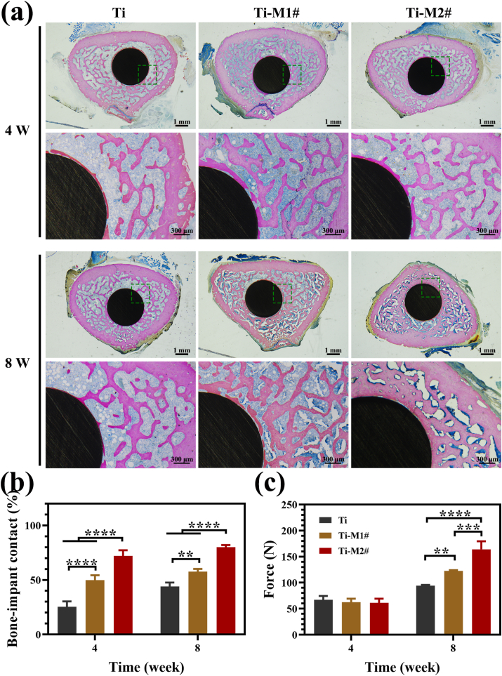
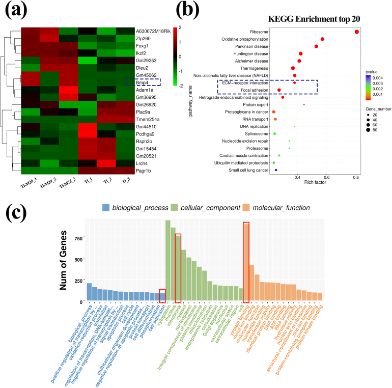
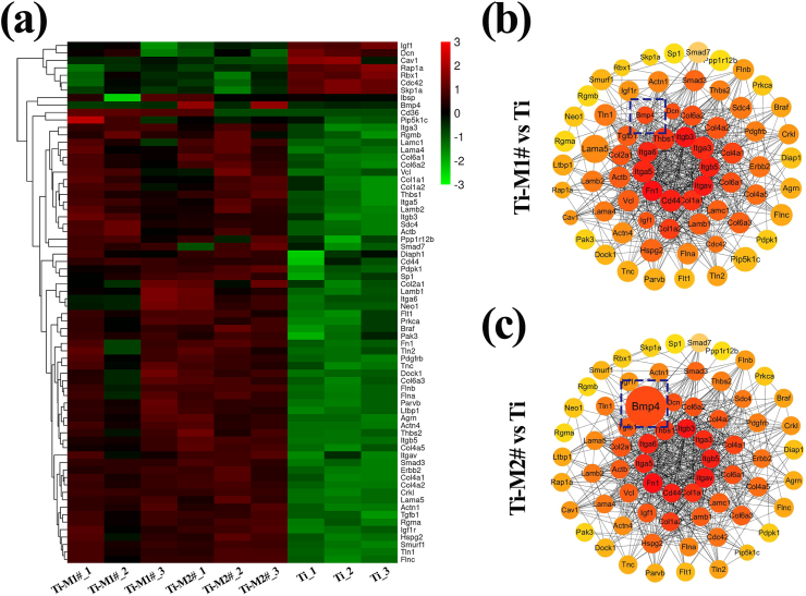
Titanium (Ti) has been the most widely used orthopedic implant in the past decades. However, their inert surface often leads to insufficient osteointegration of Ti implant. To solve this issue, two bioactive Mg(OH) films were developed on Ti surfaces using hydrothermal treatment (Ti-M1# and Ti-M2#). The Mg(OH) films showed nano-flake structures: sheets on Ti-M1# with a thickness of 14.7 ± 0.7 nm and a length of 131.5 ± 2.9 nm, and on Ti-M2# with a thickness of 13.4 ± 2.2 nm and a length of 56.9 ± 5.6 nm. Both films worked as Mg ions releasing platforms. With the gradual degradation of Mg(OH) films, weakly alkaline microenvironments will be established surrounding the modified implants. Benefiting from the sustained release of Mg ions, nanostructures, and weakly alkaline microenvironments, the as-prepared nano-Mg(OH) coated Ti showed better and osteogenesis. Notably, Ti-M2# showed better osteogenesis than Ti-M1#, which can be ascribed to its smaller nanostructure. Moreover, whole genome expression analysis was applied to study the osteogenic mechanism of nano-Mg(OH) films. For both coated samples, most of the genes related to ECM-receptor interaction, focal adhesion, and TGF-β pathways were upregulated, indicating that these signaling pathways were activated, leading to better osteogenesis. Furthermore, cells cultured on Ti-M2# showed markedly upregulated BMP-4 gene expression, suggesting that the nanostructure with Mg ion release ability can better activate BMP-4 related signaling pathways, resulting in better osteogenesis. Nano-Mg(OH) films demonstrated a superior osteogenesis and are promising surface modification strategy for orthopedic applications.
在过去几十年中,钛(Ti)一直是应用最为广泛的骨科植入物。然而,其惰性表面常常导致钛植入物的骨整合不足。为了解决这一问题,通过水热处理在钛表面制备了两种具有生物活性的Mg(OH)薄膜(Ti-M1#和Ti-M2#)。Mg(OH)薄膜呈现出纳米片状结构:Ti-M1#上的薄片厚度为14.7±0.7nm,长度为131.5±2.9nm;Ti-M2#上的薄片厚度为13.4±2.2nm,长度为56.9±5.6nm。两种薄膜均作为镁离子释放平台。随着Mg(OH)薄膜的逐渐降解,在改性植入物周围将建立弱碱性微环境。得益于镁离子的持续释放、纳米结构以及弱碱性微环境,所制备的纳米Mg(OH)涂层钛表现出更好的成骨能力。值得注意的是,Ti-M2#的成骨能力优于Ti-M1#,这可归因于其较小的纳米结构。此外,应用全基因组表达分析来研究纳米Mg(OH)薄膜的成骨机制。对于两种涂层样品,大多数与细胞外基质-受体相互作用、粘着斑和TGF-β信号通路相关的基因均上调,表明这些信号通路被激活,从而导致更好的成骨能力。此外,在Ti-M2#上培养的细胞显示BMP-4基因表达明显上调,这表明具有镁离子释放能力的纳米结构能够更好地激活与BMP-4相关的信号通路,从而产生更好的成骨能力。纳米Mg(OH)薄膜展现出卓越的成骨能力,是一种很有前景的骨科应用表面改性策略。