Alwalid Osamah, Long Xi, Xie Mingfei, Yang Jiehua, Cen Chunyuan, Liu Huan, Han Ping
Department of Radiology, Union Hospital, Tongji Medical College, Huazhong University of Science and Technology, Wuhan, China.
Hubei Province Key Laboratory of Molecular Imaging, Wuhan, China.
Front Neurol. 2021 Feb 22;12:619864. doi: 10.3389/fneur.2021.619864. eCollection 2021.
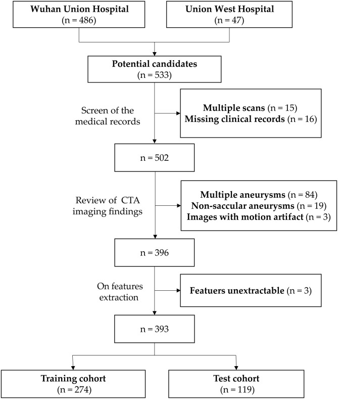
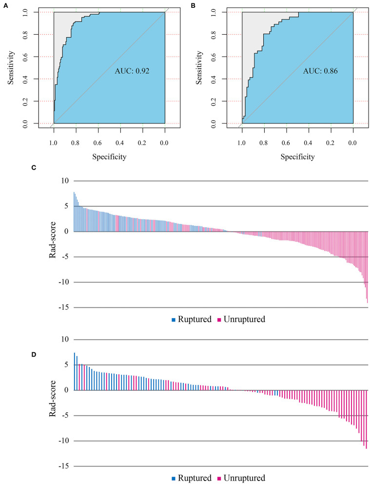
Intracranial aneurysm rupture is a devastating medical event with a high morbidity and mortality rate. Thus, timely detection and management are critical. The present study aimed to identify the aneurysm radiomics features associated with rupture and to build and evaluate a radiomics classification model of aneurysm rupture. Radiomics analysis was applied to CT angiography (CTA) images of 393 patients [152 (38.7%) with ruptured aneurysms]. Patients were divided at a ratio of 7:3 into retrospective training ( = 274) and prospective test ( = 119) cohorts. A total of 1,229 radiomics features were automatically calculated from each aneurysm. The feature number was systematically reduced, and the most important classifying features were selected. A logistic regression model was constructed using the selected features and evaluated on training and test cohorts. Radiomics score (Rad-score) was calculated for each patient and compared between ruptured and unruptured aneurysms. Nine radiomics features were selected from the CTA images and used to build the logistic regression model. The radiomics model has shown good performance in the classification of the aneurysm rupture on training and test cohorts [area under the receiver operating characteristic curve: 0.92 [95% confidence interval CI: 0.89-0.95] and 0.86 [95% CI: 0.80-0.93], respectively, < 0.001]. Rad-score showed statistically significant differences between ruptured and unruptured aneurysms (median, 2.50 vs. -1.60 and 2.35 vs. -1.01 on training and test cohorts, respectively, < 0.001). The results indicated the potential of aneurysm radiomics features for automatic classification of aneurysm rupture on CTA images.
颅内动脉瘤破裂是一种具有高发病率和死亡率的灾难性医学事件。因此,及时检测和处理至关重要。本研究旨在识别与破裂相关的动脉瘤影像组学特征,并构建和评估动脉瘤破裂的影像组学分类模型。将影像组学分析应用于393例患者的CT血管造影(CTA)图像[152例(38.7%)为破裂动脉瘤患者]。患者按7:3的比例分为回顾性训练队列(n = 274)和前瞻性测试队列(n = 119)。从每个动脉瘤中自动计算出总共1229个影像组学特征。系统地减少特征数量,并选择最重要的分类特征。使用所选特征构建逻辑回归模型,并在训练和测试队列中进行评估。为每位患者计算影像组学评分(Rad-score),并在破裂和未破裂动脉瘤之间进行比较。从CTA图像中选择了9个影像组学特征用于构建逻辑回归模型。该影像组学模型在训练和测试队列的动脉瘤破裂分类中表现出良好性能[受试者操作特征曲线下面积:分别为0.92[95%置信区间CI:0.89 - 0.95]和0.86[95%CI:0.80 - 0.93],P < 0.001]。Rad-score在破裂和未破裂动脉瘤之间显示出统计学显著差异(训练和测试队列中中位数分别为2.50对 -1.60和2.35对 -1.01,P < 0.001)。结果表明动脉瘤影像组学特征在CTA图像上对动脉瘤破裂进行自动分类的潜力。