Delcroix Olivier, Bourhis David, Keromnes Nathalie, Robin Philippe, Le Roux Pierre-Yves, Abgral Ronan, Salaun Pierre-Yves, Querellou Solène
Nuclear Medicine Department, Brest University Hospital, Brest, France.
EA 3878 GETBO IFR, Brest, France.
Front Med (Lausanne). 2021 Feb 22;8:629096. doi: 10.3389/fmed.2021.629096. eCollection 2021.
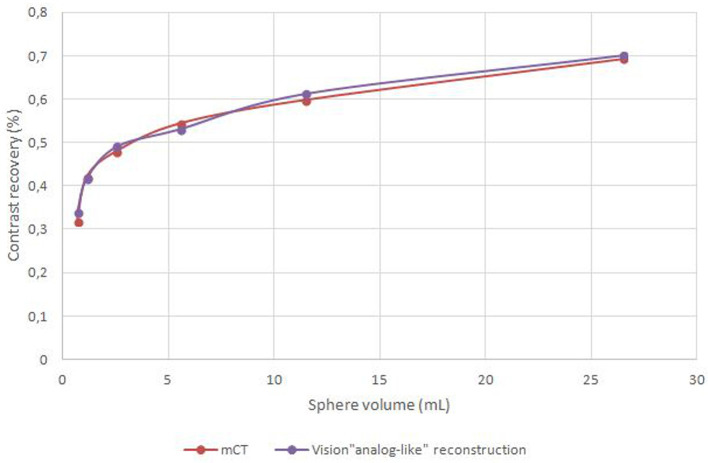
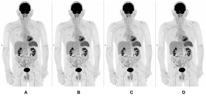
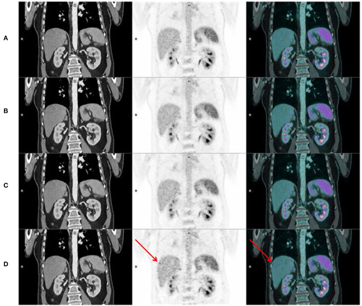
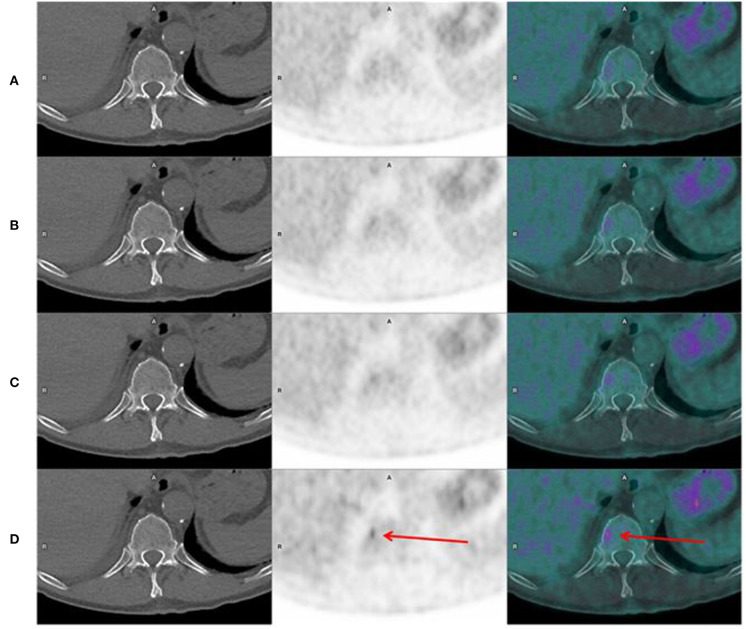
The aim of this study was to assess image quality and lesion detectability acquired with a digital Positron Emission Tomography/Computed Tomography (PET/CT) Siemens Biograph Vision 600 system. Consecutive patients who underwent a FDG PET/CT during the first week of use of a digital PET/CT (Siemens Biograph Vision 600) at the nuclear medicine department of the university hospital of Brest were analyzed. PET were realized using list mode acquisition. For all patients, 4 datasets were reconstructed. We determined, according to phantom measurements, an equivalent time acquisition/reconstruction parameters pair of the digital PET/CT corresponding to an analog PET/CT image quality ("analog-like") as reference dataset. We compared the reference dataset with 3 others digital PET/CT reconstruction parameters, allowing a decrease of emission duration: 60, 90, and 120 s per bed position. Three nuclear medicine physicians evaluated independently, for each dataset, overall image quality [Maximal Intensity Projection (MIP), noise, sharpness] using a 4-point scale. Physicians assessed also lesion detection capability by reporting new visible lesions on each digital datasets with their confidence level in comparison with analog-like dataset. Ninety-eight patients were analyzed. Image quality of MIP (IQ), sharpness (IQ), and noise (IQ) of all digital datasets (60, 90, and 120 s) were better than those evaluated with analog-like reconstruction. Moreover, digital PET/CT system improved IQ, IQ, and IQ whatever the BMI. Lesion detection capability and confidence level were higher for 60, 90, 120 s per bed position, respectively, than for analog-like images. Our study demonstrated an improvement of image quality and lesion detectability with a digital PET/CT system.
本研究的目的是评估使用数字正电子发射断层扫描/计算机断层扫描(PET/CT)西门子Biograph Vision 600系统获得的图像质量和病变可检测性。对在布雷斯特大学医院核医学科使用数字PET/CT(西门子Biograph Vision 600)的第一周内接受FDG PET/CT检查的连续患者进行了分析。PET采用列表模式采集。对所有患者重建了4个数据集。根据体模测量,我们确定了与模拟PET/CT图像质量(“类模拟”)相对应的数字PET/CT的等效时间采集/重建参数对作为参考数据集。我们将参考数据集与其他3种数字PET/CT重建参数进行了比较,这些参数允许发射持续时间减少:每个床位位置分别为60、90和120秒。三名核医学医生对每个数据集独立评估总体图像质量[最大强度投影(MIP)、噪声、清晰度],采用4分制。医生还通过报告每个数字数据集上与类模拟数据集相比新出现的可见病变及其置信水平来评估病变检测能力。共分析了9名患者。所有数字数据集(60、90和120秒)的MIP图像质量(IQ)、清晰度(IQ)和噪声(IQ)均优于类模拟重建评估的结果。此外,无论BMI如何,数字PET/CT系统均改善了IQ、IQ和IQ。每个床位位置60、90、120秒时的病变检测能力和置信水平分别高于类模拟图像。我们的研究证明了数字PET/CT系统在图像质量和病变可检测性方面的改善。 (注:原文中“Ninety-eight patients were analyzed.”疑有误,根据语境推测可能是“Ninety patients were analyzed.”,翻译时按推测内容翻译,若实际不是该内容,请根据正确原文调整。)