• 文献检索
  • 文档翻译
  • 深度研究
  • 学术资讯
  • Suppr Zotero 插件Zotero 插件
  • 邀请有礼
  • 套餐&价格
  • 历史记录
应用&插件
Suppr Zotero 插件Zotero 插件浏览器插件Mac 客户端Windows 客户端微信小程序
定价
高级版会员购买积分包购买API积分包
服务
文献检索文档翻译深度研究API 文档MCP 服务
关于我们
关于 Suppr公司介绍联系我们用户协议隐私条款
关注我们

Suppr 超能文献

核心技术专利:CN118964589B侵权必究
粤ICP备2023148730 号-1Suppr @ 2026

文献检索

告别复杂PubMed语法,用中文像聊天一样搜索,搜遍4000万医学文献。AI智能推荐,让科研检索更轻松。

立即免费搜索

文件翻译

保留排版,准确专业,支持PDF/Word/PPT等文件格式,支持 12+语言互译。

免费翻译文档

深度研究

AI帮你快速写综述,25分钟生成高质量综述,智能提取关键信息,辅助科研写作。

立即免费体验

COVID-19 患者出院后肺部残留后遗症的薄层 CT 表现及纵向变化:一项短期随访研究。

Thin-section computed tomography findings and longitudinal variations of the residual pulmonary sequelae after discharge in patients with COVID-19: a short-term follow-up study.

机构信息

Department of Radiology, National Clinical Research Center for Infectious Disease, Shenzhen Third People's Hospital, The Second Affiliated Hospital, School of Medicine Southern University of Science and Technology, 29 Bulan Road, Longgang District, Shenzhen, 518000, Guangdong, China.

Department of Infectious Disease, National Clinical Research Center for Infectious Disease, Shenzhen Third People's Hospital, The Second Affiliated Hospital, School of Medicine Southern University of Science and Technology, 29 Bulan Road, Longgang District, Shenzhen, 518000, Guangdong, China.

出版信息

Eur Radiol. 2021 Sep;31(9):7172-7183. doi: 10.1007/s00330-021-07799-9. Epub 2021 Mar 11.

DOI:10.1007/s00330-021-07799-9
PMID:33704519
原文链接:https://pmc.ncbi.nlm.nih.gov/articles/PMC7950423/
Abstract

OBJECTIVES

This study analyzed and compared CT findings and longitudinal variations after discharge between severe and non-severe coronavirus disease (COVID-19) patients who had residual pulmonary sequelae at pre-discharge.

METHODS

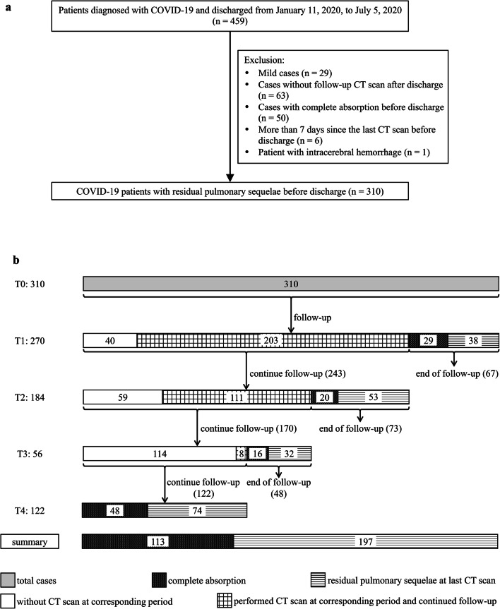
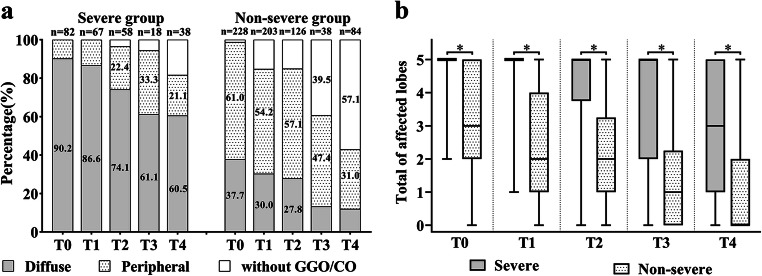
A total of 310 patients were included and stratified into severe and non-severe COVID-19 groups. Cross-sectional CT features across different time periods (T0: pre-discharge, T1: 1-4 weeks after discharge, T2: 5-8 weeks after discharge, T3: 9-12 weeks after discharge, T4: > 12 weeks after discharge) were compared, and the longitudinal variations of CT findings were analyzed and compared in both groups.

RESULTS

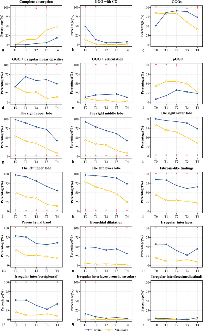
The cumulative absorption rate of fibrosis-like findings in the severe and non-severe groups at T4 was 24.3% (17/70) and 52.0% (53/102), respectively. In both groups, ground-glass opacity (GGO) with consolidation showed a clear decreasing trend at T1, after which they maintained similar lower levels. The GGO in the severe group showed an increasing trend first at T1 and then decreasing at T4; however, the incidence decreased gradually in the non-severe group. Most fibrosis-like findings showed a tendency to decrease rapidly and then remained stable. Bronchial dilatation in the severe group persisted at an intermediate level.

CONCLUSIONS

After discharge, the characteristics and changing trends of pulmonary sequelae caused by COVID-19 were significantly different between the two groups. Pulmonary sequelae were more serious and recovery was slower in patients with severe/critical disease than in patients with moderate disease. A portion of the fibrosis-like findings were completely absorbed in patients with moderate and severe/critical diseases.

KEY POINTS

• Lung sequelae were more serious and recovery was slower in severe/critical COVID-19 patients. • Complete absorption of fibrosis-like findings after a short-term follow-up was observed in at least 17/70 (24.3%) of COVID-19 patients with severe/critical disease and 53/102 (52.0%) of COVID-19 patients with moderate disease. • The most common fibrosis-like findings was a parenchymal band; irregular interface was a nonspecific sign of COVID-19, and the percentage of bronchial dilatation in patients with severe/critical disease remained at a relatively stable medium level (range, 31.6 to 47.8%) at all stages.

摘要

目的

本研究分析比较了出院时存在肺部后遗症的重症和非重症新型冠状病毒病(COVID-19)患者的 CT 表现和出院后纵向变化。

方法

共纳入 310 例患者,分为重症和非重症 COVID-19 组。比较不同时间点(T0:出院前,T1:出院后 1-4 周,T2:出院后 5-8 周,T3:出院后 9-12 周,T4:>12 周)的横断面 CT 特征,并分析比较两组 CT 表现的纵向变化。

结果

重症和非重症组在 T4 时纤维化样表现的累积吸收率分别为 24.3%(17/70)和 52.0%(53/102)。两组磨玻璃影(GGO)伴实变均在 T1 时呈明显下降趋势,之后维持在较低水平。重症组 GGO 在 T1 时先呈上升趋势,然后在 T4 时下降,但在非重症组中,发生率逐渐下降。大多数纤维化样表现呈快速下降后稳定趋势。重症组支气管扩张持续处于中等水平。

结论

出院后,COVID-19 引起的肺部后遗症的特征和变化趋势在两组间有显著差异。重症/危重症患者的肺部后遗症更严重,恢复更慢。部分纤维化样表现可在中重度 COVID-19 患者中完全吸收。

关键点

  • 重症/危重症 COVID-19 患者的肺部后遗症更严重,恢复更慢。

  • 在重症/危重症 COVID-19 患者中,至少有 17/70(24.3%)和 53/102(52.0%)纤维化样表现完全吸收,而在中症 COVID-19 患者中观察到至少 17/70(24.3%)和 53/102(52.0%)纤维化样表现完全吸收。

  • 最常见的纤维化样表现为肺实质带;界面不规则是非 COVID-19 的非特异性表现,重症/危重症患者支气管扩张的比例在各阶段均保持在相对稳定的中等水平(31.6%至 47.8%)。

https://cdn.ncbi.nlm.nih.gov/pmc/blobs/ef17/7950423/12ba46b03dda/330_2021_7799_Fig6_HTML.jpg
https://cdn.ncbi.nlm.nih.gov/pmc/blobs/ef17/7950423/60c6e78c2309/330_2021_7799_Fig1_HTML.jpg
https://cdn.ncbi.nlm.nih.gov/pmc/blobs/ef17/7950423/1ad50f9a2a12/330_2021_7799_Fig2_HTML.jpg
https://cdn.ncbi.nlm.nih.gov/pmc/blobs/ef17/7950423/dea42c20c8c6/330_2021_7799_Fig3_HTML.jpg
https://cdn.ncbi.nlm.nih.gov/pmc/blobs/ef17/7950423/7d54a2995119/330_2021_7799_Fig4_HTML.jpg
https://cdn.ncbi.nlm.nih.gov/pmc/blobs/ef17/7950423/9d459f4930ad/330_2021_7799_Fig5_HTML.jpg
https://cdn.ncbi.nlm.nih.gov/pmc/blobs/ef17/7950423/12ba46b03dda/330_2021_7799_Fig6_HTML.jpg

相似文献

1
Thin-section computed tomography findings and longitudinal variations of the residual pulmonary sequelae after discharge in patients with COVID-19: a short-term follow-up study.COVID-19 患者出院后肺部残留后遗症的薄层 CT 表现及纵向变化:一项短期随访研究。
Eur Radiol. 2021 Sep;31(9):7172-7183. doi: 10.1007/s00330-021-07799-9. Epub 2021 Mar 11.
2
The pulmonary sequalae in discharged patients with COVID-19: a short-term observational study.出院的 COVID-19 患者的肺部后遗症:一项短期观察性研究。
Respir Res. 2020 May 24;21(1):125. doi: 10.1186/s12931-020-01385-1.
3
Analysis of thin-section CT in patients with coronavirus disease (COVID-19) after hospital discharge.出院后新型冠状病毒肺炎(COVID-19)患者的薄层 CT 分析。
J Xray Sci Technol. 2020;28(3):383-389. doi: 10.3233/XST-200685.
4
Chest Computed Tomography and Clinical Follow-Up of Discharged Patients with COVID-19 in Wenzhou City, Zhejiang, China.中国浙江省温州市出院 COVID-19 患者的胸部计算机断层扫描和临床随访。
Ann Am Thorac Soc. 2020 Oct;17(10):1231-1237. doi: 10.1513/AnnalsATS.202004-324OC.
5
Prediction of the Development of Pulmonary Fibrosis Using Serial Thin-Section CT and Clinical Features in Patients Discharged after Treatment for COVID-19 Pneumonia.利用 COVID-19 肺炎治疗后出院患者的系列薄层 CT 和临床特征预测肺纤维化的发展。
Korean J Radiol. 2020 Jun;21(6):746-755. doi: 10.3348/kjr.2020.0215.
6
[Spatial and temporal distribution and predictive value of chest CT scoring in patients with COVID-19].新型冠状病毒肺炎患者胸部CT评分的时空分布及预测价值
Zhonghua Jie He He Hu Xi Za Zhi. 2021 Mar 12;44(3):230-236. doi: 10.3760/cma.j.cn112147-20200522-00626.
7
Longitudinal Radiological Findings in Patients With COVID-19 With Different Severities: From Onset to Long-Term Follow-Up After Discharge.不同严重程度的COVID-19患者的纵向影像学表现:从发病到出院后的长期随访
Front Med (Lausanne). 2021 Sep 21;8:711435. doi: 10.3389/fmed.2021.711435. eCollection 2021.
8
One-year follow-up CT findings in COVID-19 patients: A systematic review and meta-analysis.COVID-19 患者的 1 年随访 CT 表现:系统评价和荟萃分析。
Respirology. 2022 Aug;27(8):605-616. doi: 10.1111/resp.14311. Epub 2022 Jun 12.
9
Structural and Functional Pulmonary Assessment in Severe COVID-19 Survivors at 12 Months after Discharge.严重 COVID-19 幸存者出院后 12 个月的结构性和功能性肺部评估。
Tomography. 2022 Oct 13;8(5):2588-2603. doi: 10.3390/tomography8050216.
10
Analysis of Chest CT Results of Coronavirus Disease 2019 (COVID-19) Patients at First Follow-Up.分析 2019 年冠状病毒病(COVID-19)患者首次随访时的胸部 CT 结果。
Can Respir J. 2020 Nov 1;2020:5328267. doi: 10.1155/2020/5328267. eCollection 2020.

引用本文的文献

1
Matrix metalloprotease-7 is associated with post-COVID-19 persistent lung abnormalities.基质金属蛋白酶-7与新冠病毒感染后持续的肺部异常有关。
ERJ Open Res. 2024 Dec 2;10(6). doi: 10.1183/23120541.00266-2024. eCollection 2024 Nov.
2
COVID-19 Chest Manifestation on CT Scan and Associated Risk Factors for Developing Pulmonary Fibrosis.新冠病毒肺炎CT扫描的胸部表现及发生肺纤维化的相关危险因素
Cureus. 2024 Mar 21;16(3):e56616. doi: 10.7759/cureus.56616. eCollection 2024 Mar.
3
Systematic Review of the Prevalence of Long COVID.“长新冠”患病率的系统评价

本文引用的文献

1
Chest Computed Tomography and Clinical Follow-Up of Discharged Patients with COVID-19 in Wenzhou City, Zhejiang, China.中国浙江省温州市出院 COVID-19 患者的胸部计算机断层扫描和临床随访。
Ann Am Thorac Soc. 2020 Oct;17(10):1231-1237. doi: 10.1513/AnnalsATS.202004-324OC.
2
Chest lesion CT radiological features and quantitative analysis in RT-PCR turned negative and clinical symptoms resolved COVID-19 patients.RT-PCR检测呈阴性且临床症状已缓解的新冠肺炎患者胸部病变的CT影像学特征及定量分析
Quant Imaging Med Surg. 2020 Jun;10(6):1307-1317. doi: 10.21037/qims-20-531.
3
Traction Bronchiectasis/Bronchiolectasis is Associated with Interstitial Lung Abnormality Mortality.
Open Forum Infect Dis. 2023 May 3;10(7):ofad233. doi: 10.1093/ofid/ofad233. eCollection 2023 Jul.
4
Idiopathic Pulmonary Fibrosis and Post-COVID-19 Lung Fibrosis: Links and Risks.特发性肺纤维化与新冠后肺纤维化:关联与风险
Microorganisms. 2023 Mar 30;11(4):895. doi: 10.3390/microorganisms11040895.
5
Post-COVID Interstitial Lung Disease-The Tip of the Iceberg.新冠后间质性肺病——冰山一角。
Immunol Allergy Clin North Am. 2023 May;43(2):389-410. doi: 10.1016/j.iac.2023.01.004. Epub 2023 Mar 3.
6
Pulmonary and Systemic Pathology in COVID-19—Holistic Pathological Analyses.COVID-19 中的肺及全身病理——整体病理学分析。
Dtsch Arztebl Int. 2022 Jun 24;119(25):429-435. doi: 10.3238/arztebl.m2022.0231.
7
Post COVID-19 pulmonary fibrosis; a meta-analysis study.新型冠状病毒肺炎后肺纤维化:一项荟萃分析研究
Ann Med Surg (Lond). 2022 May;77:103590. doi: 10.1016/j.amsu.2022.103590. Epub 2022 Apr 6.
8
Role of antifibrotic drugs in the management of post-COVID-19 interstitial lung disease: A review of literature and report from an expert working group.抗纤维化药物在新冠后间质性肺病管理中的作用:文献综述及专家工作组报告
Lung India. 2022 Mar-Apr;39(2):177-186. doi: 10.4103/lungindia.lungindia_659_21.
9
Longitudinal Radiological Findings in Patients With COVID-19 With Different Severities: From Onset to Long-Term Follow-Up After Discharge.不同严重程度的COVID-19患者的纵向影像学表现:从发病到出院后的长期随访
Front Med (Lausanne). 2021 Sep 21;8:711435. doi: 10.3389/fmed.2021.711435. eCollection 2021.
10
Characterising long COVID: a living systematic review.描述长新冠:一项正在进行的系统综述。
BMJ Glob Health. 2021 Sep;6(9). doi: 10.1136/bmjgh-2021-005427.
牵拉性支气管扩张/支气管扩张症与间质性肺异常死亡率相关。
Eur J Radiol. 2020 Aug;129:109073. doi: 10.1016/j.ejrad.2020.109073. Epub 2020 May 18.
4
Analysis of thin-section CT in patients with coronavirus disease (COVID-19) after hospital discharge.出院后新型冠状病毒肺炎(COVID-19)患者的薄层 CT 分析。
J Xray Sci Technol. 2020;28(3):383-389. doi: 10.3233/XST-200685.
5
The pulmonary sequalae in discharged patients with COVID-19: a short-term observational study.出院的 COVID-19 患者的肺部后遗症:一项短期观察性研究。
Respir Res. 2020 May 24;21(1):125. doi: 10.1186/s12931-020-01385-1.
6
Prediction of the Development of Pulmonary Fibrosis Using Serial Thin-Section CT and Clinical Features in Patients Discharged after Treatment for COVID-19 Pneumonia.利用 COVID-19 肺炎治疗后出院患者的系列薄层 CT 和临床特征预测肺纤维化的发展。
Korean J Radiol. 2020 Jun;21(6):746-755. doi: 10.3348/kjr.2020.0215.
7
Coronavirus Disease 2019 (COVID-19) CT Findings: A Systematic Review and Meta-analysis.新型冠状病毒病 2019(COVID-19)的 CT 表现:系统评价和荟萃分析。
J Am Coll Radiol. 2020 Jun;17(6):701-709. doi: 10.1016/j.jacr.2020.03.006. Epub 2020 Mar 25.
8
CT imaging changes of corona virus disease 2019(COVID-19): a multi-center study in Southwest China.COVID-19 的 CT 影像学改变:中国西南部多中心研究。
J Transl Med. 2020 Apr 6;18(1):154. doi: 10.1186/s12967-020-02324-w.
9
Prevalence of Underlying Diseases in Hospitalized Patients with COVID-19: a Systematic Review and Meta-Analysis.新冠病毒肺炎住院患者基础疾病的患病率:一项系统评价与Meta分析
Arch Acad Emerg Med. 2020 Mar 24;8(1):e35. eCollection 2020.
10
COVID-19 and Cardiovascular Disease.新型冠状病毒肺炎与心血管疾病。
Circulation. 2020 May 19;141(20):1648-1655. doi: 10.1161/CIRCULATIONAHA.120.046941. Epub 2020 Mar 21.