Computational Oncology, Department of Epidemiology and Biostatistics, Memorial Sloan Kettering Cancer Center, New York, New York, USA.
Department of Physiology and Biophysics, Institute for Computational Biomedicine, Weill Cornell Medicine of Cornell University, New York, New York, USA.
J Magn Reson Imaging. 2021 Aug;54(2):462-471. doi: 10.1002/jmri.27599. Epub 2021 Mar 14.
A definitive diagnosis of prostate cancer requires a biopsy to obtain tissue for pathologic analysis, but this is an invasive procedure and is associated with complications.
To develop an artificial intelligence (AI)-based model (named AI-biopsy) for the early diagnosis of prostate cancer using magnetic resonance (MR) images labeled with histopathology information.
Retrospective.
Magnetic resonance imaging (MRI) data sets from 400 patients with suspected prostate cancer and with histological data (228 acquired in-house and 172 from external publicly available databases).
FIELD STRENGTH/SEQUENCE: 1.5 to 3.0 Tesla, T2-weighted image pulse sequences.
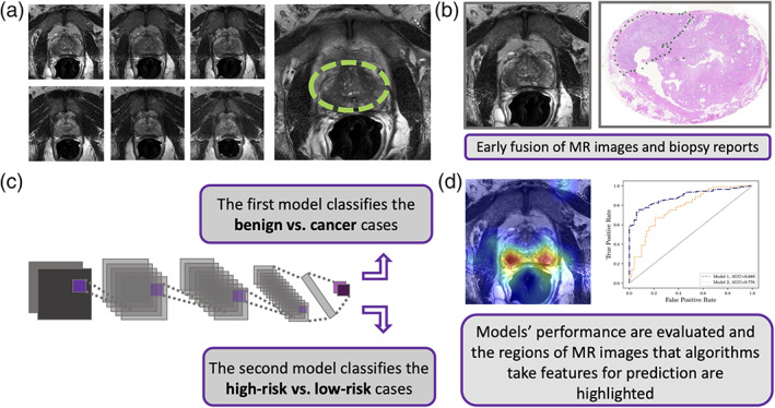
MR images reviewed and selected by two radiologists (with 6 and 17 years of experience). The patient images were labeled with prostate biopsy including Gleason Score (6 to 10) or Grade Group (1 to 5) and reviewed by one pathologist (with 15 years of experience). Deep learning models were developed to distinguish 1) benign from cancerous tumor and 2) high-risk tumor from low-risk tumor.
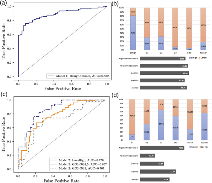
To evaluate our models, we calculated negative predictive value, positive predictive value, specificity, sensitivity, and accuracy. We also calculated areas under the receiver operating characteristic (ROC) curves (AUCs) and Cohen's kappa.
Our computational method (https://github.com/ih-lab/AI-biopsy) achieved AUCs of 0.89 (95% confidence interval [CI]: [0.86-0.92]) and 0.78 (95% CI: [0.74-0.82]) to classify cancer vs. benign and high- vs. low-risk of prostate disease, respectively.
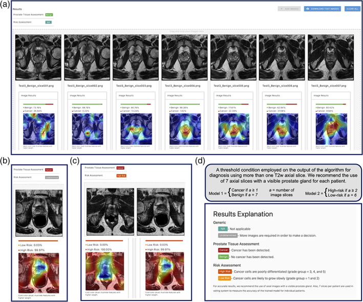
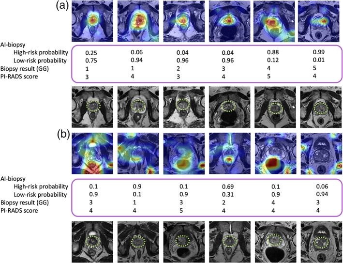
AI-biopsy provided a data-driven and reproducible way to assess cancer risk from MR images and a personalized strategy to potentially reduce the number of unnecessary biopsies. AI-biopsy highlighted the regions of MR images that contained the predictive features the algorithm used for diagnosis using the class activation map method. It is a fully automatic method with a drag-and-drop web interface (https://ai-biopsy.eipm-research.org) that allows radiologists to review AI-assessed MR images in real time.
1 TECHNICAL EFFICACY STAGE: 2.
前列腺癌的明确诊断需要活检获取组织进行病理分析,但这是一种有创性的操作,并且会引起并发症。
利用标记有组织病理学信息的磁共振(MR)图像,开发一种基于人工智能(AI)的前列腺癌早期诊断模型(命名为 AI-活检)。
回顾性研究。
来自 400 名疑似前列腺癌患者的磁共振成像(MRI)数据集,以及组织学数据(228 份为内部采集,172 份来自外部公开数据库)。
磁场强度/序列:1.5 至 3.0 特斯拉,T2 加权图像脉冲序列。
由两位具有 6 年和 17 年经验的放射科医生审查和选择 MRI 图像。患者图像由一位具有 15 年经验的病理学家进行前列腺活检标记,包括 Gleason 评分(6 至 10)或分级组(1 至 5)。开发深度学习模型以区分 1)良性肿瘤与癌性肿瘤,以及 2)高危肿瘤与低危肿瘤。
为了评估我们的模型,我们计算了阴性预测值、阳性预测值、特异性、敏感性和准确性。我们还计算了接收器操作特征(ROC)曲线下面积(AUC)和 Cohen's kappa。
我们的计算方法(https://github.com/ih-lab/AI-biopsy)在区分癌症与良性肿瘤,以及高风险与低风险前列腺疾病方面,AUC 分别为 0.89(95%置信区间 [0.86-0.92])和 0.78(95%置信区间 [0.74-0.82])。
AI-活检为从 MR 图像评估癌症风险提供了一种数据驱动且可重复的方法,并提供了一种潜在的策略来减少不必要的活检数量。AI-活检通过类激活映射方法突出了包含算法用于诊断的预测特征的 MR 图像区域。它是一种完全自动化的方法,具有拖放式网络界面(https://ai-biopsy.eipm-research.org),允许放射科医生实时审查 AI 评估的 MR 图像。
1 技术功效阶段:2。