Vascular Biology and Therapeutics Laboratory, School of Biotechnology Faculty of Science and Health, Dublin City University, Dublin, 9, Ireland.
Fraunhofer Project Centre for Embedded BioAnalytical Systems, Faculty of Science and Health, Dublin City University, Dublin, 9, Ireland.
Stem Cell Rev Rep. 2021 Oct;17(5):1713-1740. doi: 10.1007/s12015-021-10125-x. Epub 2021 Mar 17.
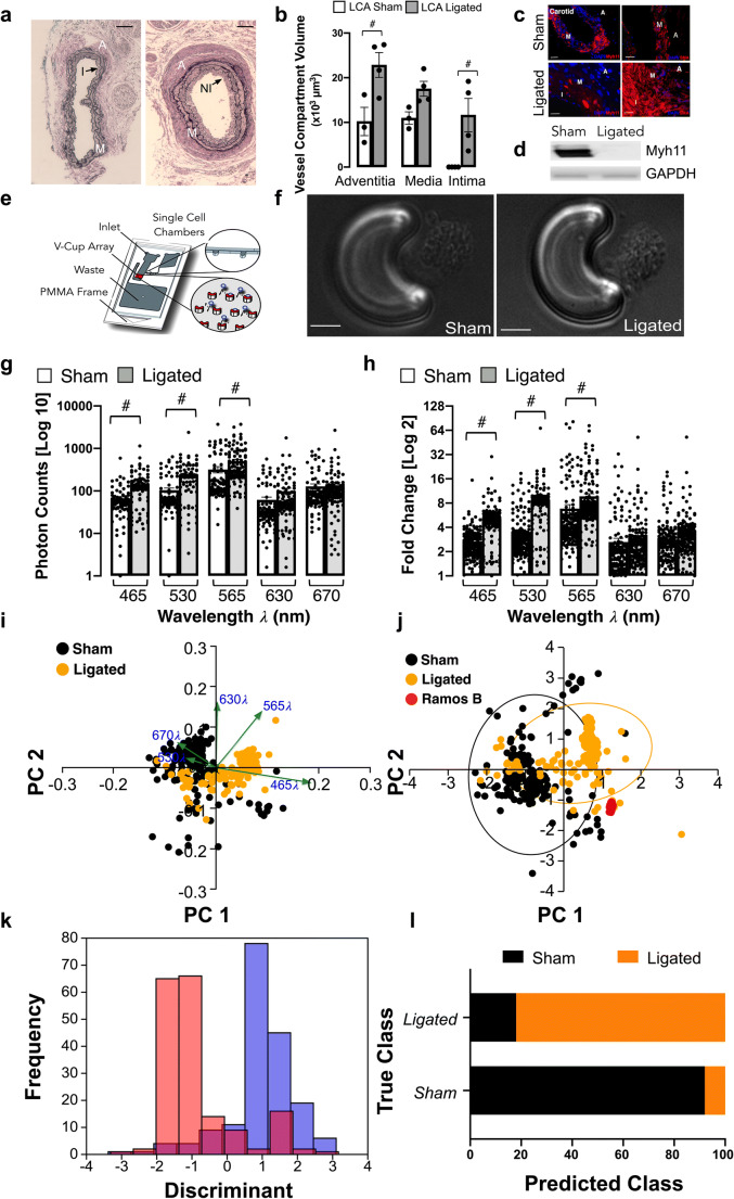
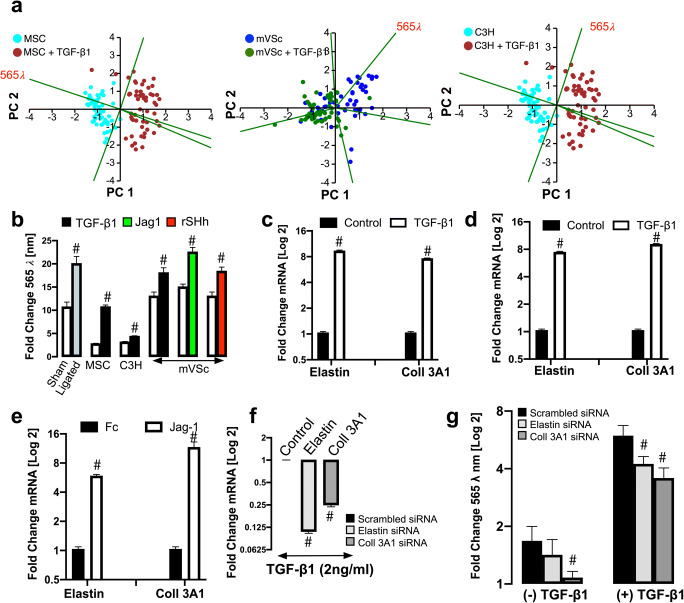
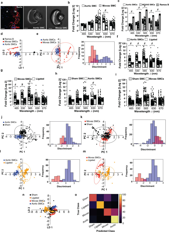
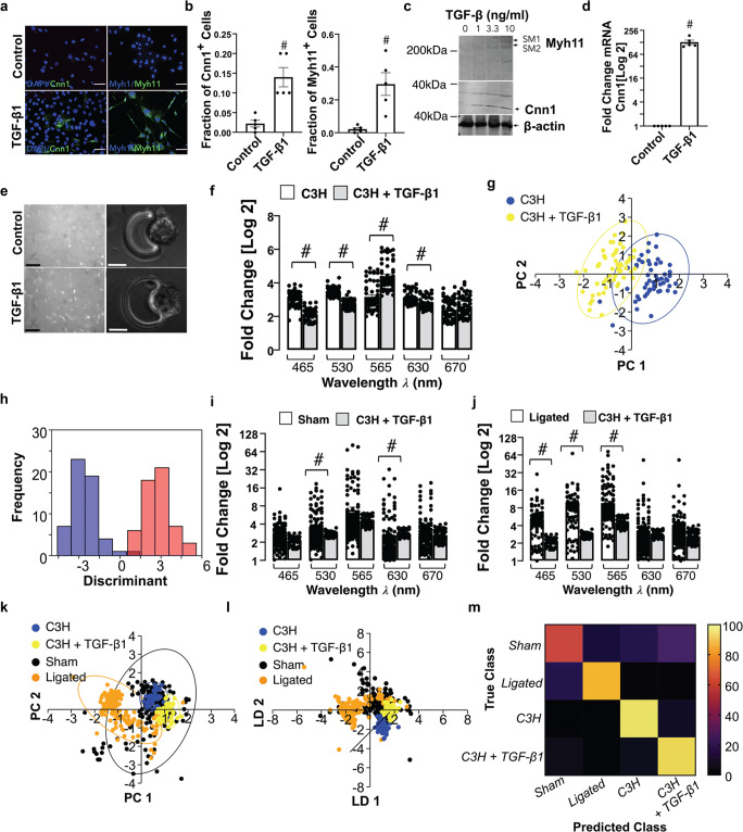
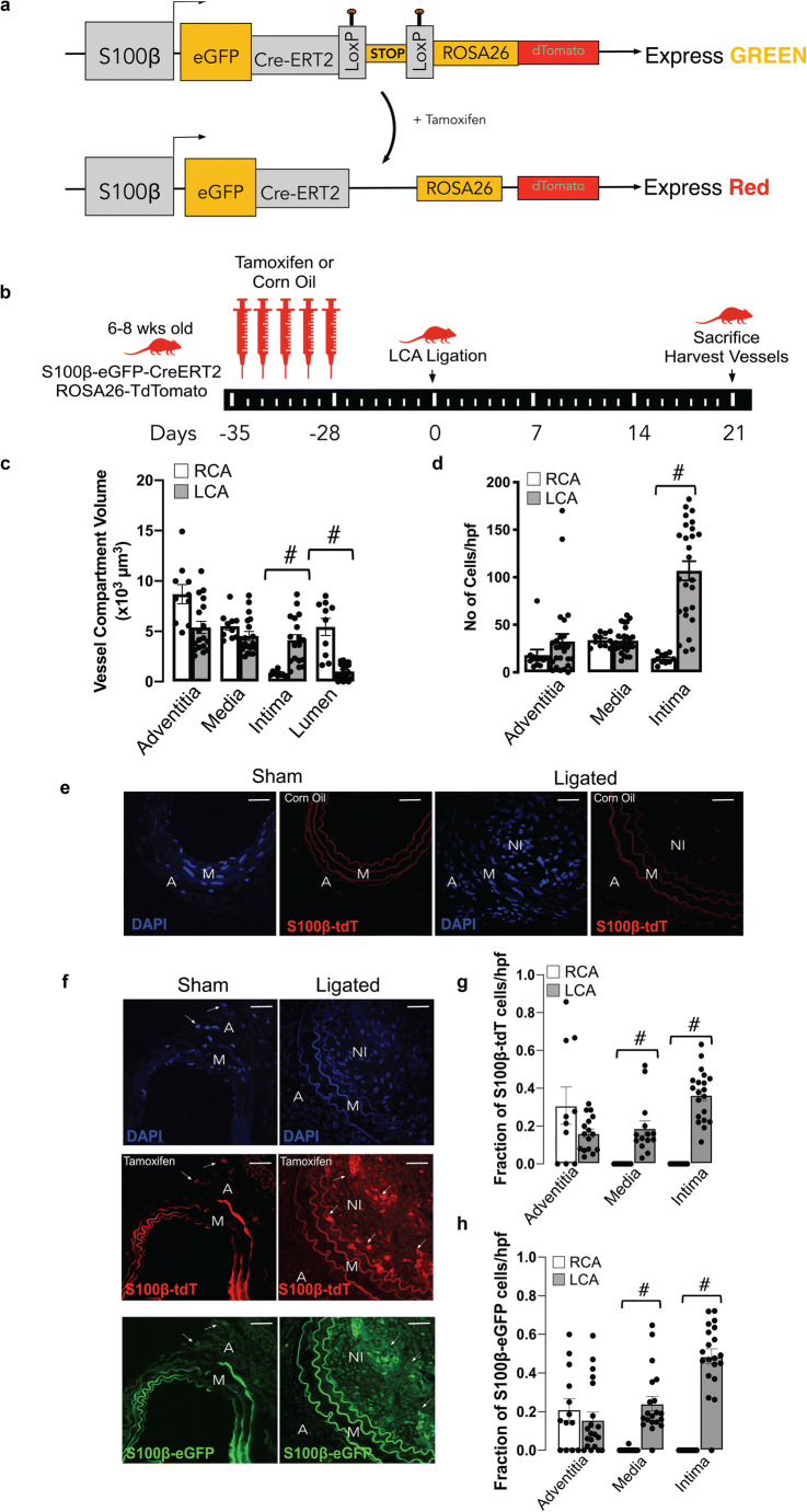
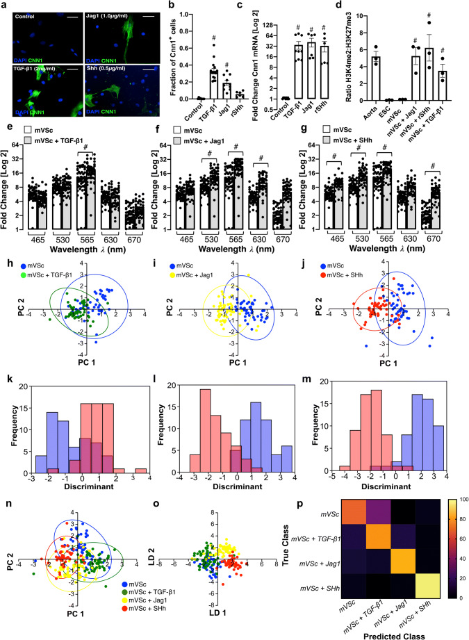
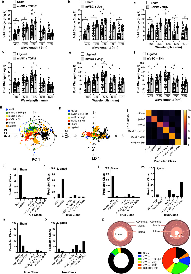
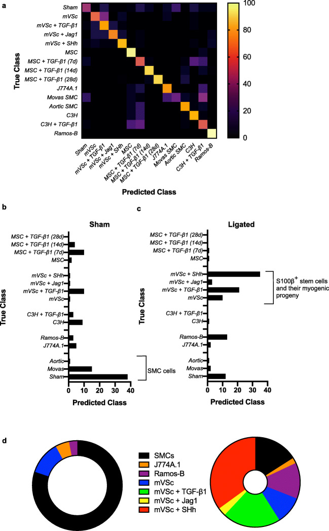
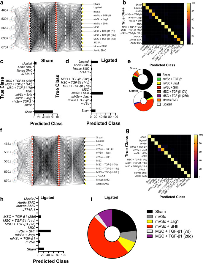
A hallmark of subclinical atherosclerosis is the accumulation of vascular smooth muscle cell (SMC)-like cells leading to intimal thickening and lesion formation. While medial SMCs contribute to vascular lesions, the involvement of resident vascular stem cells (vSCs) remains unclear. We evaluated single cell photonics as a discriminator of cell phenotype in vitro before the presence of vSC within vascular lesions was assessed ex vivo using supervised machine learning and further validated using lineage tracing analysis. Using a novel lab-on-a-Disk(Load) platform, label-free single cell photonic emissions from normal and injured vessels ex vivo were interrogated and compared to freshly isolated aortic SMCs, cultured Movas SMCs, macrophages, B-cells, S100β mVSc, bone marrow derived mesenchymal stem cells (MSC) and their respective myogenic progeny across five broadband light wavelengths (λ465 - λ670 ± 20 nm). We found that profiles were of sufficient coverage, specificity, and quality to clearly distinguish medial SMCs from different vascular beds (carotid vs aorta), discriminate normal carotid medial SMCs from lesional SMC-like cells ex vivo following flow restriction, and identify SMC differentiation of a series of multipotent stem cells following treatment with transforming growth factor beta 1 (TGF- β1), the Notch ligand Jagged1, and Sonic Hedgehog using multivariate analysis, in part, due to photonic emissions from enhanced collagen III and elastin expression. Supervised machine learning supported genetic lineage tracing analysis of S100β vSCs and identified the presence of S100βvSC-derived myogenic progeny within vascular lesions. We conclude disease-relevant photonic signatures may have predictive value for vascular disease.
动脉粥样硬化亚临床的一个标志是血管平滑肌细胞(SMC)样细胞的积累,导致内膜增厚和病变形成。虽然中膜 SMC 有助于血管病变,但驻留血管干细胞(vSCs)的参与仍不清楚。我们评估了单细胞光子学作为体外细胞表型的鉴别器,然后使用监督机器学习在血管病变中存在 vSC 之前评估其在体内的表现,并使用谱系追踪分析进一步验证。使用新型的盘上实验室(Load)平台,对离体正常和损伤血管的无标记单细胞光子发射进行了询问,并与新鲜分离的主动脉 SMC、培养的 Movas SMC、巨噬细胞、B 细胞、S100β vSC、骨髓来源的间充质干细胞(MSC)及其各自的成肌前体进行了比较,跨越五个宽带光波长(λ465-λ670±20nm)。我们发现,这些图谱具有足够的覆盖范围、特异性和质量,可以清楚地区分中膜 SMC 与不同的血管床(颈动脉与主动脉),区分正常颈动脉中膜 SMC 与血管限制后离体的 SMC 样细胞,并识别一系列多能干细胞在转化生长因子-β1(TGF-β1)、Notch 配体 Jagged1 和 Sonic Hedgehog 处理后的 SMC 分化,部分原因是增强的胶原 III 和弹性蛋白表达产生的光子发射。监督机器学习支持 S100β vSCs 的遗传谱系追踪分析,并确定了血管病变中存在 S100β vSC 衍生的成肌前体。我们得出结论,与疾病相关的光子特征可能对血管疾病具有预测价值。