• 文献检索
  • 文档翻译
  • 深度研究
  • 学术资讯
  • Suppr Zotero 插件Zotero 插件
  • 邀请有礼
  • 套餐&价格
  • 历史记录
应用&插件
Suppr Zotero 插件Zotero 插件浏览器插件Mac 客户端Windows 客户端微信小程序
定价
高级版会员购买积分包购买API积分包
服务
文献检索文档翻译深度研究API 文档MCP 服务
关于我们
关于 Suppr公司介绍联系我们用户协议隐私条款
关注我们

Suppr 超能文献

核心技术专利:CN118964589B侵权必究
粤ICP备2023148730 号-1Suppr @ 2026

文献检索

告别复杂PubMed语法,用中文像聊天一样搜索,搜遍4000万医学文献。AI智能推荐,让科研检索更轻松。

立即免费搜索

文件翻译

保留排版,准确专业,支持PDF/Word/PPT等文件格式,支持 12+语言互译。

免费翻译文档

深度研究

AI帮你快速写综述,25分钟生成高质量综述,智能提取关键信息,辅助科研写作。

立即免费体验

扩散加权磁共振成像中的时间依赖性可预测缺血性中风患者的组织转归。

Time dependence in diffusion MRI predicts tissue outcome in ischemic stroke patients.

作者信息

Lampinen Björn, Lätt Jimmy, Wasselius Johan, van Westen Danielle, Nilsson Markus

机构信息

Clinical Sciences Lund, Medical Radiation Physics, Lund University, Lund, Sweden.

Center for Medical Imaging and Physiology, Skåne University Hospital Lund, Lund, Sweden.

出版信息

Magn Reson Med. 2021 Aug;86(2):754-764. doi: 10.1002/mrm.28743. Epub 2021 Mar 23.

DOI:10.1002/mrm.28743
PMID:33755261
原文链接:https://pmc.ncbi.nlm.nih.gov/articles/PMC8445077/
Abstract

PURPOSE

Reperfusion therapy enables effective treatment of ischemic stroke presenting within 4-6 hours. However, tissue progression from ischemia to infarction is variable, and some patients benefit from treatment up until 24 hours. Improved imaging techniques are needed to identify these patients. Here, it was hypothesized that time dependence in diffusion MRI may predict tissue outcome in ischemic stroke.

METHODS

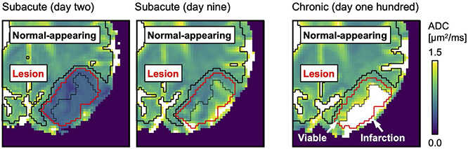
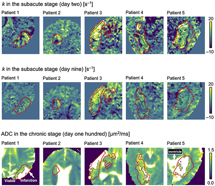
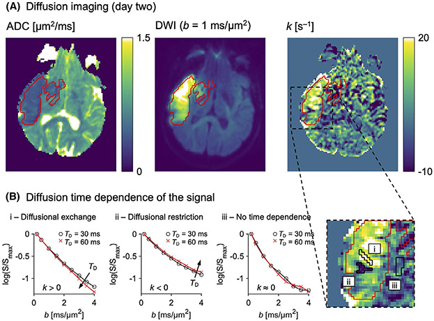
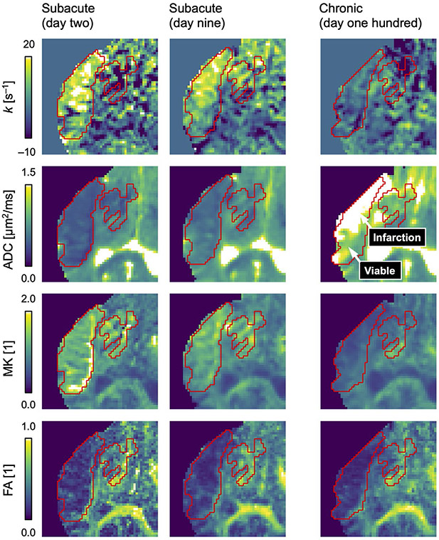
Diffusion MRI data were acquired with multiple diffusion times in five non-reperfused patients at 2, 9, and 100 days after stroke onset. Maps of "rate of kurtosis change" (k), mean kurtosis, ADC, and fractional anisotropy were derived. The ADC maps defined lesions, normal-appearing tissue, and the lesion tissue that would either be infarcted or remain viable by day 100. Diffusion parameters were compared (1) between lesions and normal-appearing tissue, and (2) between lesion tissue that would be infarcted or remain viable.

RESULTS

Positive values of k were observed within stroke lesions on day 2 (P = .001) and on day 9 (P = .023), indicating diffusional exchange. On day 100, high ADC values indicated infarction of 50 ± 20% of the lesion volumes. Tissue infarction was predicted by high k values both on day 2 (P = .026) and on day 9 (P = .046), by low mean kurtosis values on day 2 (P = .043), and by low fractional anisotropy values on day 9 (P = .029), but not by low ADC values.

CONCLUSIONS

Diffusion time dependence predicted tissue outcome in ischemic stroke more accurately than the ADC, and may be useful for predicting reperfusion benefit.

摘要

目的

再灌注疗法能够有效治疗发病4 - 6小时内的缺血性卒中。然而,组织从缺血发展到梗死的进程存在差异,部分患者在发病24小时内接受治疗仍可获益。因此需要改进成像技术来识别这些患者。在此,我们假设扩散加权磁共振成像(MRI)中的时间依赖性可以预测缺血性卒中的组织转归。

方法

对5例未接受再灌注治疗的患者在卒中发作后第2天、第9天和第100天进行了多个扩散时间点的扩散加权MRI数据采集。得出了“峰度变化率”(k)、平均峰度、表观扩散系数(ADC)和分数各向异性图。ADC图界定了梗死灶、外观正常的组织以及到第100天时会梗死或仍存活的梗死灶组织。比较了扩散参数:(1)梗死灶与外观正常组织之间;(2)会梗死或仍存活的梗死灶组织之间。

结果

在第2天(P = 0.001)和第9天(P = 0.023)的卒中病灶内观察到k值为正,提示存在扩散交换。在第100天,高ADC值提示梗死灶体积的50±20%发生了梗死。第2天(P = 0.026)和第9天(P = 0.046)的高k值、第2天的低平均峰度值(P = 0.043)以及第9天的低分数各向异性值(P = 0.029)可预测组织梗死,而低ADC值则不能。

结论

扩散时间依赖性比ADC能更准确地预测缺血性卒中的组织转归,可能有助于预测再灌注治疗的获益情况。

https://cdn.ncbi.nlm.nih.gov/pmc/blobs/1d4b/8445077/481882b9c82b/nihms-1735618-f0004.jpg
https://cdn.ncbi.nlm.nih.gov/pmc/blobs/1d4b/8445077/200098f63e53/nihms-1735618-f0001.jpg
https://cdn.ncbi.nlm.nih.gov/pmc/blobs/1d4b/8445077/643f5b8a7c92/nihms-1735618-f0002.jpg
https://cdn.ncbi.nlm.nih.gov/pmc/blobs/1d4b/8445077/aa9257ba4ce9/nihms-1735618-f0003.jpg
https://cdn.ncbi.nlm.nih.gov/pmc/blobs/1d4b/8445077/481882b9c82b/nihms-1735618-f0004.jpg

相似文献

1
Time dependence in diffusion MRI predicts tissue outcome in ischemic stroke patients.扩散加权磁共振成像中的时间依赖性可预测缺血性中风患者的组织转归。
Magn Reson Med. 2021 Aug;86(2):754-764. doi: 10.1002/mrm.28743. Epub 2021 Mar 23.
2
Probability of cortical infarction predicted by flumazenil binding and diffusion-weighted imaging signal intensity: a comparative positron emission tomography/magnetic resonance imaging study in early ischemic stroke.通过氟马西尼结合和扩散加权成像信号强度预测皮质梗死的概率:早期缺血性卒中的正电子发射断层扫描/磁共振成像对比研究
Stroke. 2004 Aug;35(8):1892-8. doi: 10.1161/01.STR.0000134746.93535.9b. Epub 2004 Jun 24.
3
Investigating potentially salvageable penumbra tissue in an in vivo model of transient ischemic stroke using sodium, diffusion, and perfusion magnetic resonance imaging.使用钠、扩散和灌注磁共振成像在短暂性脑缺血发作的体内模型中研究潜在可挽救的半暗带组织。
BMC Neurosci. 2016 Dec 7;17(1):82. doi: 10.1186/s12868-016-0316-1.
4
The role of diffusion tensor imaging in the evaluation of ischemic brain injury - a review.扩散张量成像在缺血性脑损伤评估中的作用——综述
NMR Biomed. 2002 Nov-Dec;15(7-8):561-9. doi: 10.1002/nbm.786.
5
Metabolic counterpart of decreased apparent diffusion coefficient during hyperacute ischemic stroke: a brain proton magnetic resonance spectroscopic imaging study.超急性缺血性卒中期间表观扩散系数降低的代谢对应物:一项脑质子磁共振波谱成像研究
Stroke. 2003 Jul;34(7):e82-7. doi: 10.1161/01.STR.0000078659.43423.0A. Epub 2003 Jun 19.
6
Diffusion-weighted magnetic resonance imaging in acute stroke.急性卒中的磁共振扩散加权成像
Stroke. 1998 Sep;29(9):1783-90. doi: 10.1161/01.str.29.9.1783.
7
Diffusion Kurtosis Imaging of Acute Infarction: Comparison with Routine Diffusion and Follow-up MR Imaging.弥散峰度成像在急性脑梗死中的应用:与常规弥散加权成像及随访磁共振成像的对比研究。
Radiology. 2018 May;287(2):651-657. doi: 10.1148/radiol.2017170553. Epub 2018 Mar 20.
8
Diffusional Kurtosis Imaging and Motor Outcome in Acute Ischemic Stroke.急性缺血性卒中的扩散峰度成像与运动结局
AJNR Am J Neuroradiol. 2017 Jul;38(7):1328-1334. doi: 10.3174/ajnr.A5180. Epub 2017 May 4.
9
Diffusion-Weighted MRI Stroke Volume Following Recanalization Treatment is Threshold-Dependent.血管再通治疗后扩散加权 MRI 脑卒中容积与阈值相关。
Clin Neuroradiol. 2019 Mar;29(1):135-141. doi: 10.1007/s00062-017-0634-4. Epub 2017 Oct 19.
10
Apparent diffusion coefficient thresholds do not predict the response to acute stroke thrombolysis.表观扩散系数阈值无法预测急性中风溶栓治疗的反应。
Stroke. 2005 Dec;36(12):2626-31. doi: 10.1161/01.STR.0000189688.95557.2b. Epub 2005 Nov 3.

引用本文的文献

1
Robust frequency-dependent diffusional kurtosis computation using an efficient direction scheme, axisymmetric modelling, and spatial regularization.使用高效方向方案、轴对称建模和空间正则化进行稳健的频率依赖性扩散峰度计算。
Imaging Neurosci (Camb). 2024 Jan 5;2. doi: 10.1162/imag_a_00055. eCollection 2024.
2
human neurite exchange time imaging at 500 mT/m diffusion gradients: Neurite exchange time imaging on Connectome 2.0.在500 mT/m扩散梯度下的人类神经突交换时间成像:Connectome 2.0上的神经突交换时间成像。
Imaging Neurosci (Camb). 2025;3. doi: 10.1162/imag_a_00544. Epub 2025 Apr 22.
3
Correcting pulse duration effects in the diffusional kurtosis of the multi-compartment Kärger model.

本文引用的文献

1
Tensor-valued diffusion MRI in under 3 minutes: an initial survey of microscopic anisotropy and tissue heterogeneity in intracranial tumors.张量值扩散 MRI 在 3 分钟内完成:颅内肿瘤微观各向异性和组织异质性的初步研究。
Magn Reson Med. 2020 Feb;83(2):608-620. doi: 10.1002/mrm.27959. Epub 2019 Sep 13.
2
Linear, planar and spherical tensor-valued diffusion MRI data by free waveform encoding in healthy brain, water, oil and liquid crystals.通过自由波形编码在健康大脑、水、油和液晶中获取的线性、平面和球形张量值扩散磁共振成像数据。
Data Brief. 2019 Jul 2;25:104208. doi: 10.1016/j.dib.2019.104208. eCollection 2019 Aug.
3
Monte Carlo Simulations of Water Exchange Through Myelin Wraps: Implications for Diffusion MRI.
校正多房室Kärger模型扩散峰度中的脉冲持续时间效应。
Magn Reson Med. 2025 Jun 10. doi: 10.1002/mrm.30608.
4
Diffusion MRI with double diffusion encoding and variable mixing times disentangles water exchange from transient kurtosis.采用双扩散编码和可变混合时间的扩散磁共振成像可将水交换与瞬时峰度分离。
Sci Rep. 2025 Mar 13;15(1):8747. doi: 10.1038/s41598-025-93084-4.
5
human neurite exchange imaging (NEXI) at 500 mT/m diffusion gradients.在500 mT/m扩散梯度下的人体神经突交换成像(NEXI)。
bioRxiv. 2024 Dec 17:2024.12.13.628450. doi: 10.1101/2024.12.13.628450.
6
The fuzzy MAD stroke conjecture, using Fuzzy C Means to classify multimodal apparent diffusion for ischemic stroke lesion stratification.模糊MAD卒中猜想,使用模糊C均值对缺血性卒中病变分层的多模态表观扩散进行分类。
Magn Reson Imaging. 2025 Apr;117:110294. doi: 10.1016/j.mri.2024.110294. Epub 2024 Dec 4.
7
Assessing tumor microstructure with time-dependent diffusion imaging: Considerations and feasibility on clinical MRI and MRI-Linac.利用时间依赖性扩散成像评估肿瘤微观结构:临床MRI和MRI直线加速器的考量与可行性
Med Phys. 2025 Jan;52(1):346-361. doi: 10.1002/mp.17453. Epub 2024 Oct 10.
8
In vivo mapping of cellular resolution neuropathology in brain ischemia with diffusion MRI.利用扩散磁共振成像对脑缺血中细胞分辨率神经病理学进行体内图谱绘制。
Sci Adv. 2024 Jul 19;10(29):eadk1817. doi: 10.1126/sciadv.adk1817. Epub 2024 Jul 17.
9
Compartmental anisotropy of filtered exchange imaging (FEXI) in human white matter: What is happening in FEXI?人脑白质中过滤式扩散交换成像(FEXI)的各向异性:FEXI 中发生了什么?
Magn Reson Med. 2024 Aug;92(2):660-675. doi: 10.1002/mrm.30086. Epub 2024 Mar 25.
10
Longitudinal evolution of diffusion metrics after left hemisphere ischaemic stroke.左半球缺血性中风后扩散指标的纵向演变
Brain Commun. 2023 Nov 20;5(6):fcad313. doi: 10.1093/braincomms/fcad313. eCollection 2023.
通过髓鞘包绕进行水交换的蒙特卡罗模拟:对弥散磁共振成像的启示。
IEEE Trans Med Imaging. 2019 Jun;38(6):1438-1445. doi: 10.1109/TMI.2019.2894398. Epub 2019 Feb 27.
4
Diffusion Kurtosis Imaging of Acute Infarction: Comparison with Routine Diffusion and Follow-up MR Imaging.弥散峰度成像在急性脑梗死中的应用:与常规弥散加权成像及随访磁共振成像的对比研究。
Radiology. 2018 May;287(2):651-657. doi: 10.1148/radiol.2017170553. Epub 2018 Mar 20.
5
Cumulant expansions for measuring water exchange using diffusion MRI.使用扩散 MRI 测量水交换的累积展开。
J Chem Phys. 2018 Feb 21;148(7):074109. doi: 10.1063/1.5014044.
6
Late Window Paradox.晚期窗悖论
Stroke. 2018 Mar;49(3):768-771. doi: 10.1161/STROKEAHA.117.020200. Epub 2018 Jan 24.
7
2018 Guidelines for the Early Management of Patients With Acute Ischemic Stroke: A Guideline for Healthcare Professionals From the American Heart Association/American Stroke Association.2018 急性缺血性脑卒中患者早期管理指南:美国心脏协会/美国卒中协会医疗保健专业人员指南。
Stroke. 2018 Mar;49(3):e46-e110. doi: 10.1161/STR.0000000000000158. Epub 2018 Jan 24.
8
Thrombectomy for Stroke at 6 to 16 Hours with Selection by Perfusion Imaging.6至16小时卒中的血栓切除术及灌注成像选择
N Engl J Med. 2018 Feb 22;378(8):708-718. doi: 10.1056/NEJMoa1713973. Epub 2018 Jan 24.
9
Thrombectomy 6 to 24 Hours after Stroke with a Mismatch between Deficit and Infarct.发病后 6 至 24 小时内进行取栓术治疗与缺损和梗死不匹配的脑卒中。
N Engl J Med. 2018 Jan 4;378(1):11-21. doi: 10.1056/NEJMoa1706442. Epub 2017 Nov 11.
10
Fast Versus Slow Progressors of Infarct Growth in Large Vessel Occlusion Stroke: Clinical and Research Implications.大血管闭塞性卒中梗死灶生长的快速进展者与缓慢进展者:临床及研究意义
Stroke. 2017 Sep;48(9):2621-2627. doi: 10.1161/STROKEAHA.117.017673. Epub 2017 Aug 9.