Department of Cardiology, Institute of Cardiovascular Diseases, First Affiliated Hospital of Dalian Medical University, Dalian, China.
Department of Emergency Medicine, Beijing Key Laboratory of Cardiopulmonary Cerebral Resuscitation, Beijing Chaoyang Hospital, Capital Medical University, Beijing, China.
Clin Transl Med. 2021 Mar;11(3):e374. doi: 10.1002/ctm2.374.
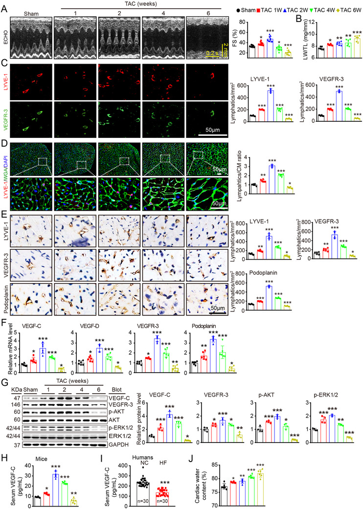
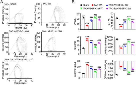
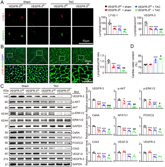
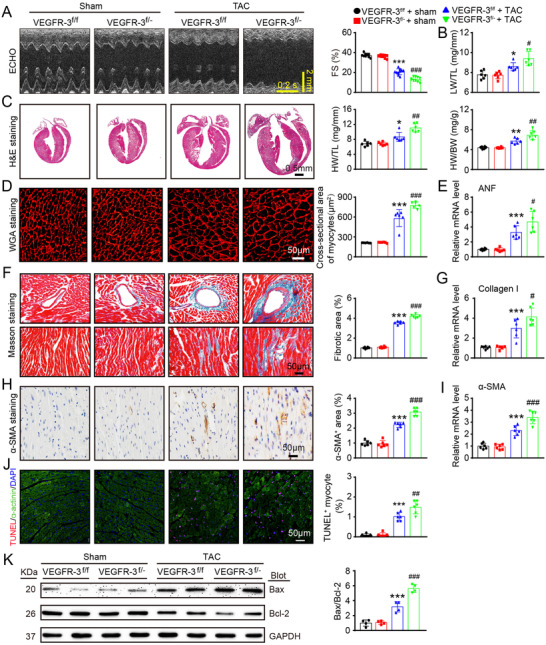
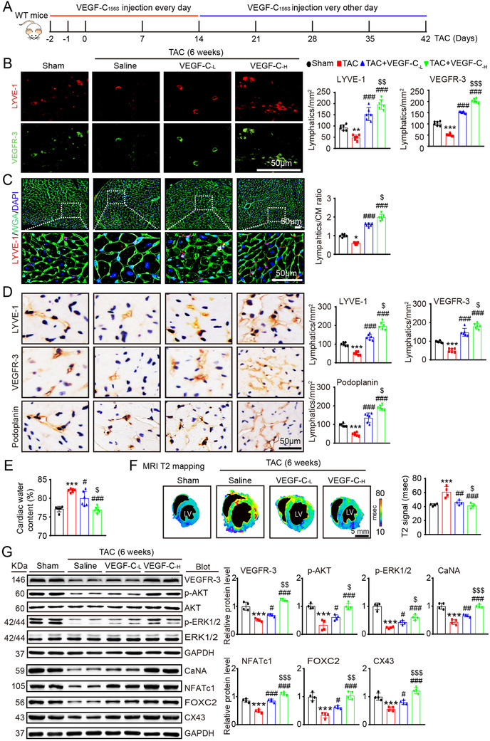
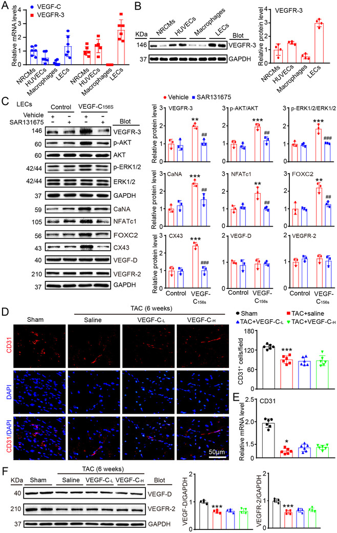
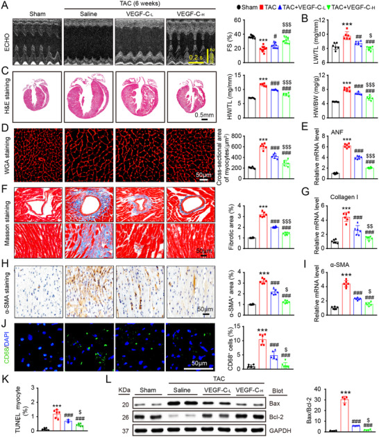
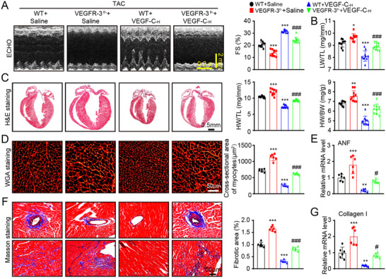
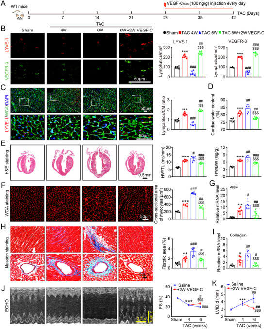
Prolonged pressure overload triggers cardiac hypertrophy and frequently leads to heart failure (HF). Vascular endothelial growth factor-C (VEGF-C) and its receptor VEGFR-3 are components of the central pathway for lymphatic vessel growth (also known as lymphangiogenesis), which has crucial functions in the maintenance of tissue fluid balance and myocardial function after ischemic injury. However, the roles of this pathway in the development of cardiac hypertrophy and dysfunction during pressure overload remain largely unknown. Eight- to 10-week-old male wild-type (WT) mice, VEGFR-3 knockdown (VEGFR-3 ) mice, and their WT littermates (VEGFR-3 ) were subjected to pressure overload induced by transverse aortic constriction (TAC) for 1-6 weeks. We found that cardiac lymphangiogenesis and the protein expression of VEGF-C and VEGFR-3 were upregulated in the early stage of cardiac hypertrophy but were markedly reduced in failing hearts. Moreover, TAC for 6 weeks significantly reduced cardiac lymphangiogenesis by inhibiting activation of VEGFR-3-mediated signals (AKT/ERK1/2, calcineurin A/NFATc1/FOXc2, and CX43), leading to increased cardiac edema, hypertrophy, fibrosis, apoptosis, inflammation, and dysfunction. These effects were further aggravated in VEGFR-3 mice and were dose-dependently attenuated by delivery of recombinant VEGF-C in WT mice. VEGF-C administration also reversed pre-established cardiac dysfunction induced by sustained pressure overload. Thus, these results demonstrate, for the first time, that activation of the VEGF-C-VEGFR-3 axis exerts a protective effect during the transition from cardiac hypertrophy to HF and highlight selective stimulation of cardiac lymphangiogenesis as a potential new therapeutic approach for hypertrophic heart diseases.
长期的压力超负荷会引发心肌肥厚,并经常导致心力衰竭(HF)。血管内皮生长因子-C(VEGF-C)及其受体 VEGFR-3 是淋巴管生长(也称为淋巴管生成)的核心途径的组成部分,在缺血性损伤后组织液平衡和心肌功能的维持中具有至关重要的作用。然而,该途径在压力超负荷引起的心肌肥厚和功能障碍发展中的作用在很大程度上仍然未知。8-10 周龄的雄性野生型(WT)小鼠、VEGFR-3 敲低(VEGFR-3 -/-)小鼠及其 WT 同窝仔鼠(WT)接受了主动脉缩窄(TAC)诱导的压力超负荷 1-6 周。我们发现,在心肌肥厚的早期阶段,心脏淋巴管生成和 VEGF-C 及 VEGFR-3 的蛋白表达上调,但在心力衰竭时明显减少。此外,TAC 持续 6 周显著减少了心脏淋巴管生成,抑制了 VEGFR-3 介导的信号(AKT/ERK1/2、钙调神经磷酸酶 A/NFATc1/FOXc2 和 CX43)的激活,导致心脏水肿、肥大、纤维化、凋亡、炎症和功能障碍增加。在 VEGFR-3 小鼠中,这些作用进一步加重,并在 WT 小鼠中通过给予重组 VEGF-C 呈剂量依赖性减轻。VEGF-C 给药还逆转了持续压力超负荷引起的预先建立的心脏功能障碍。因此,这些结果首次表明,VEGF-C-VEGFR-3 轴的激活在从心肌肥厚向 HF 的转变过程中发挥保护作用,并强调选择性刺激心脏淋巴管生成作为治疗肥厚性心脏病的一种潜在新方法。