IEEE Trans Biomed Eng. 2021 Nov;68(11):3317-3326. doi: 10.1109/TBME.2021.3070487. Epub 2021 Oct 19.
Typical SRUS images are reconstructed by localizing ultrasound microbubbles (MBs) injected in a vessel using normalized 2-dimensional cross-correlation (2DCC) between MBs signals and the point spread function of the system. However, current techniques require isolated MBs in a confined area due to inaccurate localization of densely populated MBs. To overcome this limitation, we developed the ℓ1-homotopy based compressed sensing (L1H-CS) based SRUS imaging technique which localizes densely populated MBs to visualize microvasculature in vivo.
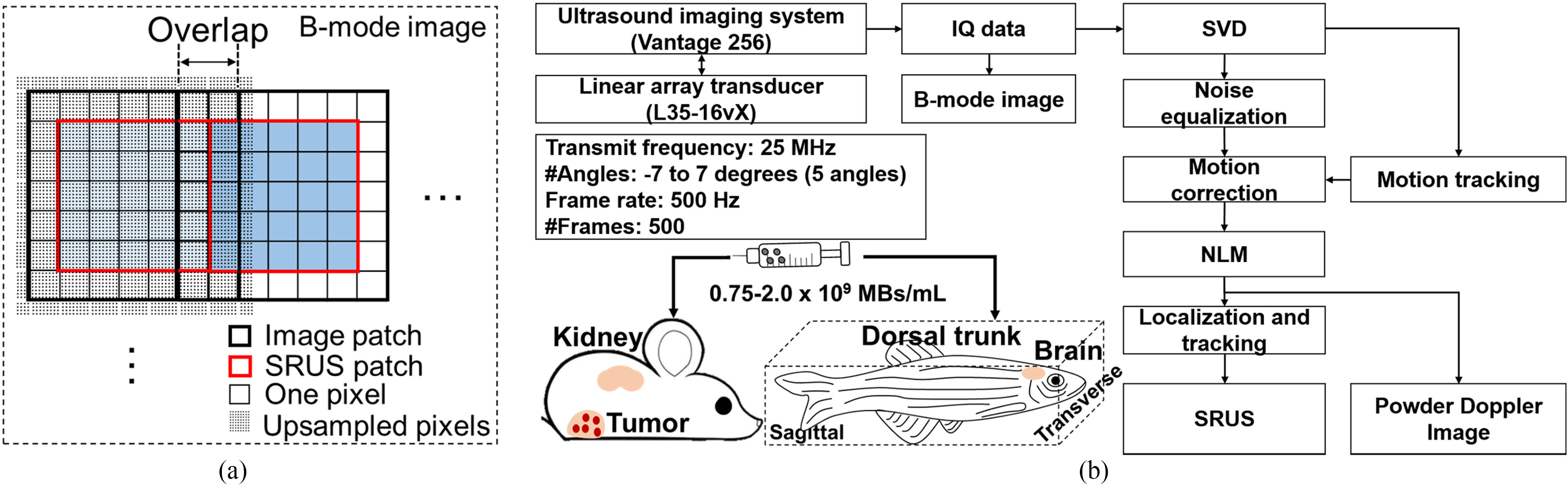
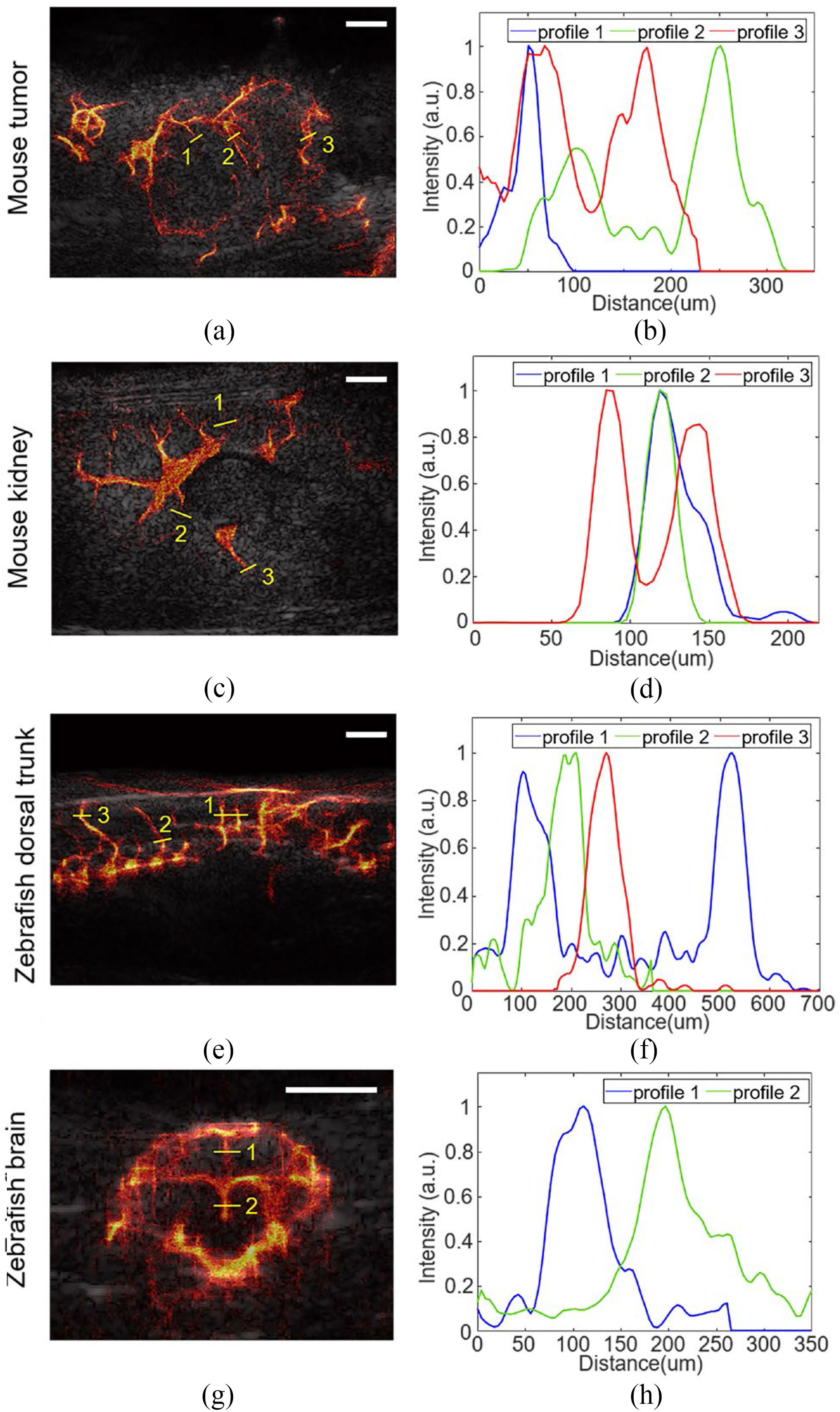
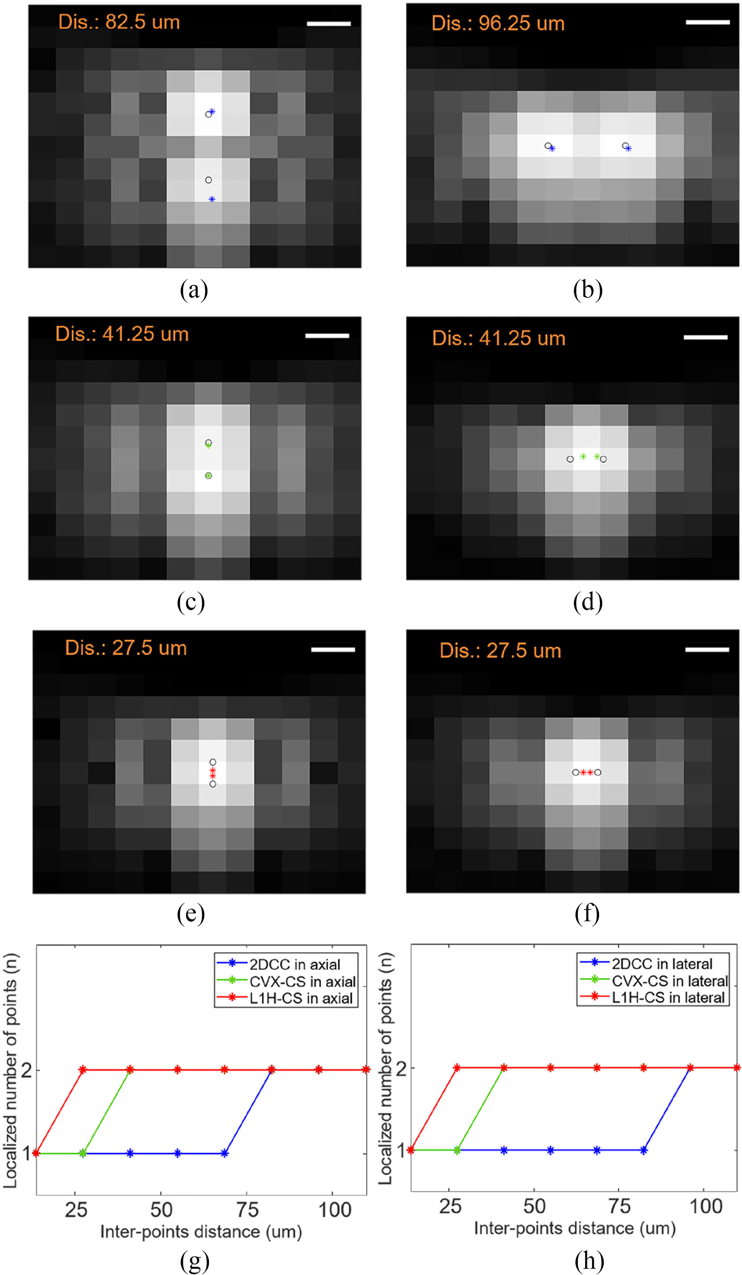
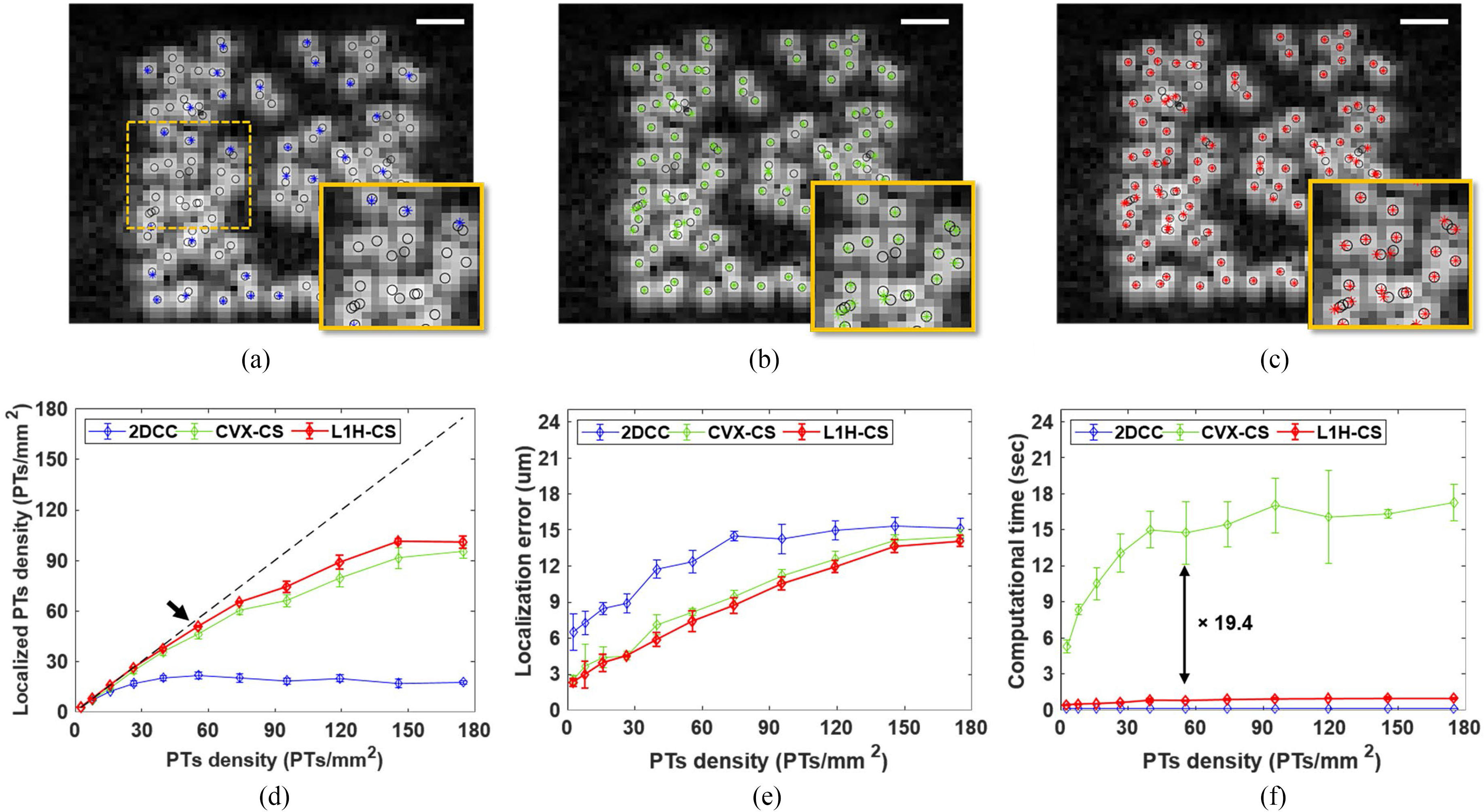
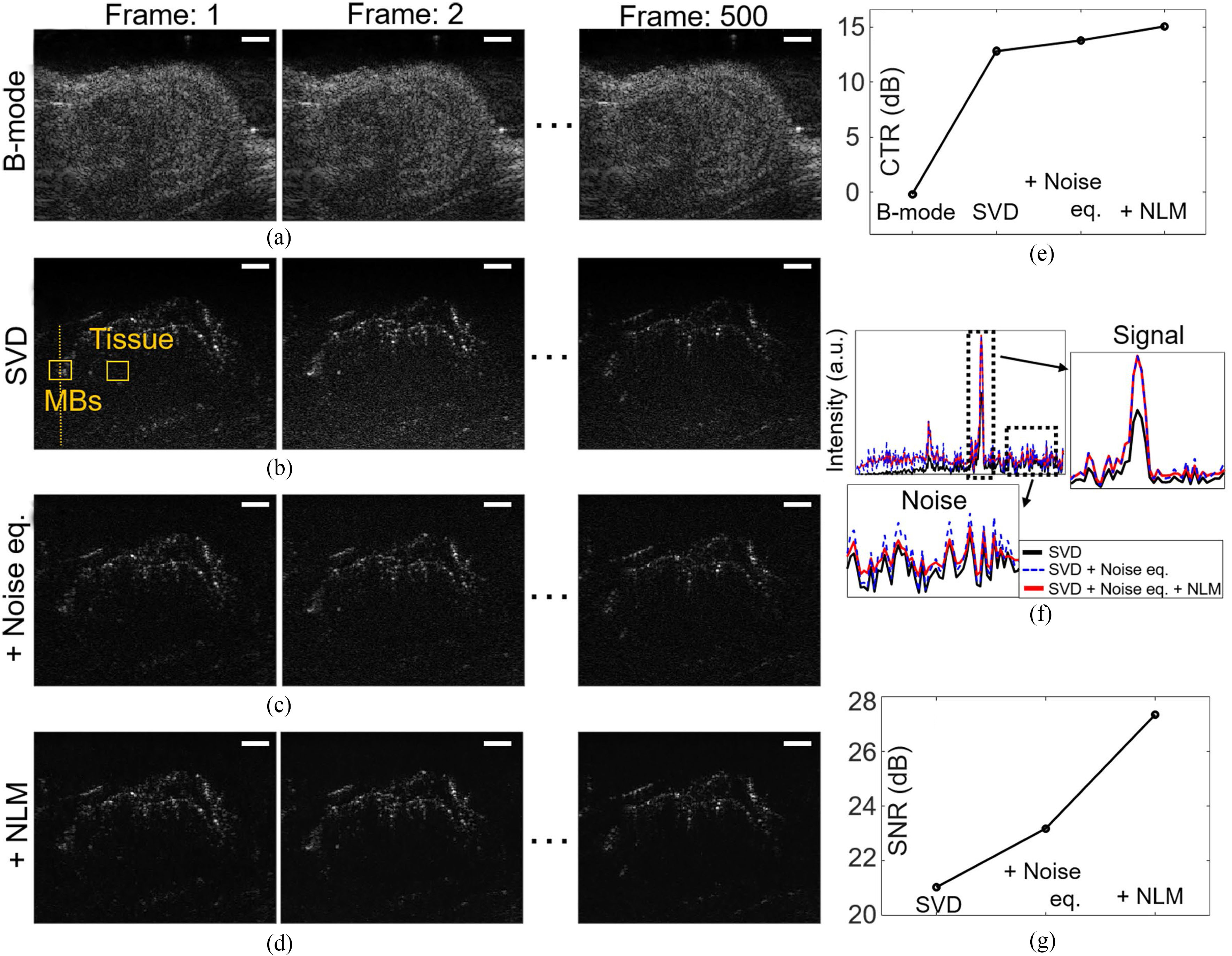
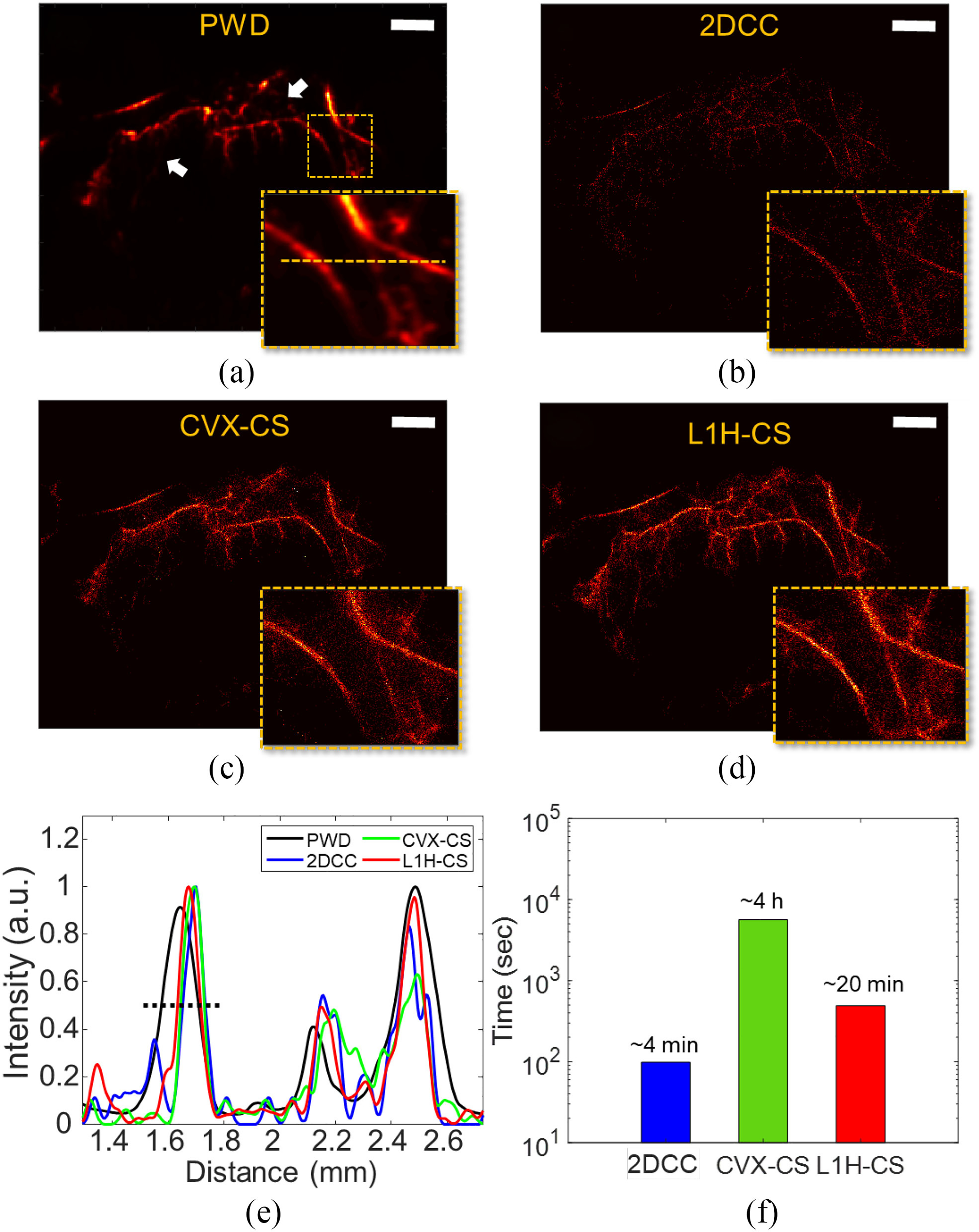
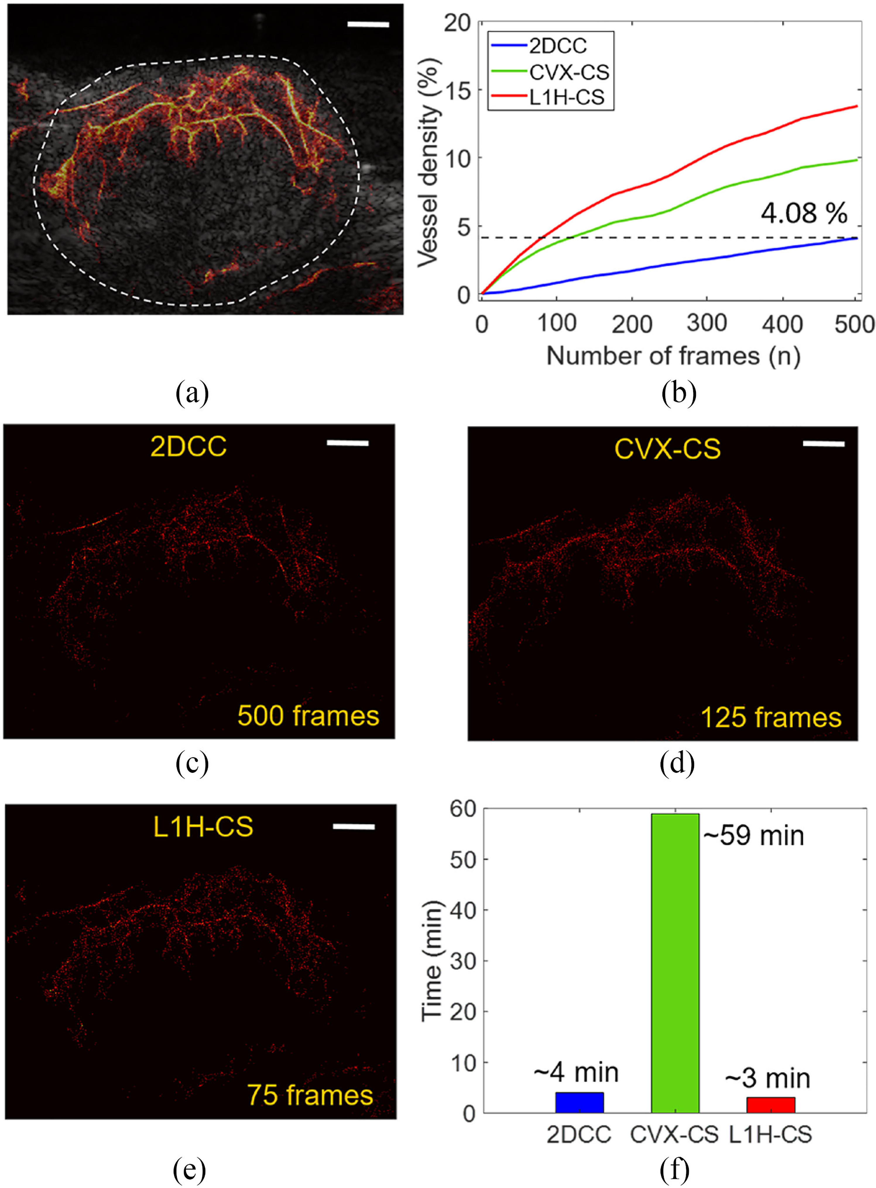
To evaluate the performance of L1H-CS, we compared the performance of 2DCC, interior-point method based compressed sensing (CVX-CS), and L1H-CS algorithms. Localization efficiency was compared using axially and laterally aligned point targets (PTs) with known distances and randomly distributed PTs generated by simulation. We developed post-processing techniques including clutter reduction, noise equalization, motion compensation, and spatiotemporal noise filtering for in vivo imaging. We then validated the capabilities of L1H-CS based SRUS imaging technique with high-density MBs in a mouse tumor model, kidney, and zebrafish dorsal trunk, and brain.
Compared to 2DCC and CVX-CS algorithms, L1H-CS achieved faster data acquisition time and considerable improvement in SRUS image quality.
These results demonstrate that the L1H-CS based SRUS imaging technique has the potential to examine microvasculature with reduced acquisition and reconstruction time to acquire enhanced SRUS image quality, which may be necessary to translate it into clinics.
典型的超声辐射力脉冲(SRUS)图像是通过对注入血管内的超声微泡(MB)进行定位来重建的,其方法是对 MB 信号与系统的点扩散函数之间的归一化二维互相关(2DCC)进行局部化。然而,由于对密集 MB 的定位不准确,当前的技术需要在受限区域中获得孤立的 MB。为了克服这一限制,我们开发了基于 L1 同伦的压缩感知(L1H-CS)的 SRUS 成像技术,该技术可以对密集的 MB 进行定位,从而可视化体内的微血管。
为了评估 L1H-CS 的性能,我们比较了 2DCC、基于内点法的压缩感知(CVX-CS)和 L1H-CS 算法的性能。使用轴向和侧向对齐的已知距离点目标(PT)和通过模拟生成的随机分布的 PT 来比较定位效率。我们开发了后处理技术,包括杂波抑制、噪声均衡、运动补偿和时空噪声滤波,用于体内成像。然后,我们在小鼠肿瘤模型、肾脏、斑马鱼背侧干线和大脑中用高密度 MB 验证了基于 L1H-CS 的 SRUS 成像技术的能力。
与 2DCC 和 CVX-CS 算法相比,L1H-CS 实现了更快的数据采集时间和 SRUS 图像质量的显著提高。
这些结果表明,基于 L1H-CS 的 SRUS 成像技术具有减少采集和重建时间以获得增强的 SRUS 图像质量的潜力,这可能是将其转化为临床应用所必需的。