Institute of Animal Reproduction and Food Research of Polish Academy of Sciences, Olsztyn, Poland.
Institute for Veterinary Medicine, Nicolaus Copernicus University, Torun, Poland.
Aging Cell. 2021 May;20(5):e13349. doi: 10.1111/acel.13349. Epub 2021 Apr 2.
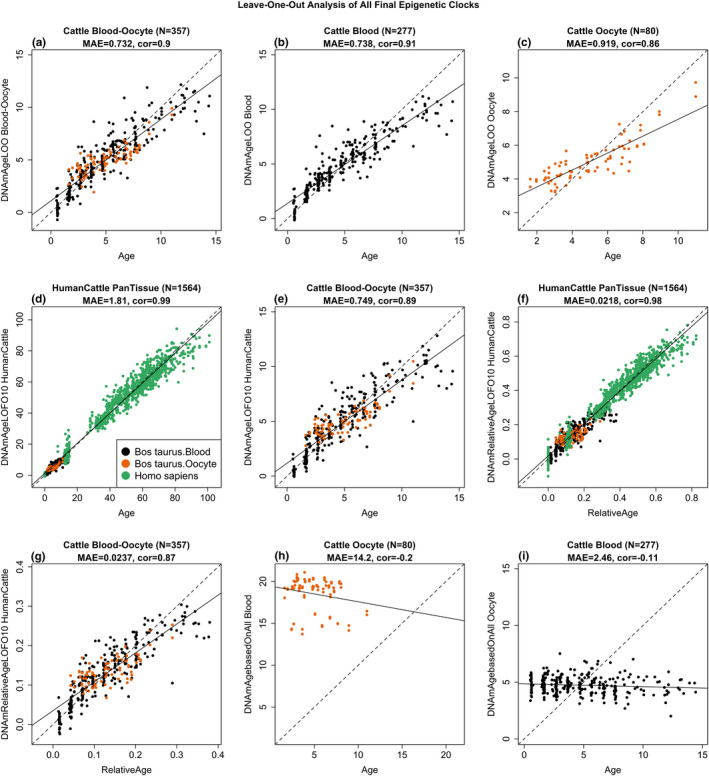
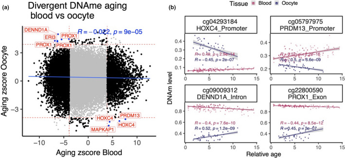
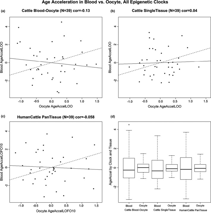
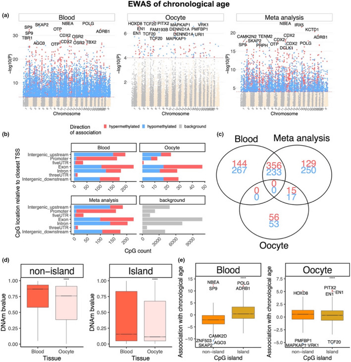
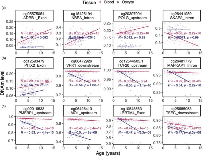
Cattle are an attractive animal model of fertility in women due to their high degree of similarity relative to follicle selection, embryo cleavage, blastocyst formation, and gestation length. To facilitate future studies of the epigenetic underpinnings of aging effects in the female reproductive axis, several DNA methylation-based biomarkers of aging (epigenetic clocks) for bovine oocytes are presented. One such clock was germane to only oocytes, while a dual-tissue clock was highly predictive of age in both oocytes and blood. Dual species clocks that apply to both humans and cattle were also developed and evaluated. These epigenetic clocks can be used to accurately estimate the biological age of oocytes. Both epigenetic clock studies and epigenome-wide association studies revealed that blood and oocytes differ substantially with respect to aging and the underlying epigenetic signatures that potentially influence the aging process. The rate of epigenetic aging was found to be slower in oocytes compared to blood; however, oocytes appeared to begin at an older epigenetic age. The epigenetic clocks for oocytes are expected to address questions in the field of reproductive aging, including the central question: how to slow aging of oocytes.
由于牛在卵泡选择、胚胎卵裂、囊胚形成和妊娠时间等方面与人类高度相似,因此它们是研究女性生育力的一种很有吸引力的动物模型。为了促进未来对女性生殖轴衰老影响的表观遗传基础的研究,本文提出了几种基于 DNA 甲基化的牛卵母细胞衰老生物标志物(表观遗传时钟)。其中一个时钟仅与卵母细胞有关,而双组织时钟在卵母细胞和血液中都能高度预测年龄。还开发并评估了适用于人类和牛的双物种时钟。这些表观遗传时钟可用于准确估计卵母细胞的生物学年龄。这两项表观遗传时钟研究和全基因组关联研究都表明,血液和卵母细胞在衰老和潜在影响衰老过程的表观遗传特征方面存在显著差异。研究发现,与血液相比,卵母细胞的表观遗传衰老速度较慢;然而,卵母细胞似乎从更老的表观遗传年龄开始。卵母细胞的表观遗传时钟有望解决生殖衰老领域的问题,包括核心问题:如何减缓卵母细胞衰老。