• 文献检索
  • 文档翻译
  • 深度研究
  • 学术资讯
  • Suppr Zotero 插件Zotero 插件
  • 邀请有礼
  • 套餐&价格
  • 历史记录
应用&插件
Suppr Zotero 插件Zotero 插件浏览器插件Mac 客户端Windows 客户端微信小程序
定价
高级版会员购买积分包购买API积分包
服务
文献检索文档翻译深度研究API 文档MCP 服务
关于我们
关于 Suppr公司介绍联系我们用户协议隐私条款
关注我们

Suppr 超能文献

核心技术专利:CN118964589B侵权必究
粤ICP备2023148730 号-1Suppr @ 2026

文献检索

告别复杂PubMed语法,用中文像聊天一样搜索,搜遍4000万医学文献。AI智能推荐,让科研检索更轻松。

立即免费搜索

文件翻译

保留排版,准确专业,支持PDF/Word/PPT等文件格式,支持 12+语言互译。

免费翻译文档

深度研究

AI帮你快速写综述,25分钟生成高质量综述,智能提取关键信息,辅助科研写作。

立即免费体验

仿生器官:在3D生物打印过程中,剪切力会降低胰岛和哺乳动物细胞的活力。

Bionic Organs: Shear Forces Reduce Pancreatic Islet and Mammalian Cell Viability during the Process of 3D Bioprinting.

作者信息

Klak Marta, Kowalska Patrycja, Dobrzański Tomasz, Tymicki Grzegorz, Cywoniuk Piotr, Gomółka Magdalena, Kosowska Katarzyna, Bryniarski Tomasz, Berman Andrzej, Dobrzyń Agnieszka, Sadowski Wojciech, Górecki Bartosz, Wszoła Michał

机构信息

Foundation of Research and Science Development, 01-793 Warsaw, Poland.

Nencki Institute of Experimental Biology, Polish Academy of Sciences, 02-093 Warsaw, Poland.

出版信息

Micromachines (Basel). 2021 Mar 14;12(3):304. doi: 10.3390/mi12030304.

DOI:10.3390/mi12030304
PMID:33799490
原文链接:https://pmc.ncbi.nlm.nih.gov/articles/PMC7999205/
Abstract

BACKGROUND

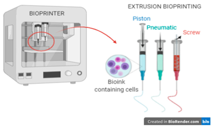
3D bioprinting is the future of constructing functional organs. Creating a bioactive scaffold with pancreatic islets presents many challenges. The aim of this paper is to assess how the 3D bioprinting process affects islet viability.

METHODS

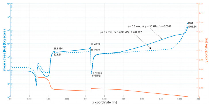
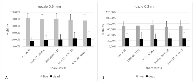
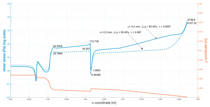
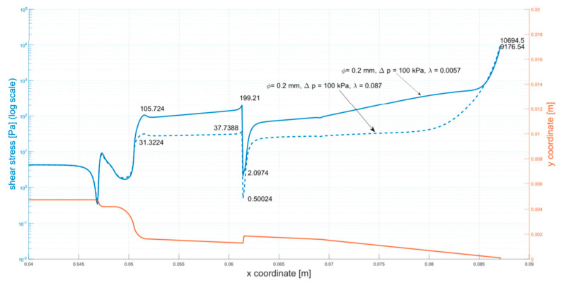
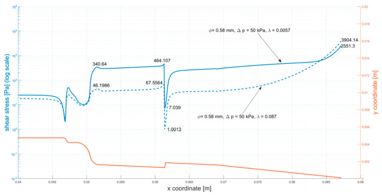
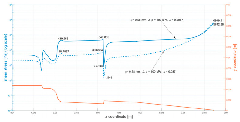
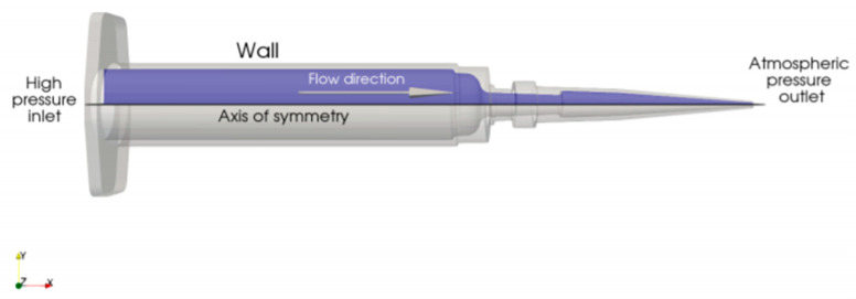
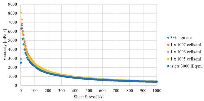
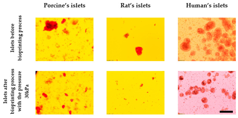
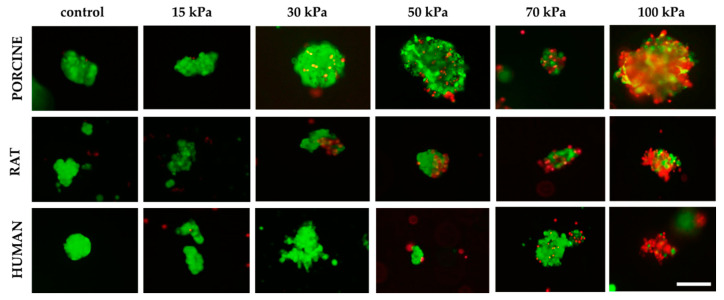
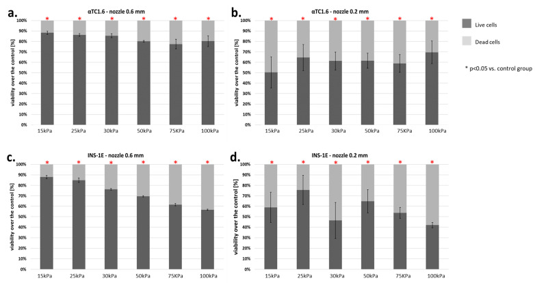
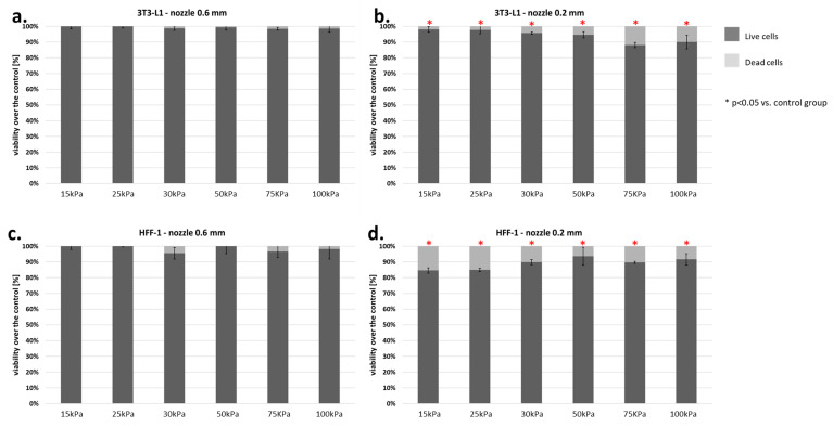
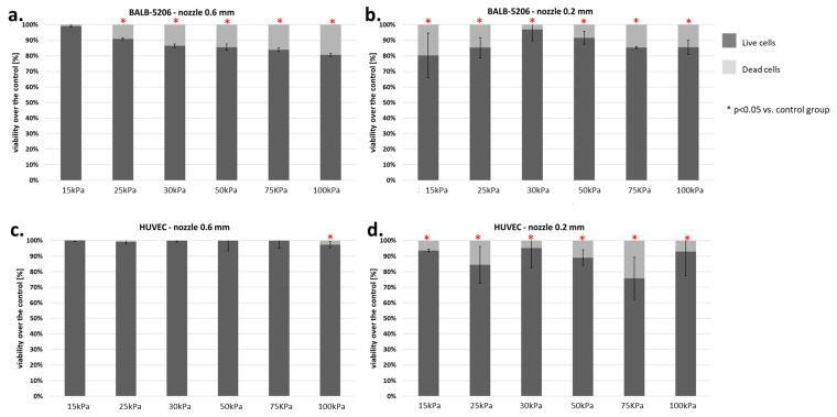
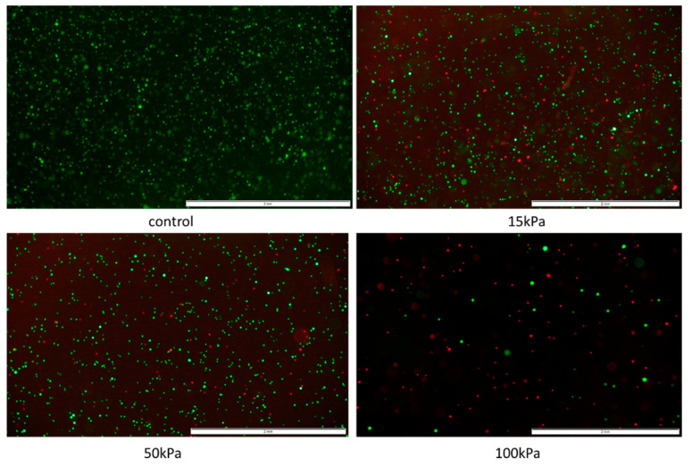
The BioX 3D printer (Cellink), 600 μm inner diameter nozzles, and 3% () alginate cell carrier solution were used with rat, porcine, and human pancreatic islets. Islets were divided into a control group (culture medium) and 6 experimental groups (each subjected to specific pressure between 15 and 100 kPa). FDA/PI staining was performed to assess the viability of islets. Analogous studies were carried out on α-cells, β-cells, fibroblasts, and endothelial cells.

RESULTS

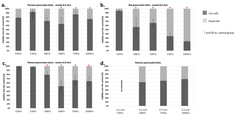
Viability of human pancreatic islets was as follows: 92% for alginate-based control and 94%, 90%, 74%, 48%, 61%, and 59% for 15, 25, 30, 50, 75, and 100 kPa, respectively. Statistically significant differences were observed between control and 50, 75, and 100 kPa, respectively. Similar observations were made for porcine and rat islets.

CONCLUSIONS

Optimal pressure during 3D bioprinting with pancreatic islets by the extrusion method should be lower than 30 kPa while using 3% () alginate as a carrier.

摘要

背景

3D生物打印是构建功能性器官的未来发展方向。利用胰岛创建具有生物活性的支架面临诸多挑战。本文旨在评估3D生物打印过程如何影响胰岛的活力。

方法

使用BioX 3D打印机(Cellink)、内径600μm的喷嘴以及3%()海藻酸盐细胞载体溶液,对大鼠、猪和人类的胰岛进行实验。胰岛被分为一个对照组(培养基)和6个实验组(每组承受15至100 kPa之间的特定压力)。采用FDA/PI染色法评估胰岛的活力。对α细胞、β细胞、成纤维细胞和内皮细胞进行了类似研究。

结果

人类胰岛的活力如下:基于海藻酸盐的对照组为92%,15、25、30、50、75和100 kPa组分别为94%、90%、74%、48%、61%和59%。对照组与50、75和100 kPa组之间分别观察到具有统计学意义的差异。对猪和大鼠胰岛也有类似观察结果。

结论

在使用3%()海藻酸盐作为载体通过挤压法进行3D生物打印胰岛时,最佳压力应低于30 kPa。

https://cdn.ncbi.nlm.nih.gov/pmc/blobs/b24a/7999205/ca2b61a1f30d/micromachines-12-00304-g014.jpg
https://cdn.ncbi.nlm.nih.gov/pmc/blobs/b24a/7999205/c5f50fb0e8a0/micromachines-12-00304-g0A1.jpg
https://cdn.ncbi.nlm.nih.gov/pmc/blobs/b24a/7999205/7a80b4446292/micromachines-12-00304-g0A2.jpg
https://cdn.ncbi.nlm.nih.gov/pmc/blobs/b24a/7999205/ddf779057634/micromachines-12-00304-g0A3.jpg
https://cdn.ncbi.nlm.nih.gov/pmc/blobs/b24a/7999205/a05861169b72/micromachines-12-00304-g0A4.jpg
https://cdn.ncbi.nlm.nih.gov/pmc/blobs/b24a/7999205/32f8c8edd55f/micromachines-12-00304-g0A5.jpg
https://cdn.ncbi.nlm.nih.gov/pmc/blobs/b24a/7999205/220f3abb06d5/micromachines-12-00304-g001.jpg
https://cdn.ncbi.nlm.nih.gov/pmc/blobs/b24a/7999205/be2f4317f0d9/micromachines-12-00304-g002.jpg
https://cdn.ncbi.nlm.nih.gov/pmc/blobs/b24a/7999205/03cfd08418ed/micromachines-12-00304-g003.jpg
https://cdn.ncbi.nlm.nih.gov/pmc/blobs/b24a/7999205/347ab840bb7a/micromachines-12-00304-g004.jpg
https://cdn.ncbi.nlm.nih.gov/pmc/blobs/b24a/7999205/08f71f7825a6/micromachines-12-00304-g005.jpg
https://cdn.ncbi.nlm.nih.gov/pmc/blobs/b24a/7999205/c58726335fbf/micromachines-12-00304-g006.jpg
https://cdn.ncbi.nlm.nih.gov/pmc/blobs/b24a/7999205/a2b39396b545/micromachines-12-00304-g007.jpg
https://cdn.ncbi.nlm.nih.gov/pmc/blobs/b24a/7999205/f3c1d7a55819/micromachines-12-00304-g008.jpg
https://cdn.ncbi.nlm.nih.gov/pmc/blobs/b24a/7999205/c85daa07549f/micromachines-12-00304-g009.jpg
https://cdn.ncbi.nlm.nih.gov/pmc/blobs/b24a/7999205/fb254b3ca2b0/micromachines-12-00304-g010.jpg
https://cdn.ncbi.nlm.nih.gov/pmc/blobs/b24a/7999205/af8e3b8444da/micromachines-12-00304-g011.jpg
https://cdn.ncbi.nlm.nih.gov/pmc/blobs/b24a/7999205/9be6ac908c79/micromachines-12-00304-g012.jpg
https://cdn.ncbi.nlm.nih.gov/pmc/blobs/b24a/7999205/e55153d3548e/micromachines-12-00304-g013.jpg
https://cdn.ncbi.nlm.nih.gov/pmc/blobs/b24a/7999205/ca2b61a1f30d/micromachines-12-00304-g014.jpg

相似文献

1
Bionic Organs: Shear Forces Reduce Pancreatic Islet and Mammalian Cell Viability during the Process of 3D Bioprinting.仿生器官:在3D生物打印过程中,剪切力会降低胰岛和哺乳动物细胞的活力。
Micromachines (Basel). 2021 Mar 14;12(3):304. doi: 10.3390/mi12030304.
2
3D Bioprinting of Functional Islets of Langerhans in an Alginate/Methylcellulose Hydrogel Blend.3D 生物打印海藻酸钠/甲基纤维素水凝胶混合物中的功能性胰岛。
Adv Healthc Mater. 2019 Apr;8(7):e1801631. doi: 10.1002/adhm.201801631. Epub 2019 Mar 5.
3
Optimising Bioprinting Nozzles through Computational Modelling and Design of Experiments.通过计算建模和实验设计优化生物打印喷嘴
Biomimetics (Basel). 2024 Jul 29;9(8):460. doi: 10.3390/biomimetics9080460.
4
Bioprinting an Artificial Pancreas for Type 1 Diabetes.生物打印人工胰腺治疗 1 型糖尿病。
Curr Diab Rep. 2019 Jul 4;19(8):53. doi: 10.1007/s11892-019-1166-x.
5
Bioprinted 3D Bionic Scaffolds with Pancreatic Islets as a New Therapy for Type 1 Diabetes-Analysis of the Results of Preclinical Studies on a Mouse Model.具有胰岛的生物打印3D仿生支架作为1型糖尿病的新疗法——小鼠模型临床前研究结果分析
J Funct Biomater. 2023 Jul 14;14(7):371. doi: 10.3390/jfb14070371.
6
Bioprinting endothelial cells with alginate for 3D tissue constructs.使用藻酸盐生物打印内皮细胞以构建3D组织
J Biomech Eng. 2009 Nov;131(11):111002. doi: 10.1115/1.3128729.
7
Viability and Functionality of Neonatal Porcine Islet-like Cell Clusters Bioprinted in Alginate-Based Bioinks.在基于藻酸盐的生物墨水中生物打印的新生猪胰岛样细胞簇的活力和功能
Biomedicines. 2022 Jun 15;10(6):1420. doi: 10.3390/biomedicines10061420.
8
Development of a Coaxial 3D Printing Platform for Biofabrication of Implantable Islet-Containing Constructs.用于可植入胰岛包含构建体生物制造的同轴 3D 打印平台的开发。
Adv Healthc Mater. 2019 Apr;8(7):e1801181. doi: 10.1002/adhm.201801181. Epub 2019 Jan 11.
9
Cell reprogramming by 3D bioprinting of human fibroblasts in polyurethane hydrogel for fabrication of neural-like constructs.通过在聚氨酯水凝胶中 3D 生物打印人成纤维细胞来进行细胞重编程,用于制造类神经结构。
Acta Biomater. 2018 Apr 1;70:57-70. doi: 10.1016/j.actbio.2018.01.044. Epub 2018 Feb 7.
10
Enhanced rheological behaviors of alginate hydrogels with carrageenan for extrusion-based bioprinting.藻酸盐水凝胶与卡拉胶协同增强挤出式生物打印的流变性能。
J Mech Behav Biomed Mater. 2019 Oct;98:187-194. doi: 10.1016/j.jmbbm.2019.06.014. Epub 2019 Jun 22.

引用本文的文献

1
Oxygenation and function of endocrine bioartificial pancreatic tissue constructs under flow for preclinical optimization.流动状态下内分泌生物人工胰腺组织构建物的氧合作用及功能,用于临床前优化。
J Tissue Eng. 2025 Jan 23;16:20417314241284826. doi: 10.1177/20417314241284826. eCollection 2025 Jan-Dec.
2
3D Bioprinting in Limb Salvage Surgery.肢体挽救手术中的3D生物打印
J Funct Biomater. 2024 Dec 19;15(12):383. doi: 10.3390/jfb15120383.
3
Graphene Oxide (GO)-Based Bioink with Enhanced 3D Printability and Mechanical Properties for Tissue Engineering Applications.

本文引用的文献

1
Novel Strategies in Artificial Organ Development: What Is the Future of Medicine?人工器官发展的新策略:医学的未来是什么?
Micromachines (Basel). 2020 Jun 30;11(7):646. doi: 10.3390/mi11070646.
2
3D Bioprinting of Functional Islets of Langerhans in an Alginate/Methylcellulose Hydrogel Blend.3D 生物打印海藻酸钠/甲基纤维素水凝胶混合物中的功能性胰岛。
Adv Healthc Mater. 2019 Apr;8(7):e1801631. doi: 10.1002/adhm.201801631. Epub 2019 Mar 5.
3
A Comparative Study of a 3D Bioprinted Gelatin-Based Lattice and Rectangular-Sheet Structures.
用于组织工程应用的具有增强3D打印性和机械性能的氧化石墨烯(GO)基生物墨水。
Nanomaterials (Basel). 2024 Apr 26;14(9):760. doi: 10.3390/nano14090760.
4
SliceChip: a benchtop fluidic platform for organotypic culture and serial assessment of human and rodent pancreatic slices.SliceChip:一种台式流体平台,用于器官型培养和对人及啮齿动物胰腺切片的连续评估。
Lab Chip. 2024 Mar 12;24(6):1557-1572. doi: 10.1039/d3lc00850a.
5
Bioprinted 3D Bionic Scaffolds with Pancreatic Islets as a New Therapy for Type 1 Diabetes-Analysis of the Results of Preclinical Studies on a Mouse Model.具有胰岛的生物打印3D仿生支架作为1型糖尿病的新疗法——小鼠模型临床前研究结果分析
J Funct Biomater. 2023 Jul 14;14(7):371. doi: 10.3390/jfb14070371.
6
Bioinks of Natural Biomaterials for Printing Tissues.用于打印组织的天然生物材料生物墨水
Bioengineering (Basel). 2023 Jun 10;10(6):705. doi: 10.3390/bioengineering10060705.
7
Methodology for characterizing the printability of hydrogels.水凝胶可印刷性的表征方法。
Int J Bioprint. 2023 Jan 10;9(2):667. doi: 10.18063/ijb.v9i2.667. eCollection 2023.
8
Non-Invasive Three-Dimensional Cell Analysis in Bioinks by Raman Imaging.基于拉曼成像的生物墨水无创三维细胞分析。
ACS Appl Mater Interfaces. 2022 Jul 13;14(27):30455-30465. doi: 10.1021/acsami.1c24463. Epub 2022 Jul 1.
9
Viability and Functionality of Neonatal Porcine Islet-like Cell Clusters Bioprinted in Alginate-Based Bioinks.在基于藻酸盐的生物墨水中生物打印的新生猪胰岛样细胞簇的活力和功能
Biomedicines. 2022 Jun 15;10(6):1420. doi: 10.3390/biomedicines10061420.
10
Three-Dimensional Bioprinting of Decellularized Extracellular Matrix-Based Bioinks for Tissue Engineering.三维打印脱细胞细胞外基质基生物墨水用于组织工程。
Molecules. 2022 May 26;27(11):3442. doi: 10.3390/molecules27113442.
基于明胶的3D生物打印晶格结构与矩形片结构的对比研究
Gels. 2018 Sep 4;4(3):73. doi: 10.3390/gels4030073.
4
Bioprinting functional tissues.生物打印功能组织。
Acta Biomater. 2019 Sep 1;95:32-49. doi: 10.1016/j.actbio.2019.01.009. Epub 2019 Jan 11.
5
Pancreatic Islet Transplantation in Humans: Recent Progress and Future Directions.人类胰岛移植:最新进展与未来方向。
Endocr Rev. 2019 Apr 1;40(2):631-668. doi: 10.1210/er.2018-00154.
6
Endoscopic Islet Autotransplantation Into Gastric Submucosa-1000-Day Follow-up of Patients.内镜下胰岛自体移植至胃黏膜下层——患者1000天随访
Transplant Proc. 2018 Sep;50(7):2119-2123. doi: 10.1016/j.transproceed.2018.02.138. Epub 2018 Mar 14.
7
Islets Allotransplantation Into Gastric Submucosa in a Patient with Portal Hypertension: 4-year Follow-up.门静脉高压患者胰岛同种异体移植至胃黏膜下层:4年随访
Transplant Proc. 2018 Jul-Aug;50(6):1910-1913. doi: 10.1016/j.transproceed.2018.02.170. Epub 2018 Mar 13.
8
A Novel Subcutaneous Site of Islet Transplantation Superior to the Liver.胰岛移植的新皮下部位优于肝脏。
Transplantation. 2018 Jun;102(6):945-952. doi: 10.1097/TP.0000000000002162.
9
Bioprinting and Cellular Therapies for Type 1 Diabetes.生物打印和细胞疗法治疗 1 型糖尿病。
Trends Biotechnol. 2017 Nov;35(11):1025-1034. doi: 10.1016/j.tibtech.2017.07.006. Epub 2017 Aug 5.
10
Effect of shear stress on iPSC-derived human brain microvascular endothelial cells (dhBMECs).切应力对诱导多能干细胞源性人脑微血管内皮细胞(dhBMECs)的影响。
Fluids Barriers CNS. 2017 Aug 4;14(1):20. doi: 10.1186/s12987-017-0068-z.