Department of Physiology, Anatomy and Genetics, University of Oxford, Sherrington Building, Parks Road, Oxford, OX1 3PT, UK.
School of Medicine's Cardiovascular Medicine Research Center, Yale University, 300 George Street, New Haven, CT, 06511, USA.
Sci Rep. 2021 Apr 8;11(1):7802. doi: 10.1038/s41598-021-87186-y.
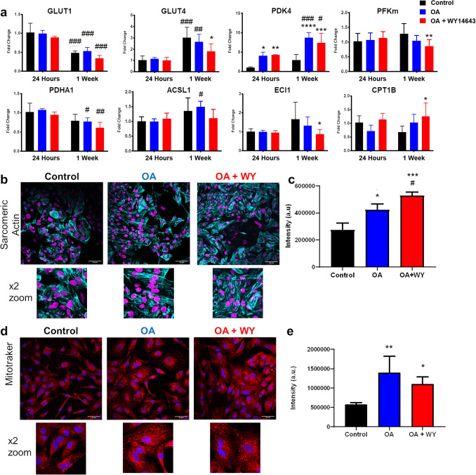
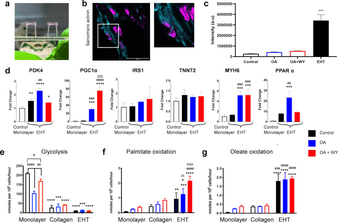
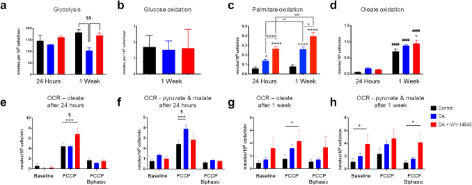
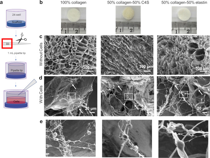
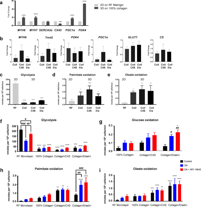
Human induced pluripotent stem cell-derived cardiomyocytes (hiPSC-CMs) enable human cardiac cells to be studied in vitro, although they use glucose as their primary metabolic substrate and do not recapitulate the properties of adult cardiomyocytes. Here, we have explored the interplay between maturation by stimulation of fatty acid oxidation and by culture in 3D. We have investigated substrate metabolism in hiPSC-CMs grown as a monolayer and in 3D, in porous collagen-derived scaffolds and in engineered heart tissue (EHT), by measuring rates of glycolysis and glucose and fatty acid oxidation (FAO), and changes in gene expression and mitochondrial oxygen consumption. FAO was stimulated by activation of peroxisome proliferator-activated receptor alpha (PPARα), using oleate and the agonist WY-14643, which induced an increase in FAO in monolayer hiPSC-CMs. hiPSC-CMs grown in 3D on collagen-derived scaffolds showed reduced glycolysis and increased FAO compared with monolayer cells. Activation of PPARα further increased FAO in cells on collagen/elastin scaffolds but not collagen or collagen/chondroitin-4-sulphate scaffolds. In EHT, FAO was significantly higher than in monolayer cells or those on static scaffolds and could be further increased by culture with oleate and WY-14643. In conclusion, a more mature metabolic phenotype can be induced by culture in 3D and FAO can be incremented by pharmacological stimulation.
人诱导多能干细胞衍生的心肌细胞 (hiPSC-CMs) 使人们能够在体外研究人心肌细胞,尽管它们使用葡萄糖作为主要代谢底物,并且不能再现成人心肌细胞的特性。在这里,我们探讨了刺激脂肪酸氧化和 3D 培养之间的相互作用。我们通过测量糖酵解和葡萄糖和脂肪酸氧化 (FAO) 的速率以及基因表达和线粒体耗氧量的变化,研究了在单层和 3D 培养、多孔胶原衍生支架和工程心脏组织 (EHT) 中生长的 hiPSC-CMs 的底物代谢。通过激活过氧化物酶体增殖物激活受体α (PPARα) 用油酸和激动剂 WY-14643 刺激 FAO,这导致单层 hiPSC-CMs 中的 FAO 增加。与单层细胞相比,在胶原衍生支架上 3D 培养的 hiPSC-CMs 显示出降低的糖酵解和增加的 FAO。在胶原/弹性蛋白支架上的细胞中,PPARα 的激活进一步增加了 FAO,但在胶原或胶原/硫酸软骨素 4 支架上则没有。在 EHT 中,FAO 明显高于单层细胞或静态支架上的细胞,并且可以通过用油酸和 WY-14643 培养进一步增加。总之,3D 培养可以诱导更成熟的代谢表型,并且可以通过药物刺激增加 FAO。