Zeng Dongqiang, Wang Miaohong, Wu Jiani, Lin Siheng, Ye Zilan, Zhou Rui, Wang Gaofeng, Wu Jianhua, Sun Huiying, Bin Jianping, Liao Yulin, Li Nailin, Shi Min, Liao Wangjun
Department of Oncology, Nanfang Hospital, Southern Medical University, Guangzhou, China.
Department of Dermatology, Johns Hopkins School of Medicine, Baltimore, MD, United States.
Front Oncol. 2021 Mar 23;11:620688. doi: 10.3389/fonc.2021.620688. eCollection 2021.
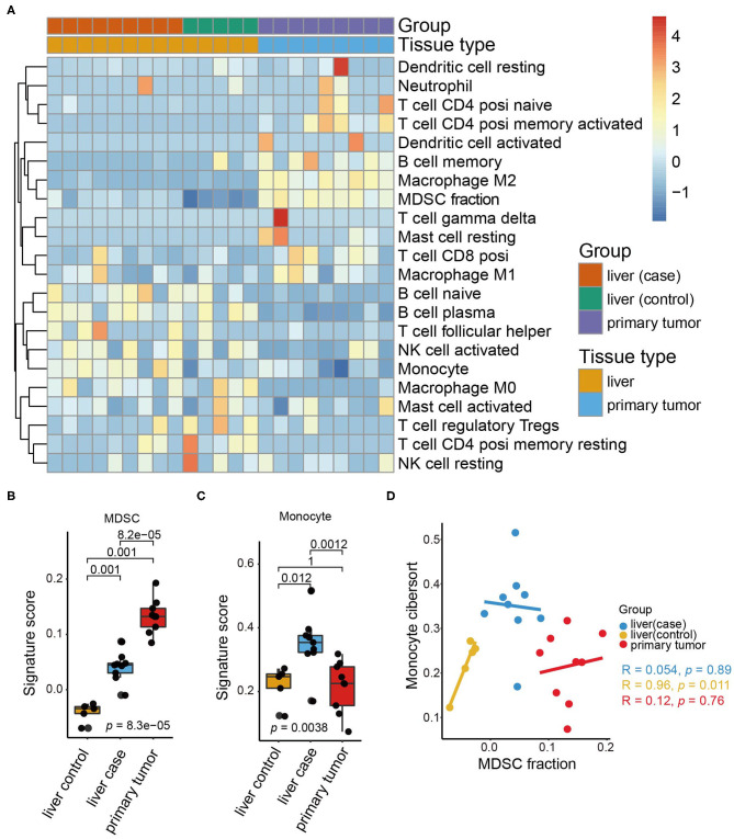
Colorectal cancer, the fourth leading cause of cancer mortality, is prone to metastasis, especially to the liver. The pre-metastatic microenvironment comprising various resident stromal cells and immune cells is essential for metastasis. However, how the dynamic evolution of immune components facilitates pre-metastatic niche formation remains unclear. Utilizing RNA-seq data from our orthotopic colorectal cancer mouse model, we applied single sample gene set enrichment analysis and Cell type Identification By Estimating Relative Subsets Of RNA Transcripts to investigate the tumor microenvironment landscape of pre-metastatic liver, and define the exact role of myeloid-derived suppressor cells (MDSCs) acting in the regulation of infiltrating immune cells and gene pathways activation. Flow cytometry analysis was conducted to quantify the MDSCs levels in human and mice samples. In the current work, based on the high-throughput transcriptome data, we depicted the immune cell infiltration pattern of pre-metastatic liver and highlighted MDSCs as the dominant altered cell type. Notably, flow cytometry analysis showed that high frequencies of MDSCs, was detected in the pre-metastatic liver of orthotopic colorectal cancer tumor-bearing mice, and in the peripheral blood of patients with stage I-III colorectal cancer. MDSCs accumulation in the liver drove immunosuppressive factors secretion and immune checkpoint score upregulation, consequently shaping the pre-metastatic niche with sustained immune suppression. Metabolic reprogramming such as upregulated glycolysis/gluconeogenesis and HIF-1 signaling pathways in the primary tumor was also demonstrated to correlate with MDSCs infiltration in the pre-metastatic liver. Some chemokines were identified as a potential mechanism for MDSCs recruitment. Collectively, our study elucidates the alterations of MDSCs during pre-metastatic niche transformation, and illuminates the latent biological mechanism by which primary tumors impact MDSC aggregation in the targeted liver.
结直肠癌是癌症死亡的第四大主要原因,易于发生转移,尤其是肝转移。由各种驻留基质细胞和免疫细胞组成的转移前微环境对转移至关重要。然而,免疫成分的动态演变如何促进转移前生态位的形成仍不清楚。利用我们的原位结直肠癌小鼠模型的RNA测序数据,我们应用单样本基因集富集分析和通过估计RNA转录本的相对子集进行细胞类型鉴定,以研究转移前肝脏的肿瘤微环境景观,并确定骨髓来源的抑制细胞(MDSCs)在调节浸润免疫细胞和基因通路激活中的确切作用。进行流式细胞术分析以量化人和小鼠样本中的MDSCs水平。在当前的工作中,基于高通量转录组数据,我们描绘了转移前肝脏的免疫细胞浸润模式,并突出了MDSCs作为主要改变的细胞类型。值得注意的是,流式细胞术分析表明,在原位结直肠癌荷瘤小鼠的转移前肝脏以及I-III期结直肠癌患者的外周血中检测到高频率的MDSCs。肝脏中MDSCs的积累促使免疫抑制因子分泌和免疫检查点评分上调,从而形成具有持续免疫抑制作用的转移前生态位。原发性肿瘤中糖酵解/糖异生和HIF-1信号通路等代谢重编程也被证明与转移前肝脏中MDSCs的浸润相关。一些趋化因子被确定为MDSCs募集的潜在机制。总的来说,我们的研究阐明了转移前生态位转变过程中MDSCs的变化,并揭示了原发性肿瘤影响靶向肝脏中MDSC聚集的潜在生物学机制。