Translational Research Laboratory, Gastroenterology and Hepatology, Ente Ospedaliero Cantonale, Università Della Svizzera Italiana, Lugano, Switzerland.
Department of Hepatology & Gastroenterology, Charité Universitätsmedizin Berlin, Berlin, Germany.
Gut Microbes. 2021 Jan-Dec;13(1):1-20. doi: 10.1080/19490976.2021.1911534.
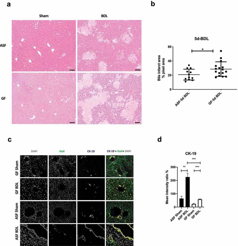
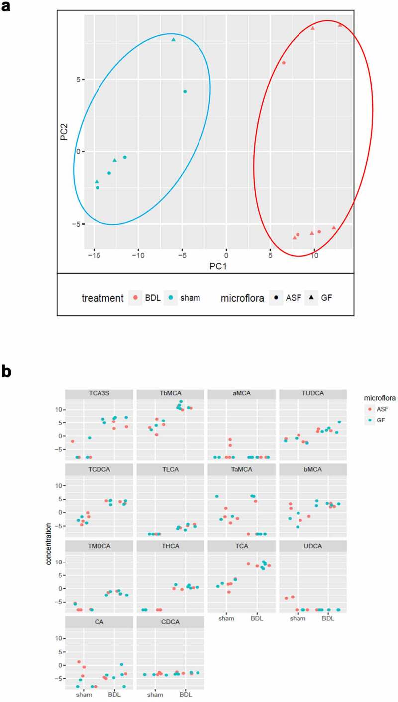
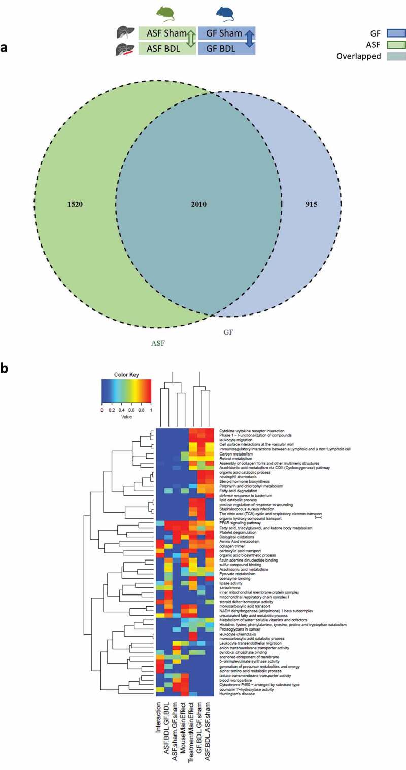
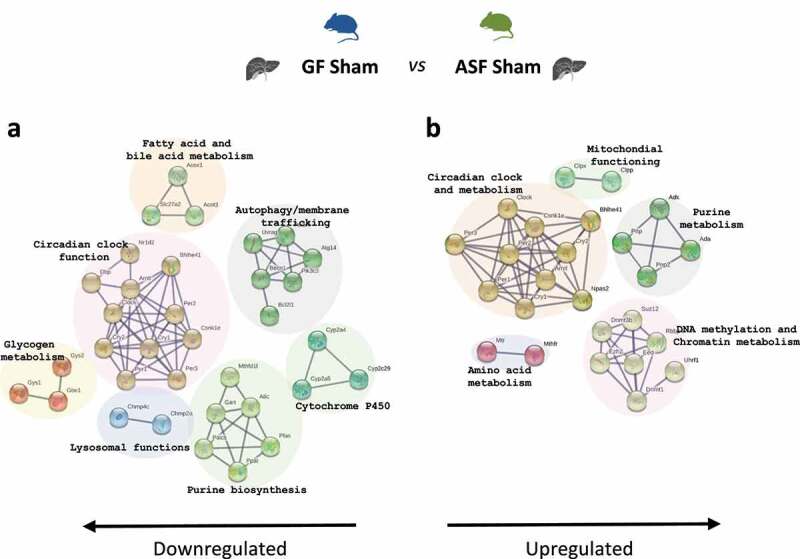
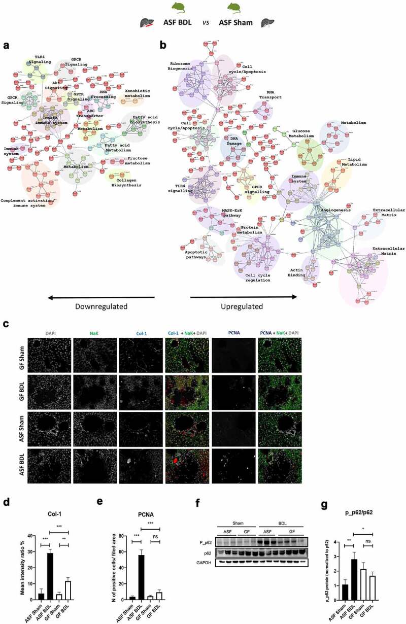
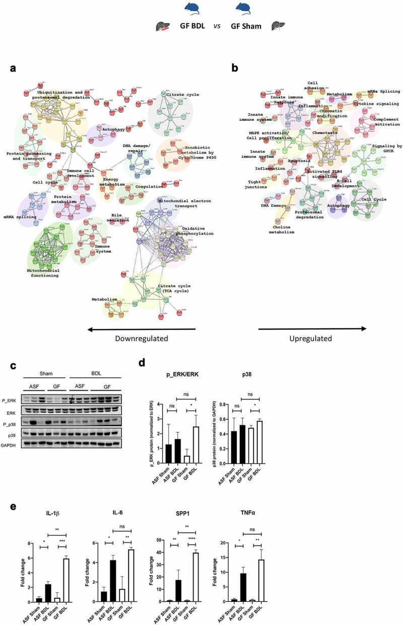
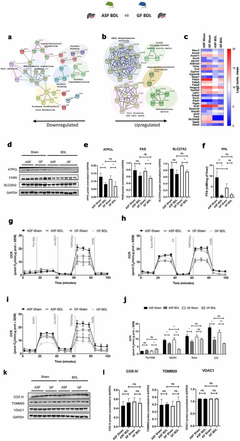
Intestinal microbiota regulates multiple host metabolic and immunological processes. Consequently, any difference in its qualitative and quantitative composition is susceptible to exert significant effects, in particular along the gut-liver axis. Indeed, recent findings suggest that such changes modulate the severity and the evolution of a wide spectrum of hepatobiliary disorders. However, the mechanisms linking intestinal microbiota and the pathogenesis of liver disease remain largely unknown. In this work, we investigated how a distinct composition of the intestinal microbiota, in comparison with germ-free conditions, may lead to different outcomes in an experimental model of acute cholestasis. Acute cholestasis was induced in germ-free (GF) and altered Schaedler's flora (ASF) colonized mice by common bile duct ligation (BDL). Studies were performed 5 days after BDL and hepatic histology, gene expression, inflammation, lipids metabolism, and mitochondrial functioning were evaluated in normal and cholestatic mice. Differences in plasma concentration of bile acids (BA) were evaluated by UHPLC-HRMS. The absence of intestinal microbiota was associated with significant aggravation of hepatic bile infarcts after BDL. At baseline, we found the absence of gut microbiota induced altered expression of genes involved in the metabolism of fatty and amino acids. In contrast, acute cholestasis induced altered expression of genes associated with extracellular matrix, cell cycle, autophagy, activation of MAPK, inflammation, metabolism of lipids, and mitochondrial functioning pathways. Ductular reactions, cell proliferation, deposition of collagen 1 and autophagy were increased in the presence of microbiota after BDL whereas GF mice were more susceptible to hepatic inflammation as evidenced by increased gene expression levels of osteopontin, interleukin (IL)-1β and activation of the ERK/MAPK pathway as compared to ASF colonized mice. Additonally, we found that the presence of microbiota provided partial protection to the mitochondrial functioning and impairment in the fatty acid metabolism after BDL. The concentration of the majority of BA markedly increased after BDL in both groups without remarkable differences according to the hygiene status of the mice. In conclusion, acute cholestasis induced more severe liver injury in GF mice compared to mice with limited intestinal bacterial colonization. This protective effect was associated with different hepatic gene expression profiles mostly related to tissue repair, metabolic and immune functions. Our findings suggest that microbial-induced differences may impact the course of cholestasis and modulate liver injury, offering a background for novel therapies based on the modulation of the intestinal microbiota.
肠道微生物群调节多种宿主代谢和免疫过程。因此,其定性和定量组成的任何差异都容易产生显著的影响,特别是沿着肠道-肝脏轴。事实上,最近的发现表明,这种变化可以调节广泛的肝胆疾病的严重程度和演变。然而,将肠道微生物群与肝病发病机制联系起来的机制在很大程度上仍然未知。在这项工作中,我们研究了与无菌条件相比,肠道微生物群的不同组成如何导致急性胆汁淤积实验模型中的不同结果。通过胆总管结扎(BDL)在无菌(GF)和改变的 Schaedler 菌群(ASF)定植的小鼠中诱导急性胆汁淤积。BDL 后 5 天进行研究,并在正常和胆汁淤积的小鼠中评估肝组织学、基因表达、炎症、脂质代谢和线粒体功能。通过 UHPLC-HRMS 评估血浆中胆汁酸(BA)的浓度差异。肠道微生物群缺失与 BDL 后肝内胆汁淤积的严重程度显著加重有关。在基线时,我们发现肠道微生物群缺失导致参与脂肪酸和氨基酸代谢的基因表达发生改变。相反,急性胆汁淤积诱导与细胞外基质、细胞周期、自噬、MAPK 激活、炎症、脂质代谢和线粒体功能途径相关的基因表达发生改变。BDL 后,微生物存在时胆管反应、细胞增殖、胶原 1 沉积和自噬增加,而 GF 小鼠比 ASF 定植的小鼠更容易发生肝炎症,这表现为骨桥蛋白、白细胞介素(IL)-1β和 ERK/MAPK 途径的基因表达水平增加。此外,我们发现,微生物的存在对 BDL 后线粒体功能和脂肪酸代谢的损伤提供了部分保护。两组小鼠在 BDL 后大多数 BA 的浓度都明显增加,但根据小鼠的卫生状况没有明显差异。总之,与有限的肠道细菌定植的小鼠相比,急性胆汁淤积在 GF 小鼠中引起更严重的肝损伤。这种保护作用与主要与组织修复、代谢和免疫功能相关的不同肝基因表达谱有关。我们的研究结果表明,微生物诱导的差异可能影响胆汁淤积的过程并调节肝损伤,为基于肠道微生物群调节的新型疗法提供了背景。