Oden Institute for Computational Engineering and Sciences, The University of Texas at Austin, Austin, 201 E. 24th Street, POB 4.102, 1 University Station (C0200), Austin, TX, 78712-1229, USA.
Livestrong Cancer Institutes, The University of Texas at Austin, Austin, Austin, TX, USA.
Sci Rep. 2021 Apr 19;11(1):8520. doi: 10.1038/s41598-021-87887-4.
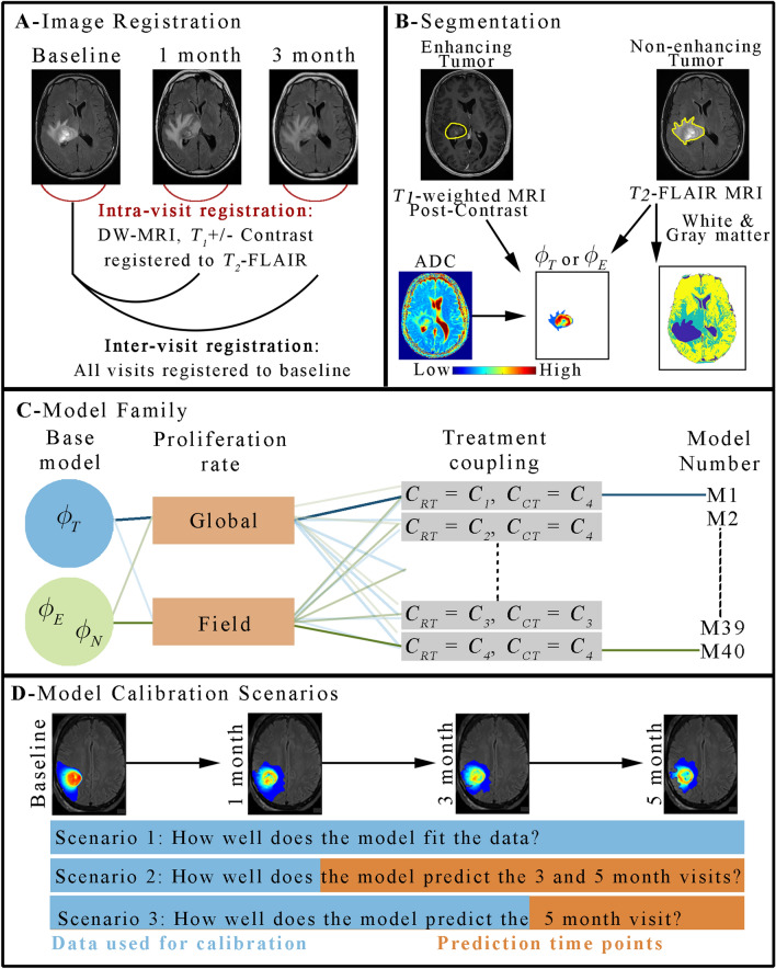
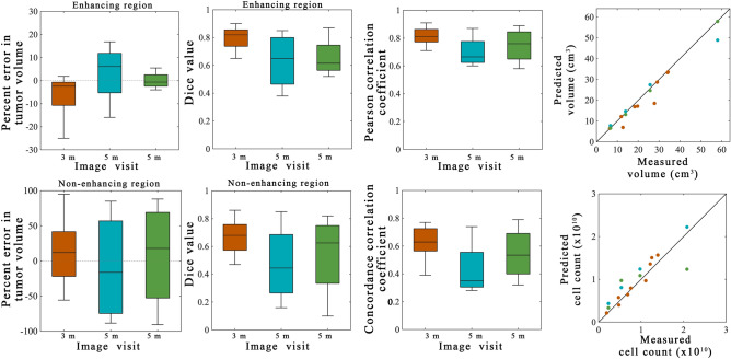
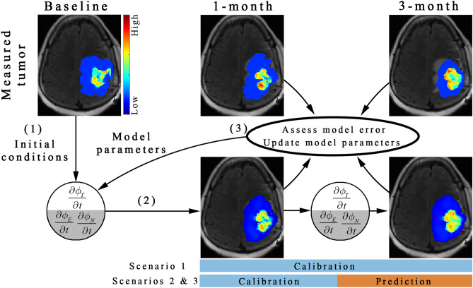
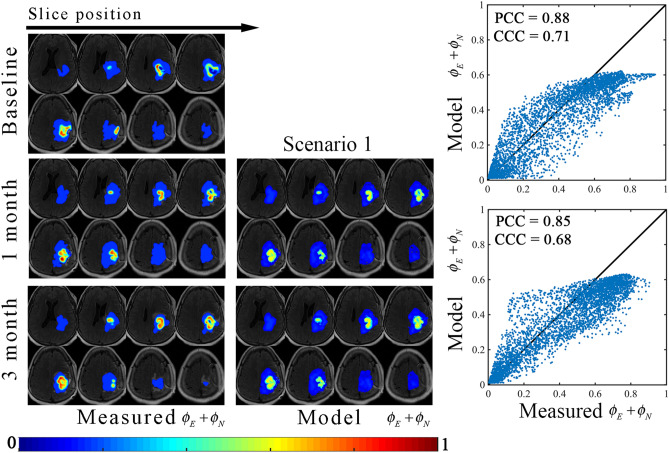
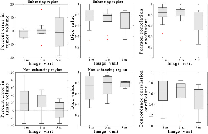
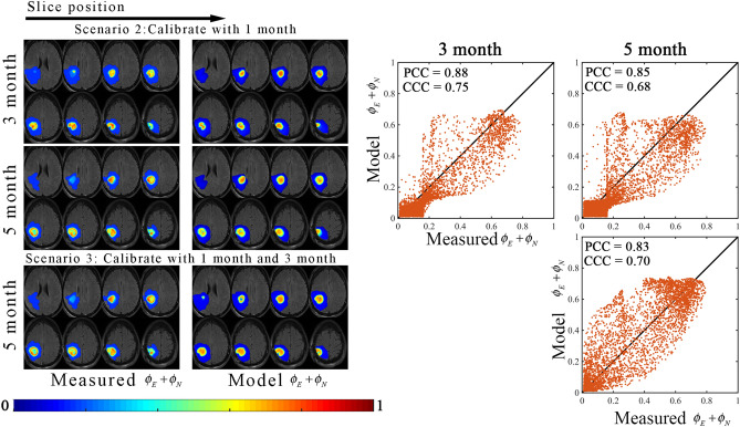
High-grade gliomas are an aggressive and invasive malignancy which are susceptible to treatment resistance due to heterogeneity in intratumoral properties such as cell proliferation and density and perfusion. Non-invasive imaging approaches can measure these properties, which can then be used to calibrate patient-specific mathematical models of tumor growth and response. We employed multiparametric magnetic resonance imaging (MRI) to identify tumor extent (via contrast-enhanced T-weighted, and T-FLAIR) and capture intratumoral heterogeneity in cell density (via diffusion-weighted imaging) to calibrate a family of mathematical models of chemoradiation response in nine patients with unresected or partially resected disease. The calibrated model parameters were used to forecast spatially-mapped individual tumor response at future imaging visits. We then employed the Akaike information criteria to select the most parsimonious member from the family, a novel two-species model describing the enhancing and non-enhancing components of the tumor. Using this model, we achieved low error in predictions of the enhancing volume (median: - 2.5%, interquartile range: 10.0%) and a strong correlation in total cell count (Kendall correlation coefficient 0.79) at 3-months post-treatment. These preliminary results demonstrate the plausibility of using multiparametric MRI data to inform spatially-informative, biologically-based predictive models of tumor response in the setting of clinical high-grade gliomas.
高级别神经胶质瘤是一种侵袭性和浸润性的恶性肿瘤,由于肿瘤内特性(如细胞增殖和密度以及灌注)的异质性,容易产生治疗抵抗。非侵入性成像方法可以测量这些特性,然后可以用于校准患者特定的肿瘤生长和反应的数学模型。我们使用多参数磁共振成像(MRI)来识别肿瘤范围(通过对比增强 T 加权和 T-FLAIR),并通过扩散加权成像捕获肿瘤内细胞密度的异质性,以校准 9 名未切除或部分切除疾病患者的化学放射治疗反应的一系列数学模型。校准后的模型参数用于预测未来影像学检查时的空间映射个体肿瘤反应。然后,我们使用赤池信息量准则从模型族中选择最简约的成员,这是一种描述肿瘤增强和非增强部分的双物种模型。使用该模型,我们在治疗后 3 个月实现了增强体积预测的低误差(中位数:-2.5%,四分位距:10.0%)和总细胞计数的强相关性(肯德尔相关系数 0.79)。这些初步结果表明,使用多参数 MRI 数据来为临床高级别神经胶质瘤中肿瘤反应的空间信息生物基础预测模型提供信息是合理的。