Chen Xun, Wei Qing, Sun Hongmei, Zhang Xiaobo, Yang Changrong, Tao Ying, Nong Guangmin
Department of Pediatrics, The First Affiliated Hospital of Guangxi Medical University, Guangxi, China.
Int J Stem Cells. 2021 Aug 30;14(3):331-340. doi: 10.15283/ijsc20156.
To investigate the effect and the underlying mechanism of exosomes secreted by human umbilical cord mesenchymal stem cells (hUCMSCs) on diffuse alveolar hemorrhage (DAH) in murine lupus.
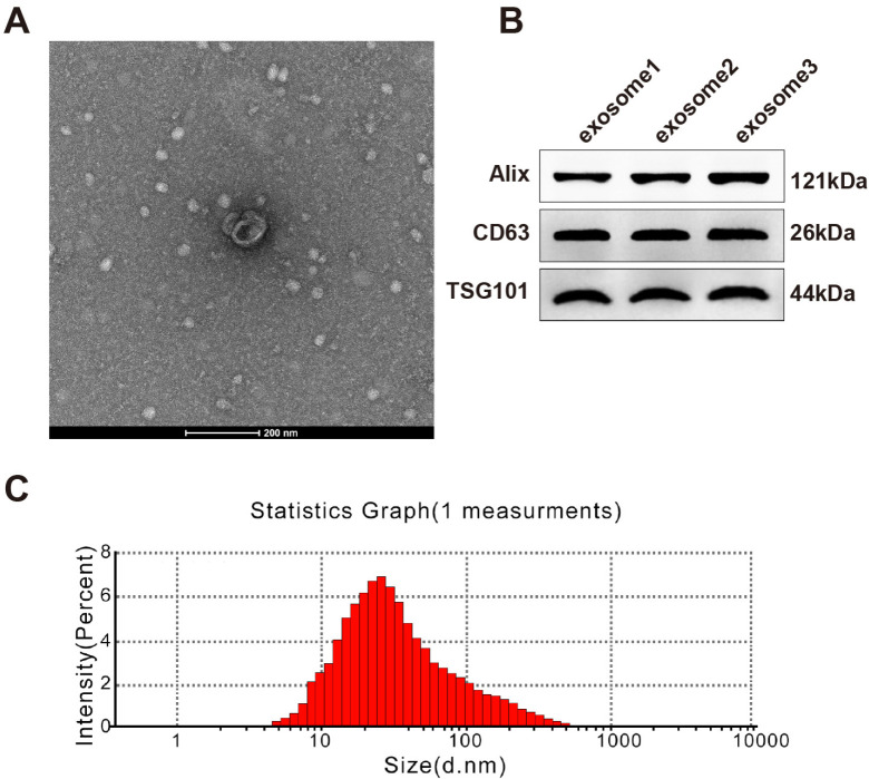
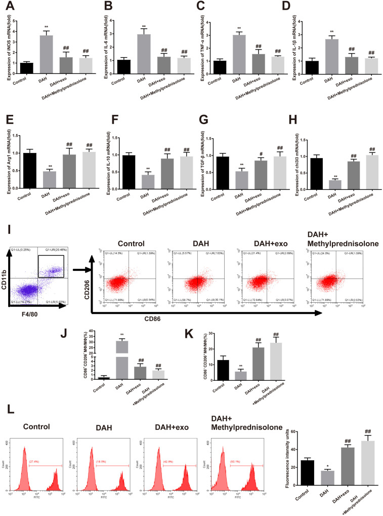
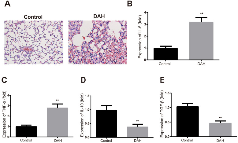
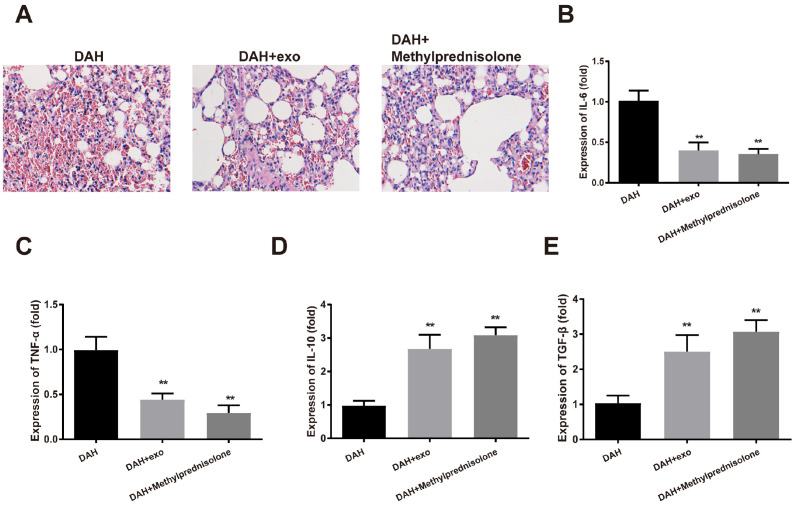
Exosomes were extracted from cultured hUCMSCs by ultracentrifugation. The expressions of exosome markers (Alix, CD63 and TSG101) were measured for identification of hUCMSC-derived exosomes (hUCMSC-exosomes). The alveolar hemorrhage of DAH mice was revealed by H&E staining. The primary alveolar macrophages were isolated from bronchoalveolar lavage fluid (BALF) of DAH mice. The expressions of M1 macrophage markers (iNOS, IL-6, TNF- and IL-1) and M2 macrophage markers (Arg1, IL-10, TGF- and chi3l3) were detected. Flow cytometry measured the ratio of M1/M2 macrophages. ELISA measured the secretion of pro-inflammatory cytokines (IL-6 and TNF-) and anti-inflammatory cytokines (IL-10 and TGF-). DAH mice had hemorrhage and small-vessel vasculitis in the lung, with neutrophil and monocyte infiltration observed around the capillary and small artery. Furthermore, increases of IL-6 and TNF-, and decreases of IL-10 and TGF- were detected in the BALF of DAH mice. M1 makers were overexpressed in alveolar macrophages of DAH mice while M2 makers were lowly expressed. DAH mice had a higher proportion of M1 macrophages than M2 macrophages. After hUCMSC-exosome or methylprednisolone treatment in DAH mice, the alveolar injuries and inflammatory responses were attenuated, and the proportion of M2 macrophages was increased.
hUCMSC-exosomes attenuate DAH-induced inflammatory responses and alveolar hemorrhage by regulating macrophage polarization.
研究人脐带间充质干细胞(hUCMSCs)分泌的外泌体对小鼠狼疮性弥漫性肺泡出血(DAH)的影响及其潜在机制。
通过超速离心从培养的hUCMSCs中提取外泌体。检测外泌体标志物(Alix、CD63和TSG101)的表达以鉴定hUCMSC来源的外泌体(hUCMSC-外泌体)。用苏木精-伊红染色显示DAH小鼠的肺泡出血情况。从DAH小鼠的支气管肺泡灌洗液(BALF)中分离出原代肺泡巨噬细胞。检测M1巨噬细胞标志物(iNOS、IL-6、TNF-α和IL-1β)和M2巨噬细胞标志物(Arg1、IL-10、TGF-β和chi3l3)的表达。流式细胞术检测M1/M2巨噬细胞的比例。酶联免疫吸附测定法检测促炎细胞因子(IL-6和TNF-α)和抗炎细胞因子(IL-10和TGF-β)的分泌。DAH小鼠肺部有出血和小血管血管炎,在毛细血管和小动脉周围观察到中性粒细胞和单核细胞浸润。此外,在DAH小鼠的BALF中检测到IL-6和TNF-α升高,IL-10和TGF-β降低。DAH小鼠肺泡巨噬细胞中M1标志物过表达而M2标志物低表达。DAH小鼠中M1巨噬细胞的比例高于M2巨噬细胞。在DAH小鼠中给予hUCMSC-外泌体或甲基强的松龙治疗后,肺泡损伤和炎症反应减轻,M2巨噬细胞的比例增加。
hUCMSC-外泌体通过调节巨噬细胞极化减轻DAH诱导的炎症反应和肺泡出血。