Computers and Systems Department, Faculty of Engineering, Mansoura University, Mansoura 35511, Egypt.
BioImaging Laboratory, Bioengineering Department, University of Louisville, Louisville, KY 40292, USA.
Sensors (Basel). 2021 Apr 7;21(8):2586. doi: 10.3390/s21082586.
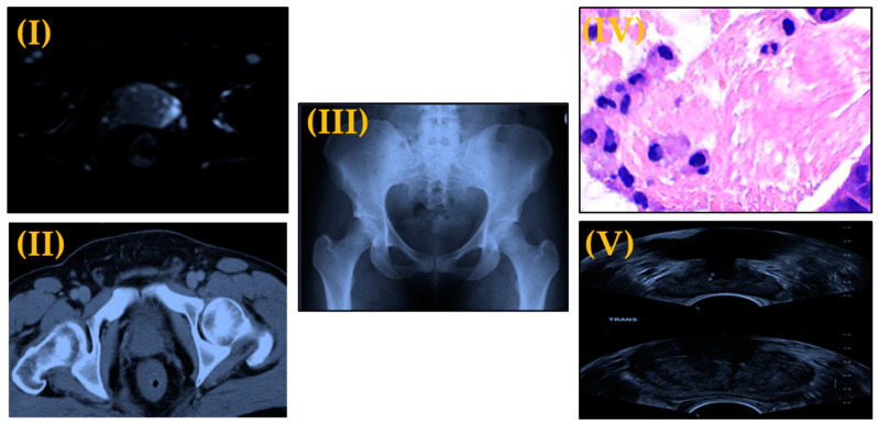
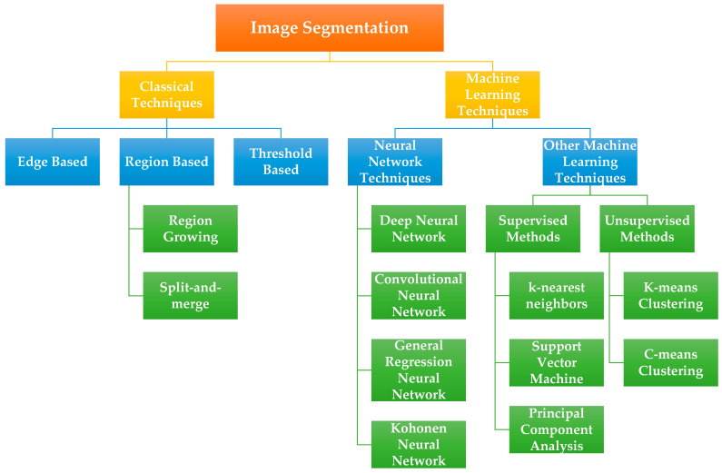
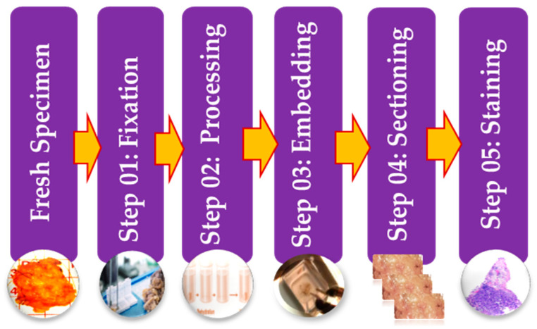
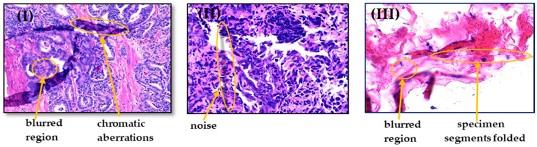
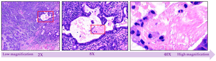
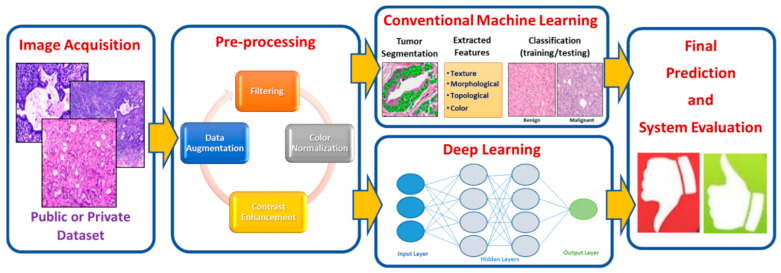
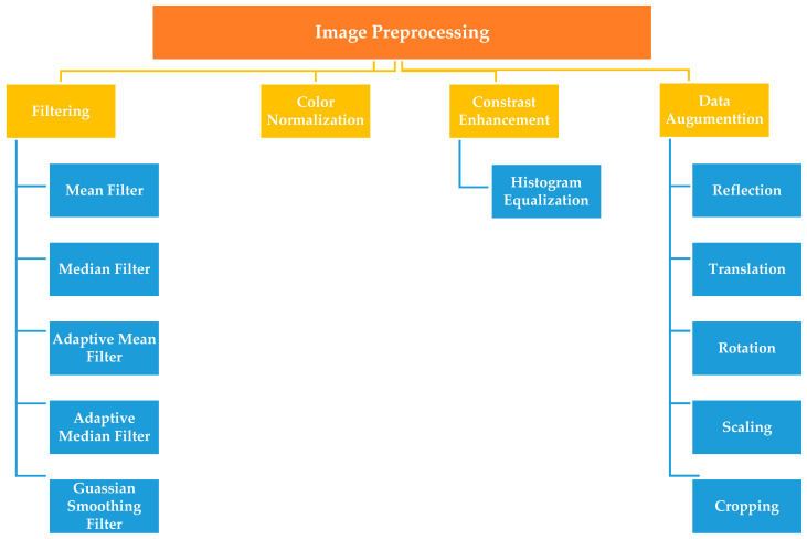
Prostate cancer is one of the most identified cancers and second most prevalent among cancer-related deaths of men worldwide. Early diagnosis and treatment are substantial to stop or handle the increase and spread of cancer cells in the body. Histopathological image diagnosis is a gold standard for detecting prostate cancer as it has different visual characteristics but interpreting those type of images needs a high level of expertise and takes too much time. One of the ways to accelerate such an analysis is by employing artificial intelligence (AI) through the use of computer-aided diagnosis (CAD) systems. The recent developments in artificial intelligence along with its sub-fields of conventional machine learning and deep learning provide new insights to clinicians and researchers, and an abundance of research is presented specifically for histopathology images tailored for prostate cancer. However, there is a lack of comprehensive surveys that focus on prostate cancer using histopathology images. In this paper, we provide a very comprehensive review of most, if not all, studies that handled the prostate cancer diagnosis using histopathological images. The survey begins with an overview of histopathological image preparation and its challenges. We also briefly review the computing techniques that are commonly applied in image processing, segmentation, feature selection, and classification that can help in detecting prostate malignancies in histopathological images.
前列腺癌是全球男性癌症相关死亡中最常见的癌症之一,也是第二大常见癌症。早期诊断和治疗对于阻止或控制体内癌细胞的增长和扩散至关重要。组织病理学图像诊断是检测前列腺癌的金标准,因为它具有不同的视觉特征,但解释这类图像需要高度的专业知识,并且需要花费太多时间。加速这种分析的一种方法是通过使用人工智能 (AI) 并利用计算机辅助诊断 (CAD) 系统。人工智能及其传统机器学习和深度学习等子领域的最新发展为临床医生和研究人员提供了新的见解,并且专门针对前列腺癌的组织病理学图像提出了大量研究。然而,缺乏使用组织病理学图像针对前列腺癌的全面调查。在本文中,我们对使用组织病理学图像诊断前列腺癌的大多数(如果不是全部)研究进行了非常全面的回顾。调查首先概述了组织病理学图像的准备及其挑战。我们还简要回顾了图像处理、分割、特征选择和分类中常用的计算技术,这些技术有助于在组织病理学图像中检测前列腺恶性肿瘤。