Sivakumar Shivan, Abu-Shah Enas, Ahern David J, Arbe-Barnes Edward H, Jainarayanan Ashwin K, Mangal Nagina, Reddy Srikanth, Rendek Aniko, Easton Alistair, Kurz Elke, Silva Michael, Soonawalla Zahir, Heij Lara R, Bashford-Rogers Rachael, Middleton Mark R, Dustin Michael L
Department of Oncology, University of Oxford, Oxford OX3 7DQ, UK.
Kennedy Institute of Rheumatology, University of Oxford, Oxford OX3 7FY, UK.
Cancers (Basel). 2021 Apr 8;13(8):1776. doi: 10.3390/cancers13081776.
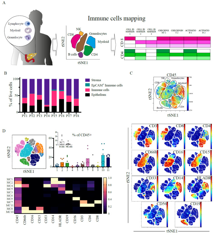
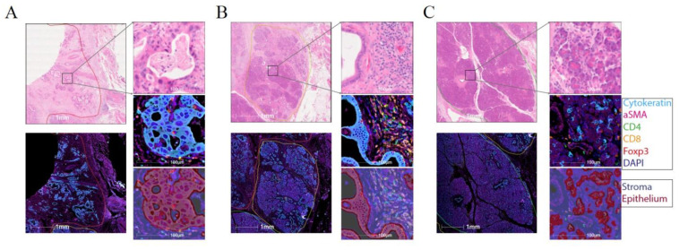
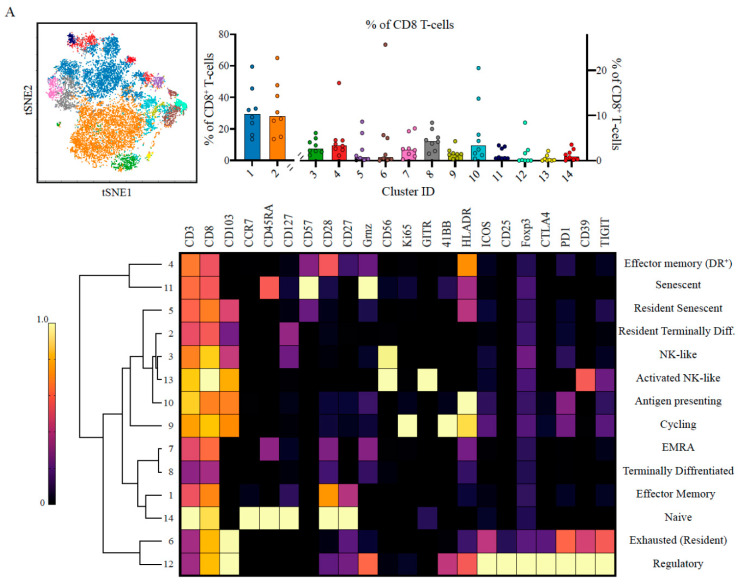
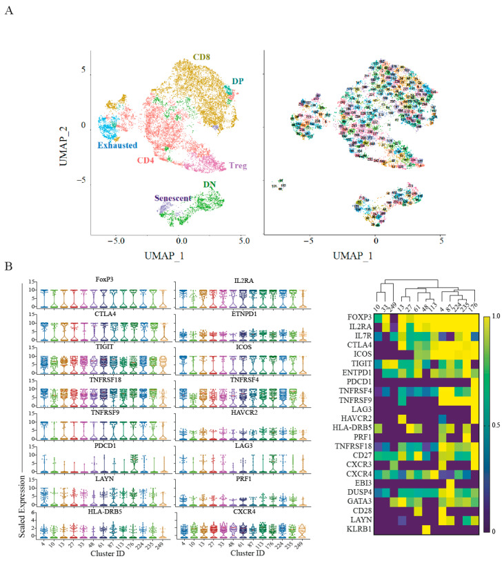
Pancreatic cancer has one of the worst prognoses of any human malignancy and leukocyte infiltration is a major prognostic marker of the disease. As current immunotherapies confer negligible survival benefits, there is a need to better characterise leukocytes in pancreatic cancer to identify better therapeutic strategies. In this study, we analysed 32 human pancreatic cancer patients from two independent cohorts. A multi-parameter mass-cytometry analysis was performed on 32,000 T-cells from eight patients. Single-cell RNA sequencing dataset analysis was performed on a cohort of 24 patients. Multiplex immunohistochemistry imaging and spatial analysis were performed to map immune infiltration into the tumour microenvironment. Regulatory T-cell populations demonstrated highly immunosuppressive states with high TIGIT, ICOS and CD39 expression. CD8 T-cells were found to be either in senescence or an exhausted state. The exhausted CD8 T-cells had low PD-1 expression but high TIGIT and CD39 expression. These findings were corroborated in an independent pancreatic cancer single-cell RNA dataset. These data suggest that T-cells are major players in the suppressive microenvironment of pancreatic cancer. Our work identifies multiple novel therapeutic targets that should form the basis for rational design of a new generation of clinical trials in pancreatic ductal adenocarcinoma.
胰腺癌是所有人类恶性肿瘤中预后最差的疾病之一,白细胞浸润是该疾病的主要预后标志物。由于目前的免疫疗法对生存率的提升微乎其微,因此有必要更好地描述胰腺癌中的白细胞,以确定更好的治疗策略。在本研究中,我们分析了来自两个独立队列的32例人类胰腺癌患者。对8例患者的32000个T细胞进行了多参数质谱流式细胞术分析。对24例患者的队列进行了单细胞RNA测序数据集分析。进行了多重免疫组化成像和空间分析,以绘制免疫细胞浸润到肿瘤微环境中的情况。调节性T细胞群体表现出高度免疫抑制状态,TIGIT、ICOS和CD39表达较高。发现CD8 T细胞处于衰老或耗竭状态。耗竭的CD8 T细胞PD-1表达较低,但TIGIT和CD39表达较高。这些发现在一个独立的胰腺癌单细胞RNA数据集中得到了证实。这些数据表明,T细胞是胰腺癌抑制性微环境中的主要参与者。我们的工作确定了多个新的治疗靶点,这些靶点应为合理设计新一代胰腺癌导管腺癌临床试验奠定基础。