Department of Regenerative Medicine, State Research Institute Centre for Innovative Medicine, LT-08406, Vilnius, Lithuania.
Department of Sensory and Motor System Medicine, Department of Oral-maxillofacial Surgery, Dentistry and Orthodontics, Graduate School of Medicine, The University of Tokyo, Hongo 7-3-1, Bunkyo-ku, Tokyo, 113-8655, Japan.
Stem Cell Res Ther. 2021 Apr 29;12(1):251. doi: 10.1186/s13287-021-02286-w.
Due to its low capacity for self-repair, articular cartilage is highly susceptible to damage and deterioration, which leads to the development of degenerative joint diseases such as osteoarthritis (OA). Menstrual blood-derived mesenchymal stem/stromal cells (MenSCs) are much less characterized, as compared to bone marrow mesenchymal stem/stromal cells (BMMSCs). However, MenSCs seem an attractive alternative to classical BMMSCs due to ease of access and broader differentiation capacity. The aim of this study was to evaluate chondrogenic differentiation potential of MenSCs and BMMSCs stimulated with transforming growth factor β (TGF-β3) and activin A.
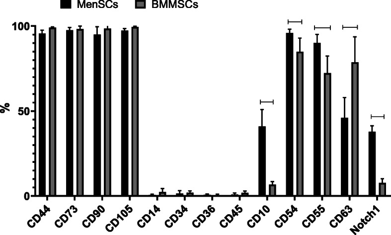
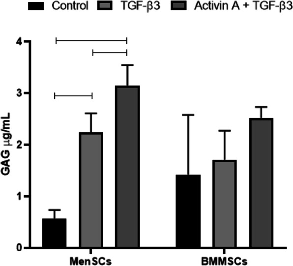
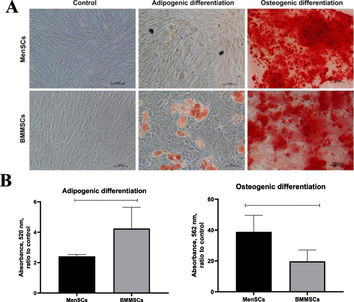
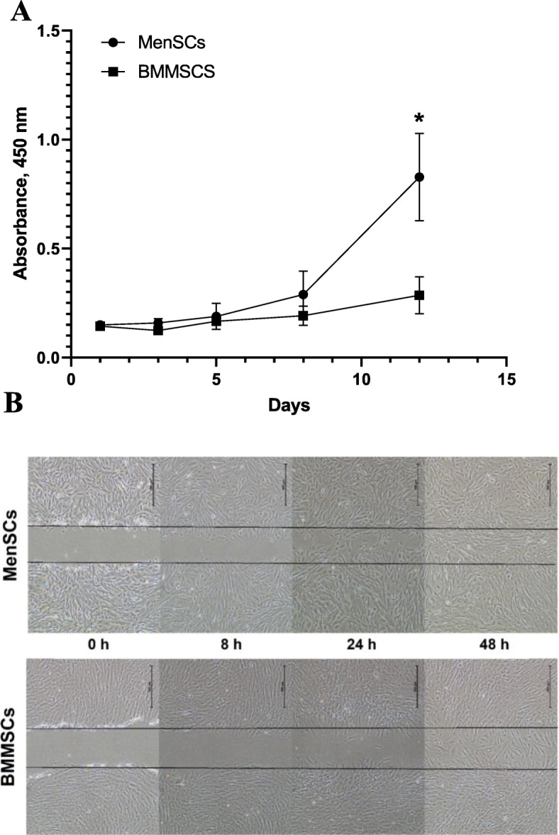
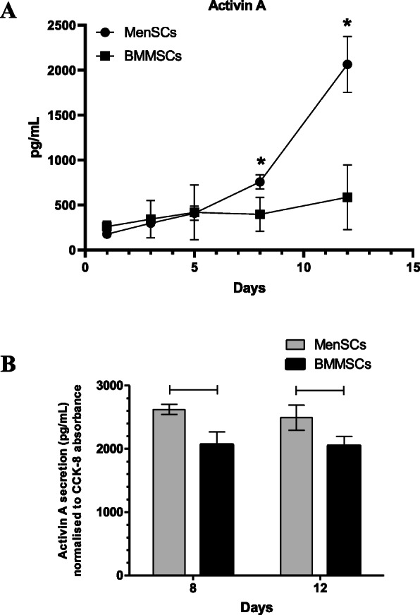
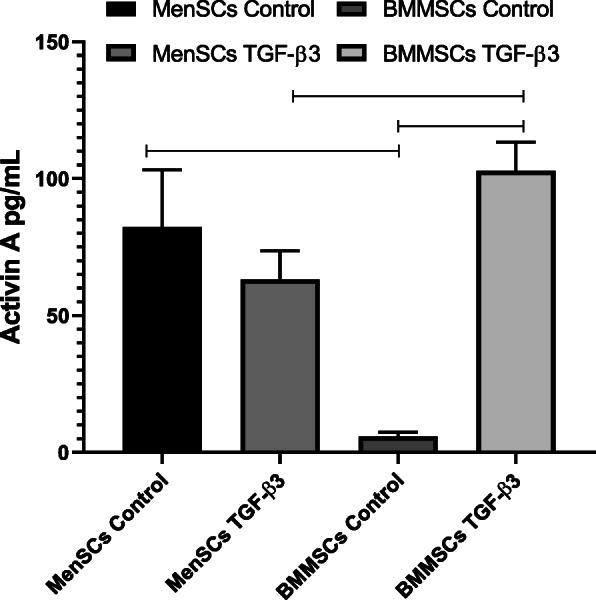
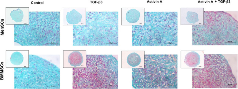
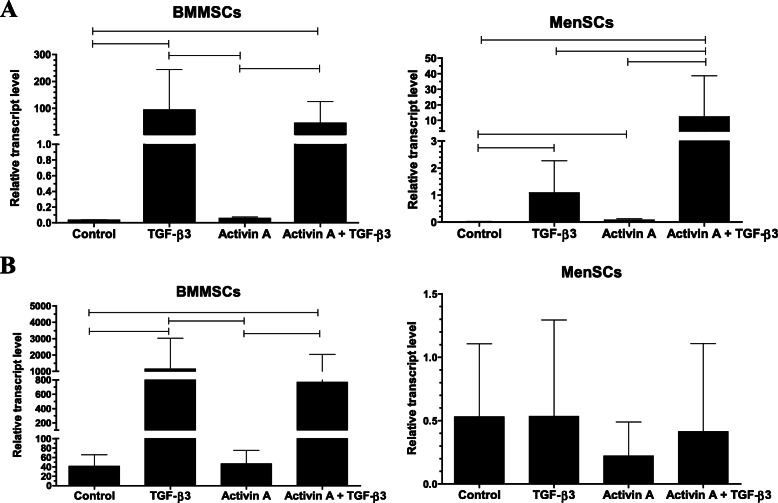
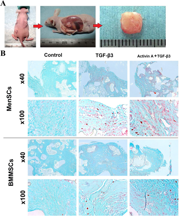
MenSCs (n = 6) and BMMSCs (n = 5) were isolated from different healthy donors. Expression of cell surface markers CD90, CD73, CD105, CD44, CD45, CD14, CD36, CD55, CD54, CD63, CD106, CD34, CD10, and Notch1 was analyzed by flow cytometry. Cell proliferation capacity was determined using CCK-8 proliferation kit and cell migration ability was evaluated by scratch assay. Adipogenic differentiation capacity was evaluated according to Oil-Red staining and osteogenic differentiation according to Alizarin Red staining. Chondrogenic differentiation (activin A and TGF-β3 stimulation) was investigated in vitro and in vivo (subcutaneous scaffolds in nude BALB/c mice) by expression of chondrogenic genes (collagen type II, aggrecan), GAG assay and histologically. Activin A protein production was evaluated by ELISA during chondrogenic differentiation in monolayer culture.
MenSCs exhibited a higher proliferation rate, as compared to BMMSCs, and a different expression profile of several cell surface markers. Activin A stimulated collagen type II gene expression and glycosaminoglycan synthesis in TGF-β3 treated MenSCs but not in BMMSCs, both in vitro and in vivo, although the effects of TGF-β3 alone were more pronounced in BMMSCs in vitro.
These data suggest that activin A exerts differential effects on the induction of chondrogenic differentiation in MenSCs vs. BMMSCs, which implies that different mechanisms of chondrogenic regulation are activated in these cells. Following further optimization of differentiation protocols and the choice of growth factors, potentially including activin A, MenSCs may turn out to be a promising population of stem cells for the development of cell-based therapies with the capacity to stimulate cartilage repair and regeneration in OA and related osteoarticular disorders.
由于其自我修复能力较低,关节软骨极易受到损伤和恶化,从而导致骨关节炎(OA)等退行性关节疾病的发生。与骨髓间充质干细胞(BMMSCs)相比,经血来源的间充质干细胞(MenSCs)的特征要少得多。然而,由于易于获得和更广泛的分化能力,MenSCs 似乎是经典 BMMSCs 的一个有吸引力的替代品。本研究旨在评估转化生长因子β(TGF-β3)和激活素 A 刺激的 MenSCs 和 BMMSCs 的软骨分化潜力。
从不同的健康供体中分离出 MenSCs(n=6)和 BMMSCs(n=5)。通过流式细胞术分析细胞表面标志物 CD90、CD73、CD105、CD44、CD45、CD14、CD36、CD55、CD54、CD63、CD106、CD34、CD10 和 Notch1 的表达。使用 CCK-8 增殖试剂盒测定细胞增殖能力,划痕试验评估细胞迁移能力。根据油红 O 染色评估脂肪生成分化能力,根据茜素红染色评估成骨分化能力。通过体外和体内(裸鼠 BALB/c 皮下支架)研究软骨分化(激活素 A 和 TGF-β3 刺激),通过表达软骨基因(Ⅱ型胶原、聚集蛋白聚糖)、GAG 测定和组织学评估。在单层培养的软骨分化过程中通过 ELISA 评估激活素 A 蛋白的产生。
MenSCs 的增殖率高于 BMMSCs,并且几种细胞表面标志物的表达模式也不同。激活素 A 刺激 TGF-β3 处理的 MenSCs 中胶原 II 基因表达和糖胺聚糖合成,但不刺激 BMMSCs,无论是在体外还是体内,尽管 TGF-β3 单独在体外对 BMMSCs 的作用更为明显。
这些数据表明,激活素 A 对 MenSCs 与 BMMSCs 诱导软骨分化的作用不同,这意味着这些细胞中激活了不同的软骨调节机制。在进一步优化分化方案和选择生长因子(可能包括激活素 A)后,MenSCs 可能成为一种很有前途的干细胞群体,用于开发细胞疗法,以刺激 OA 和相关骨关节炎疾病中的软骨修复和再生。