Division of Inflammation and Infection/Rheumatology, Department of Biomedical and Clinical Sciences, Linköping University, Linköping, Sweden.
Department of Clinical Physiology, University Hospital and Department of Health, Medicine and Caring Sciences, Linköping University, Linköping, Sweden.
Lupus. 2021 Jul;30(8):1244-1253. doi: 10.1177/09612033211013898. Epub 2021 May 6.
The progress of accelerated atherosclerosis in systemic lupus erythematosus (SLE) is incompletely understood. Circulating osteopontin (OPN) is increased in autoimmune conditions, e.g. SLE, and its serum concentration was recently reported to associate with subclinical atherosclerosis in SLE, as measured by carotid intima-media thickness. The aim of this study was to investigate whether OPN may be used as a surrogate biomarker of subclinical atherosclerosis in SLE patients with different disease phenotypes.
We recruited 60 well-characterised SLE cases and 60 age- and sex-matched healthy controls. The SLE cases were divided into three different disease phenotypes: SLE with antiphospholipid syndrome (APS), lupus nephritis, and isolated skin and joint involvement. Plasma OPN was detected by ELISA (Quantikine®, R&D Systems). Common carotid arteries intima media thickness was compared between the studied groups in relation to OPN levels and risk factors for vascular changes. Intima media thickness of common carotid arteries was measured by using a sensitive ultrasound technique (LOGIQ™ E9 ultrasound, GE Healthcare).
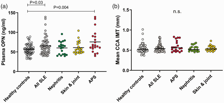
OPN levels were significantly higher among the entire SLE group ( = 60) compared to the healthy controls ( = 0.03). SLE cases with concomitant APS ( = 20) showed higher OPN levels than the controls ( = 0.004), whereas none of the other two subgroups differed significantly from the healthy controls. OPN and intima media thickness were correlated to several traditional risk factors of atherosclerosis, as well as to SLE-related factors. Yet, no significant correlation was observed between OPN levels and ultrasound findings of the common carotid arteries.
In line with previous studies, we observed increased OPN levels among SLE patients as compared to matched controls. However, the OPN concentrations did not correlate with intima media thickness of the common carotid arteries. Based on our findings, the use of OPN as a surrogate biomarker of subclinical atherosclerosis in SLE subjects, regardless of clinical phenotypes, cannot be recommended.
系统性红斑狼疮(SLE)患者的动脉粥样硬化加速进展的机制尚不完全清楚。在自身免疫性疾病(如 SLE)中,循环骨桥蛋白(OPN)增加,最近有研究报道其血清浓度与 SLE 患者的亚临床动脉粥样硬化相关,可通过颈动脉内膜中层厚度(CIMT)进行测量。本研究旨在探讨 OPN 是否可作为不同疾病表型的 SLE 患者亚临床动脉粥样硬化的替代生物标志物。
我们招募了 60 名特征明确的 SLE 患者和 60 名年龄和性别匹配的健康对照者。将 SLE 患者分为三种不同的疾病表型:伴有抗磷脂综合征(APS)的 SLE、狼疮肾炎和单纯皮肤及关节受累。通过 ELISA(Quantikine®,R&D Systems)检测血浆 OPN。比较研究组间 OPN 水平与血管变化的危险因素之间的 CIMT 差异。使用敏感的超声技术(LOGIQ™ E9 超声,GE Healthcare)测量颈总动脉内膜中层厚度。
与健康对照组相比,整个 SLE 组(n=60)的 OPN 水平显著升高( =0.03)。同时伴有 APS 的 SLE 患者(n=20)的 OPN 水平高于对照组( =0.004),而其他两个亚组与健康对照组之间无显著差异。OPN 与内膜中层厚度与动脉粥样硬化的多种传统危险因素以及与 SLE 相关的因素相关。然而,OPN 水平与颈总动脉超声结果之间未观察到显著相关性。
与之前的研究一致,我们观察到与匹配的对照组相比,SLE 患者的 OPN 水平升高。然而,OPN 浓度与颈总动脉内膜中层厚度不相关。基于我们的研究结果,不能推荐 OPN 作为 SLE 患者亚临床动脉粥样硬化的替代生物标志物,无论其临床表型如何。