Beijing City Key Lab for Medical Physics and Engineering, Institute of Heavy Ion Physics, School of Physics, Peking University, Beijing, China.
Center for MRI Research, Academy for Advanced Interdisciplinary Studies, Peking University, Beijing, China.
Hum Brain Mapp. 2021 Aug 1;42(11):3667-3679. doi: 10.1002/hbm.25461. Epub 2021 May 7.
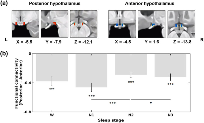
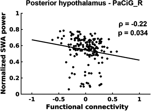
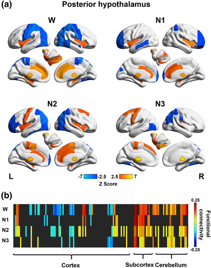
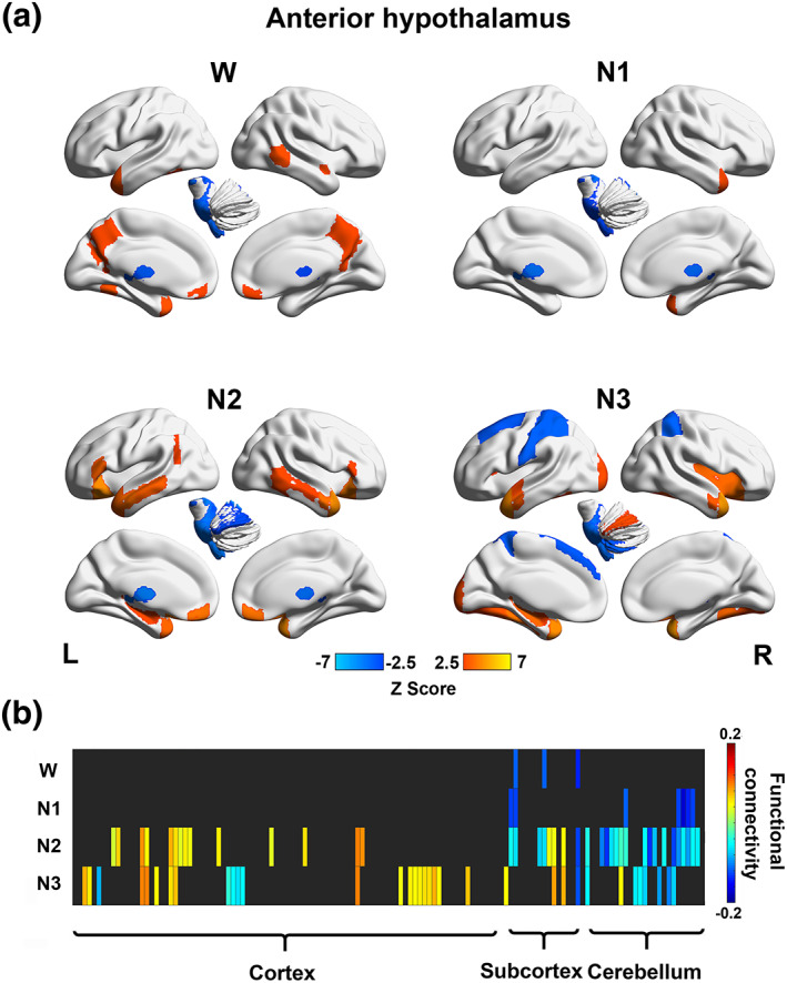
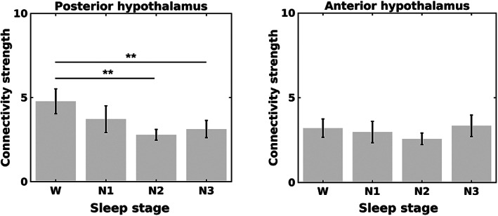
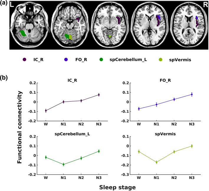
Animal experiments indicate that the hypothalamus plays an essential role in regulating the sleep-wake cycle. A recent neuroimaging study conducted under resting wakefulness conditions suggested the presence of a wake-promoting region and a sleep-promoting region in the human posterior hypothalamus and anterior hypothalamus, respectively, and interpreted their anticorrelated organization in resting-state functional networks as evidence for their opposing roles in sleep-wake regulation. However, whether and how the functional networks of the two hypothalamic regions reorganize according to their wake- or sleep-promoting roles during sleep are unclear. Here, we constructed functional networks of the posterior and anterior hypothalamus during wakefulness and nonrapid eye movement (NREM) sleep using simultaneous electroencephalography and functional magnetic resonance imaging data collected from 62 healthy participants. The functional networks of the posterior and anterior hypothalamus exhibited inversely correlated organizations during both wakefulness and NREM sleep. The connectivity strength of the posterior hypothalamic functional network was stronger during wakefulness than during stable sleep. From wakefulness to sleep, the anterior cingulate gyrus, paracingulate gyrus, insular cortex, and fontal operculum cortex showed decreased positive connectivity, while the precentral gyrus and postcentral gyrus showed decreased negative connectivity with the posterior hypothalamus. Additionally, the insular cortex and frontal operculum cortex showed negative connectivity during wakefulness and positive connectivity during sleep with the anterior hypothalamus, exhibiting an increasing trend. These findings provide insights into the correspondence between the functional network organizations and hypothalamic sleep-wake regulation in humans.
动物实验表明,下丘脑在调节睡眠-觉醒周期中起着至关重要的作用。最近一项在静息清醒状态下进行的神经影像学研究表明,人类下丘脑的后部和前部分别存在促进觉醒和促进睡眠的区域,并将其在静息态功能网络中的反相关组织解释为其在睡眠-觉醒调节中对立作用的证据。然而,在睡眠期间,这两个下丘脑区域的功能网络是否以及如何根据其促进觉醒或睡眠的作用进行重组尚不清楚。在这里,我们使用同时采集的 62 名健康参与者的脑电图和功能磁共振成像数据,构建了清醒和非快速眼动(NREM)睡眠期间下丘脑后部和前部的功能网络。在清醒和 NREM 睡眠期间,下丘脑后部和前部的功能网络均呈现出反向相关的组织。在清醒状态下,下丘脑后部功能网络的连接强度强于稳定睡眠状态。从清醒到睡眠,扣带回前回、扣带回旁回、脑岛和额盖皮质的正连接性降低,而中央前回和中央后回的负连接性降低。此外,脑岛和额盖皮质在清醒时与下丘脑前部呈现负连接,在睡眠时呈现正连接,呈递增趋势。这些发现为人类下丘脑睡眠-觉醒调节的功能网络组织之间的对应关系提供了深入的了解。