• 文献检索
  • 文档翻译
  • 深度研究
  • 学术资讯
  • Suppr Zotero 插件Zotero 插件
  • 邀请有礼
  • 套餐&价格
  • 历史记录
应用&插件
Suppr Zotero 插件Zotero 插件浏览器插件Mac 客户端Windows 客户端微信小程序
定价
高级版会员购买积分包购买API积分包
服务
文献检索文档翻译深度研究API 文档MCP 服务
关于我们
关于 Suppr公司介绍联系我们用户协议隐私条款
关注我们

Suppr 超能文献

核心技术专利:CN118964589B侵权必究
粤ICP备2023148730 号-1Suppr @ 2026

文献检索

告别复杂PubMed语法,用中文像聊天一样搜索,搜遍4000万医学文献。AI智能推荐,让科研检索更轻松。

立即免费搜索

文件翻译

保留排版,准确专业,支持PDF/Word/PPT等文件格式,支持 12+语言互译。

免费翻译文档

深度研究

AI帮你快速写综述,25分钟生成高质量综述,智能提取关键信息,辅助科研写作。

立即免费体验

糖尿病性视网膜病变中的光学相干断层扫描高反射性视网膜病灶:一项半自动检测比较研究。

OCT Hyperreflective Retinal Foci in Diabetic Retinopathy: A Semi-Automatic Detection Comparative Study.

作者信息

Midena Edoardo, Torresin Tommaso, Velotta Erika, Pilotto Elisabetta, Parrozzani Raffaele, Frizziero Luisa

机构信息

Department of Ophthalmology, University of Padova, Padova, Italy.

IRCCS-Fondazione Bietti, Rome, Italy.

出版信息

Front Immunol. 2021 Apr 22;12:613051. doi: 10.3389/fimmu.2021.613051. eCollection 2021.

DOI:10.3389/fimmu.2021.613051
PMID:33968016
原文链接:https://pmc.ncbi.nlm.nih.gov/articles/PMC8100046/
Abstract

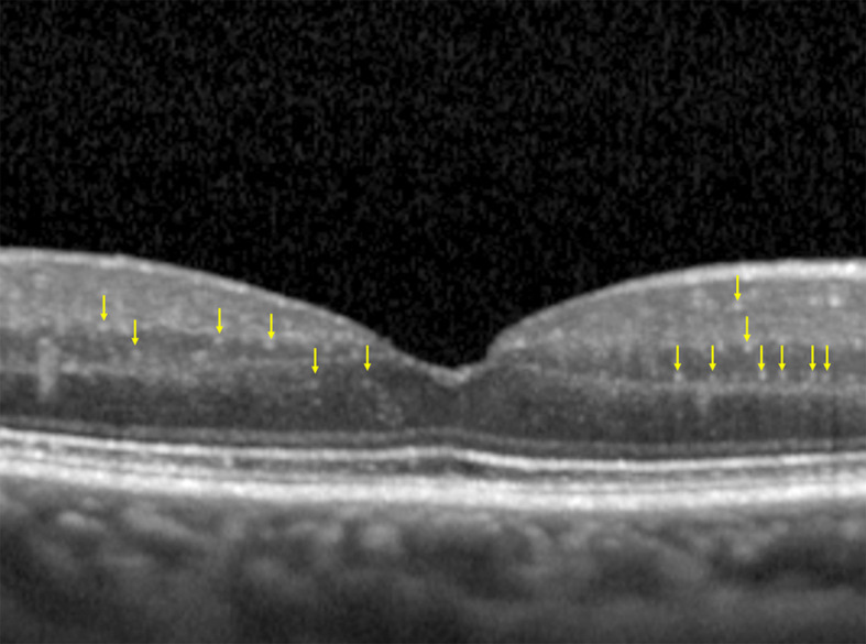
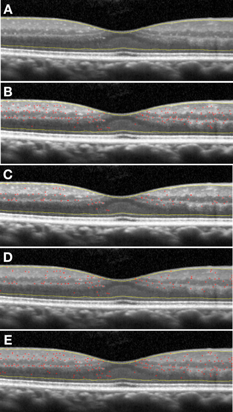
Optical coherence tomography (OCT) allows us to identify, into retinal layers, new morphological entities, which can be considered clinical biomarkers of retinal diseases. According to the literature, solitary, small (<30 µm), medium level hyperreflective (similar to retinal fiber layer) retinal foci (HRF) may represent aggregates of activated microglial cells and an biomarker of retinal inflammation. The identification and quantification of this imaging biomarker allows for estimating the level and possibly the amount of intraretinal inflammation in major degenerative retinal disorders, whose inflammatory component has already been demonstrated (diabetic retinopathy, age-related macular degeneration, radiation retinopathy). Currently, diabetic retinopathy (DR) probably represents the best clinical model to apply this analysis in the definition of this clinical biomarker. However, the main limitation to the clinical use of HRF is related to the technical difficulty of counting them: a time-consuming methodology, which also needs trained examiners. To contribute to solve this limitation, we developed and validated a new method for the semi-automatic detection of HRF in OCT scans. OCT scans of patients affected by DR, were analyzed. HRF were manually counted in High Resolution spectral domain OCT images. Then, the same OCT scans underwent semi-automatic HRF counting, using an ImageJ software with four different settings profiles. Statistical analysis showed an excellent intraclass correlation coefficient (ICC) between the manual count and each of the four semi-automated methods. The use of the second setting profile allows to obtain at the Bland-Altman graph a of -0.2 foci and a limit of agreement of ±16.3 foci. This validation approach opens the way not only to the reliable and daily clinical applicable quantification of HRF, but also to a better knowledge of the inflammatory component-including its progression and regression changes-of diabetic retinopathy.

摘要

光学相干断层扫描(OCT)使我们能够在视网膜各层中识别出新的形态学实体,这些实体可被视为视网膜疾病的临床生物标志物。根据文献,孤立的、小的(<30 µm)、中等水平高反射性(类似于视网膜纤维层)的视网膜病灶(HRF)可能代表活化小胶质细胞的聚集体,是视网膜炎症的生物标志物。对这种成像生物标志物的识别和定量有助于估计主要退行性视网膜疾病中视网膜内炎症的水平以及可能的炎症量,这些疾病的炎症成分已得到证实(糖尿病性视网膜病变、年龄相关性黄斑变性、放射性视网膜病变)。目前,糖尿病性视网膜病变(DR)可能是应用这种分析来定义这种临床生物标志物的最佳临床模型。然而,HRF临床应用的主要限制与计数的技术难度有关:这是一种耗时的方法,还需要训练有素的检查人员。为了有助于解决这一限制,我们开发并验证了一种在OCT扫描中半自动检测HRF的新方法。对患有DR的患者的OCT扫描进行了分析。在高分辨率光谱域OCT图像中手动计数HRF。然后,使用具有四种不同设置配置文件的ImageJ软件对相同的OCT扫描进行半自动HRF计数。统计分析显示手动计数与四种半自动方法中的每一种之间都有出色的组内相关系数(ICC)。使用第二种设置配置文件在布兰德-奥特曼图上可得到-0.2个病灶的偏差和±16.3个病灶的一致性界限。这种验证方法不仅为HRF的可靠且日常临床适用的定量分析开辟了道路,也为更好地了解糖尿病性视网膜病变的炎症成分(包括其进展和消退变化)开辟了道路。

https://cdn.ncbi.nlm.nih.gov/pmc/blobs/4652/8100046/dad5df2aea96/fimmu-12-613051-g003.jpg
https://cdn.ncbi.nlm.nih.gov/pmc/blobs/4652/8100046/9922be8158be/fimmu-12-613051-g001.jpg
https://cdn.ncbi.nlm.nih.gov/pmc/blobs/4652/8100046/9e10318154d3/fimmu-12-613051-g002.jpg
https://cdn.ncbi.nlm.nih.gov/pmc/blobs/4652/8100046/dad5df2aea96/fimmu-12-613051-g003.jpg
https://cdn.ncbi.nlm.nih.gov/pmc/blobs/4652/8100046/9922be8158be/fimmu-12-613051-g001.jpg
https://cdn.ncbi.nlm.nih.gov/pmc/blobs/4652/8100046/9e10318154d3/fimmu-12-613051-g002.jpg
https://cdn.ncbi.nlm.nih.gov/pmc/blobs/4652/8100046/dad5df2aea96/fimmu-12-613051-g003.jpg

相似文献

1
OCT Hyperreflective Retinal Foci in Diabetic Retinopathy: A Semi-Automatic Detection Comparative Study.糖尿病性视网膜病变中的光学相干断层扫描高反射性视网膜病灶:一项半自动检测比较研究。
Front Immunol. 2021 Apr 22;12:613051. doi: 10.3389/fimmu.2021.613051. eCollection 2021.
2
Fast and Automated Hyperreflective Foci Segmentation Based on Image Enhancement and Improved 3D U-Net in SD-OCT Volumes with Diabetic Retinopathy.基于图像增强和改进3D U-Net的糖尿病视网膜病变SD-OCT体积中快速自动高反射灶分割
Transl Vis Sci Technol. 2020 Apr 13;9(2):21. doi: 10.1167/tvst.9.2.21. eCollection 2020 Apr.
3
Hyper-reflective foci segmentation in SD-OCT retinal images with diabetic retinopathy using deep convolutional neural networks.基于深度卷积神经网络的糖尿病视网膜病变 SD-OCT 视网膜图像高反射病灶分割
Med Phys. 2019 Oct;46(10):4502-4519. doi: 10.1002/mp.13728. Epub 2019 Aug 22.
4
Decorrelation Signal of Diabetic Hyperreflective Foci on Optical Coherence Tomography Angiography.糖尿病性高反射病灶的相干光断层扫描血管造影去相关信号。
Sci Rep. 2018 Jun 11;8(1):8798. doi: 10.1038/s41598-018-27192-9.
5
Hyper-reflective dots in optical coherence tomography imaging and inflammation markers in diabetic retinopathy.光学相干断层扫描成像中的高反射点与糖尿病视网膜病变中的炎症标志物。
J Anat. 2023 Oct;243(4):697-705. doi: 10.1111/joa.13889. Epub 2023 May 24.
6
Hyperreflective intraretinal spots in diabetics without and with nonproliferative diabetic retinopathy: an in vivo study using spectral domain OCT.糖尿病患者无和伴非增生性糖尿病视网膜病变的视网膜内高反射性斑点:应用谱域 OCT 的活体研究。
J Diabetes Res. 2013;2013:491835. doi: 10.1155/2013/491835. Epub 2013 Dec 9.
7
Livelayer: a semi-automatic software program for segmentation of layers and diabetic macular edema in optical coherence tomography images.Livelayer:一种用于光学相干断层扫描图像中分层和糖尿病性黄斑水肿自动分割的半自动软件程序。
Sci Rep. 2021 Jul 2;11(1):13794. doi: 10.1038/s41598-021-92713-y.
8
A computer-aided diagnostic system for detecting diabetic retinopathy in optical coherence tomography images.一种用于在光学相干断层扫描图像中检测糖尿病视网膜病变的计算机辅助诊断系统。
Med Phys. 2017 Mar;44(3):914-923. doi: 10.1002/mp.12071.
9
Semi-Automated Quantification of Retinal and Choroidal Biomarkers in Retinal Vascular Diseases: Agreement of Spectral-Domain Optical Coherence Tomography with and without Enhanced Depth Imaging Mode.视网膜血管疾病中视网膜和脉络膜生物标志物的半自动定量分析:谱域光学相干断层扫描在有无增强深度成像模式下的一致性
Diagnostics (Basel). 2022 Jan 27;12(2):333. doi: 10.3390/diagnostics12020333.
10
Ratiometric analysis of optical coherence tomography-measured in vivo retinal layer thicknesses for the detection of early diabetic retinopathy.用于检测早期糖尿病视网膜病变的光学相干断层扫描测量的体内视网膜层厚度的比率分析。
J Biophotonics. 2017 Nov;10(11):1430-1441. doi: 10.1002/jbio.201600282. Epub 2017 Jun 21.

引用本文的文献

1
Retinal Hyperreflective Foci Are Biomarkers of Ocular Disease: A Scoping Review With Evidence From Humans and Insights From Animal Models.视网膜高反射灶是眼部疾病的生物标志物:一项基于人类证据和动物模型见解的范围综述。
J Ophthalmol. 2025 May 29;2025:9573587. doi: 10.1155/joph/9573587. eCollection 2025.
2
Prospective evaluation of soluble CD14 as a biomarker following five aflibercept treatments in diabetic macular edema.在糖尿病性黄斑水肿患者接受五次阿柏西普治疗后,将可溶性CD14作为生物标志物的前瞻性评估。
Sci Rep. 2025 May 16;15(1):17080. doi: 10.1038/s41598-025-93472-w.
3
Hyperreflective Retinal Foci (HRF): Definition and Role of an Invaluable OCT Sign.

本文引用的文献

1
Hyperreflective Foci in the Retina of Active Relapse-Onset Multiple Sclerosis.活动性复发型多发性硬化症视网膜中的高反射灶
Ophthalmology. 2020 Dec;127(12):1774-1776. doi: 10.1016/j.ophtha.2020.03.024. Epub 2020 Mar 29.
2
Neuroretinal Imaging Inflammatory Biomarkers Anticipating Radiation-Induced Macular Edema.神经视网膜成像中的炎症生物标志物可预测放射性黄斑水肿。
Retina. 2019 Oct;39(10):e45-e46. doi: 10.1097/IAE.0000000000002647.
3
Automatic segmentation of hyperreflective foci in OCT images.自动分割 OCT 图像中的高反射焦点。
高反射性视网膜病灶(HRF):一种重要的光学相干断层扫描(OCT)征象的定义及作用
J Clin Med. 2025 Apr 27;14(9):3021. doi: 10.3390/jcm14093021.
4
Letter to the Editor Regarding "Intraretinal Hyper-Reflective Foci Are Almost Universally Present and Co-Localize With Intraretinal Fluid in Diabetic Macular Edema".致编辑的信:关于“糖尿病性黄斑水肿中视网膜内高反射灶几乎普遍存在且与视网膜内液共定位”
Invest Ophthalmol Vis Sci. 2025 Feb 3;66(2):59. doi: 10.1167/iovs.66.2.59.
5
Longitudinal Optical Coherence Tomography Imaging Reveals Hyperreflective Foci Characteristics in Relapsing-Remitting Multiple Sclerosis Patients.纵向光学相干断层扫描成像揭示复发缓解型多发性硬化症患者的高反射灶特征。
J Clin Med. 2024 Aug 26;13(17):5056. doi: 10.3390/jcm13175056.
6
Are the Hypo-Reflective Clumps Associated With Age-Related Macular Degeneration in Adaptive Optics Ophthalmoscopy Autofluorescent?自适应光学眼底镜检查中与年龄相关性黄斑变性相关的低反射团块是否为自发荧光?
Invest Ophthalmol Vis Sci. 2024 Aug 1;65(10):28. doi: 10.1167/iovs.65.10.28.
7
Automatic exudate and aneurysm segmentation in OCT images using UNET++ and hyperreflective-foci feature based bagged tree ensemble.基于 UNET++和基于高亮反射点特征的袋装树集成的 OCT 图像自动渗出物和动脉瘤分割。
PLoS One. 2024 May 24;19(5):e0304146. doi: 10.1371/journal.pone.0304146. eCollection 2024.
8
A Prospective Randomized Study Comparing 27-Gauge Vitrectomy to 23-Gauge Vitrectomy for Epiretinal Membranes and Full-Thickness Macular Holes.一项比较27G玻璃体切除术与23G玻璃体切除术治疗视网膜前膜和全层黄斑裂孔的前瞻性随机研究。
J Curr Ophthalmol. 2024 Mar 29;35(3):259-266. doi: 10.4103/joco.joco_318_22. eCollection 2023 Jul-Sep.
9
Radiation retinopathy following episcleral brachytherapy for intraocular tumors: Current treatment options.眼内肿瘤巩膜外近距离放射治疗后的放射性视网膜病变:当前的治疗选择。
J Contemp Brachytherapy. 2023 Oct;15(5):372-382. doi: 10.5114/jcb.2023.132398. Epub 2023 Oct 26.
10
Frequency-aware optical coherence tomography image super-resolution via conditional generative adversarial neural network.通过条件生成对抗神经网络实现频率感知光学相干断层扫描图像超分辨率
Biomed Opt Express. 2023 Sep 11;14(10):5148-5161. doi: 10.1364/BOE.494557. eCollection 2023 Oct 1.
Comput Methods Programs Biomed. 2019 Sep;178:91-103. doi: 10.1016/j.cmpb.2019.06.019. Epub 2019 Jun 17.
4
Hyper-reflective foci segmentation in SD-OCT retinal images with diabetic retinopathy using deep convolutional neural networks.基于深度卷积神经网络的糖尿病视网膜病变 SD-OCT 视网膜图像高反射病灶分割
Med Phys. 2019 Oct;46(10):4502-4519. doi: 10.1002/mp.13728. Epub 2019 Aug 22.
5
Early retinal and choroidal OCT and OCT angiography signs of inflammation after uncomplicated cataract surgery.单纯白内障手术后炎症的早期视网膜和脉络膜 OCT 和 OCT 血管造影表现。
Br J Ophthalmol. 2019 Jul;103(7):1001-1007. doi: 10.1136/bjophthalmol-2018-312461. Epub 2018 Aug 20.
6
Artificial intelligence in retina.视网膜中的人工智能。
Prog Retin Eye Res. 2018 Nov;67:1-29. doi: 10.1016/j.preteyeres.2018.07.004. Epub 2018 Aug 1.
7
Association Between Soluble CD14 in the Aqueous Humor and Hyperreflective Foci on Optical Coherence Tomography in Patients With Diabetic Macular Edema.房水中可溶性 CD14 与糖尿病性黄斑水肿患者光学相干断层扫描上的高反射病灶之间的关系。
Invest Ophthalmol Vis Sci. 2018 Feb 1;59(2):715-721. doi: 10.1167/iovs.17-23042.
8
Assessment of Neurotrophins and Inflammatory Mediators in Vitreous of Patients With Diabetic Retinopathy.糖尿病视网膜病变患者玻璃体内神经营养因子和炎症介质的评估
Invest Ophthalmol Vis Sci. 2017 Oct 1;58(12):5594-5603. doi: 10.1167/iovs.17-21973.
9
Automatic detection of Hyperreflective Foci in optical coherence tomography B-scans using Morphological Component Analysis.使用形态成分分析在光学相干断层扫描B扫描中自动检测高反射灶
Annu Int Conf IEEE Eng Med Biol Soc. 2017 Jul;2017:1497-1500. doi: 10.1109/EMBC.2017.8037119.
10
OCT Biomarkers as Functional Outcome Predictors in Diabetic Macular Edema Treated with Dexamethasone Implant.OCT 生物标志物作为经地塞米松植入物治疗的糖尿病性黄斑水肿的功能预后预测指标。
Ophthalmology. 2018 Feb;125(2):267-275. doi: 10.1016/j.ophtha.2017.08.031. Epub 2017 Sep 19.