Xie Sha, Okuwobi Idowu Paul, Li Mingchao, Zhang Yuhan, Yuan Songtao, Chen Qiang
School of Computer Science and Engineering, Nanjing University of Science and Technology, Nanjing, China.
Department of Ophthalmology, First Affiliated Hospital with Nanjing Medical University, Nanjing, China.
Transl Vis Sci Technol. 2020 Apr 13;9(2):21. doi: 10.1167/tvst.9.2.21. eCollection 2020 Apr.
To design a robust and automated hyperreflective foci (HRF) segmentation framework for spectral-domain optical coherence tomography (SD-OCT) volumes, especially volumes with low HRF-background contrast.
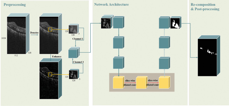
HRF in retinal SD-OCT volumes appear with low-contrast characteristics that results in the difficulty of HRF segmentation. Therefore to effectively segment the HRF we proposed a fully automated method for HRF segmentation in SD-OCT volumes with diabetic retinopathy (DR). First, we generated the enhanced SD-OCT images from the denoised SD-OCT images with an enhancement method. Then the enhanced images were cascaded with the denoised images as the two-channel input to the network against the low-contrast HRF. Finally, we replaced the standard convolution with slice-wise dilated convolution in the last layer of the encoder path of 3D U-Net to obtain long-range information.
We evaluated our method using two-fold cross-validation on 33 SD-OCT volumes from 27 patients. The average dice similarity coefficient was 70.73%, which was higher than that of the existing methods with significant difference ( < 0.01).
Experimental results demonstrated that the proposed method is faster and achieves more reliable segmentation results than the current HRF segmentation algorithms. We expect that this method will contribute to clinical diagnosis and disease surveillance.
Our framework for the automated HRF segmentation of SD-OCT volumes may improve the clinical diagnosis of DR.
为光谱域光学相干断层扫描(SD-OCT)容积数据设计一个强大的、自动化的高反射灶(HRF)分割框架,尤其是针对HRF与背景对比度低的容积数据。
视网膜SD-OCT容积数据中的HRF具有低对比度特征,这导致HRF分割困难。因此,为了有效分割HRF,我们提出了一种用于糖尿病视网膜病变(DR)的SD-OCT容积数据中HRF分割的全自动方法。首先,我们使用一种增强方法从去噪后的SD-OCT图像生成增强后的SD-OCT图像。然后,将增强后的图像与去噪后的图像级联,作为针对低对比度HRF的网络的双通道输入。最后,我们在3D U-Net编码器路径的最后一层用逐切片扩张卷积替换标准卷积,以获取远距离信息。
我们使用27例患者的33个SD-OCT容积数据进行了两折交叉验证来评估我们的方法。平均骰子相似系数为70.73%,高于现有方法,差异有统计学意义(<0.01)。
实验结果表明,所提出的方法比当前的HRF分割算法更快,并且能获得更可靠的分割结果。我们期望该方法将有助于临床诊断和疾病监测。
我们用于SD-OCT容积数据自动HRF分割的框架可能会改善DR的临床诊断。