• 文献检索
  • 文档翻译
  • 深度研究
  • 学术资讯
  • Suppr Zotero 插件Zotero 插件
  • 邀请有礼
  • 套餐&价格
  • 历史记录
应用&插件
Suppr Zotero 插件Zotero 插件浏览器插件Mac 客户端Windows 客户端微信小程序
定价
高级版会员购买积分包购买API积分包
服务
文献检索文档翻译深度研究API 文档MCP 服务
关于我们
关于 Suppr公司介绍联系我们用户协议隐私条款
关注我们

Suppr 超能文献

核心技术专利:CN118964589B侵权必究
粤ICP备2023148730 号-1Suppr @ 2026

文献检索

告别复杂PubMed语法,用中文像聊天一样搜索,搜遍4000万医学文献。AI智能推荐,让科研检索更轻松。

立即免费搜索

文件翻译

保留排版,准确专业,支持PDF/Word/PPT等文件格式,支持 12+语言互译。

免费翻译文档

深度研究

AI帮你快速写综述,25分钟生成高质量综述,智能提取关键信息,辅助科研写作。

立即免费体验

基于双自动编码器和奇异值分解的特征优化在 MRI 图像脑部肿瘤分割中的应用。

A dual autoencoder and singular value decomposition based feature optimization for the segmentation of brain tumor from MRI images.

机构信息

Noorul Islam Centre for Higher Education, Kumrancoil, Tamil Nadu, India.

, Malappuram, India.

出版信息

BMC Med Imaging. 2021 May 13;21(1):82. doi: 10.1186/s12880-021-00614-3.

DOI:10.1186/s12880-021-00614-3
PMID:33985449
原文链接:https://pmc.ncbi.nlm.nih.gov/articles/PMC8117624/
Abstract

BACKGROUND

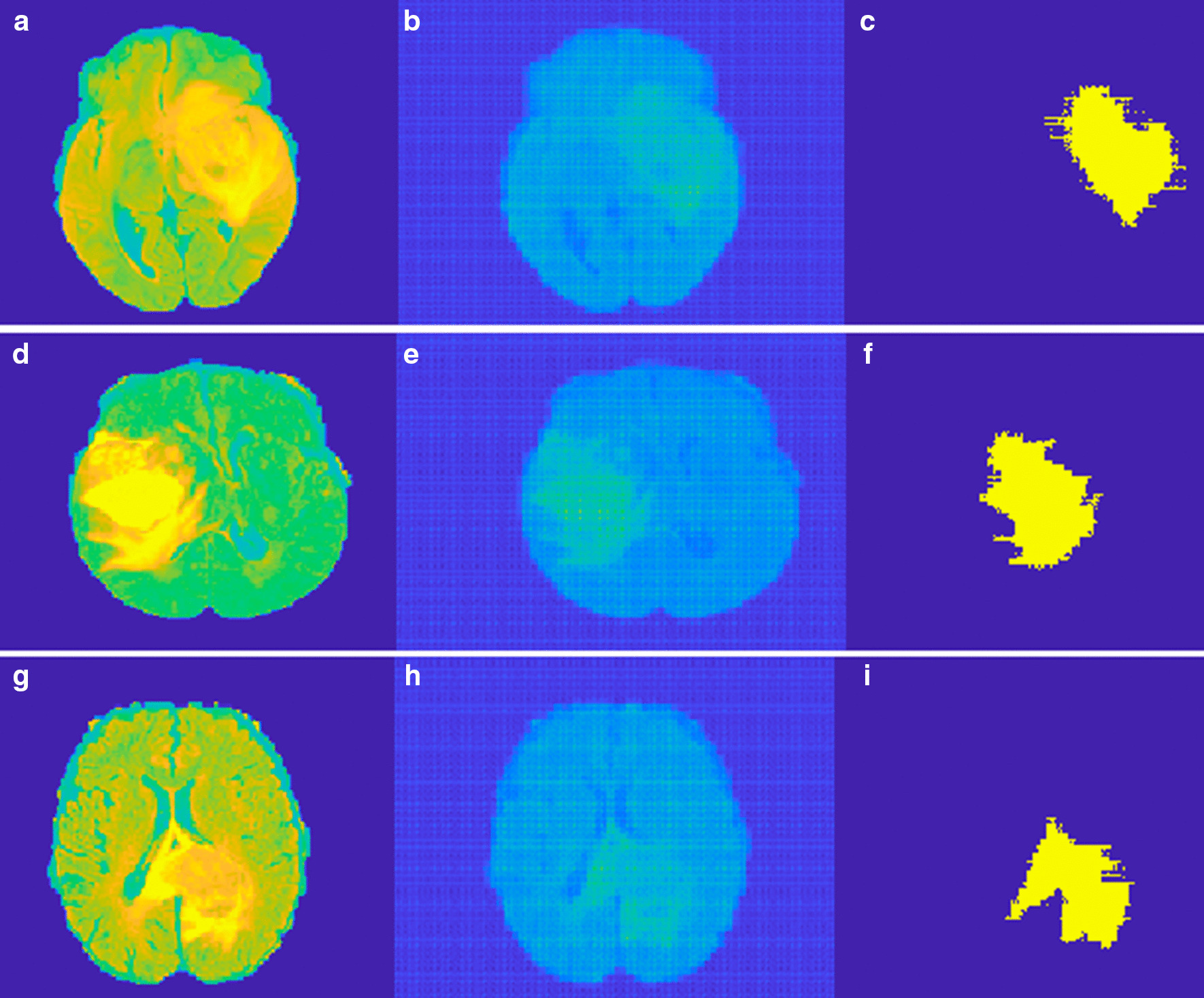
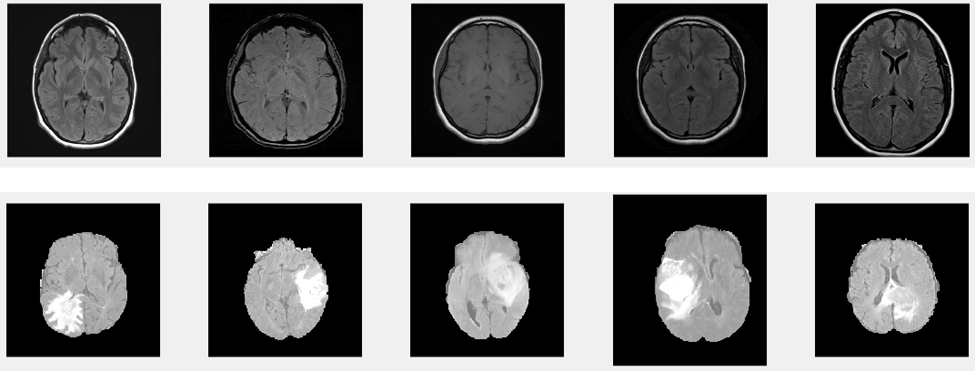
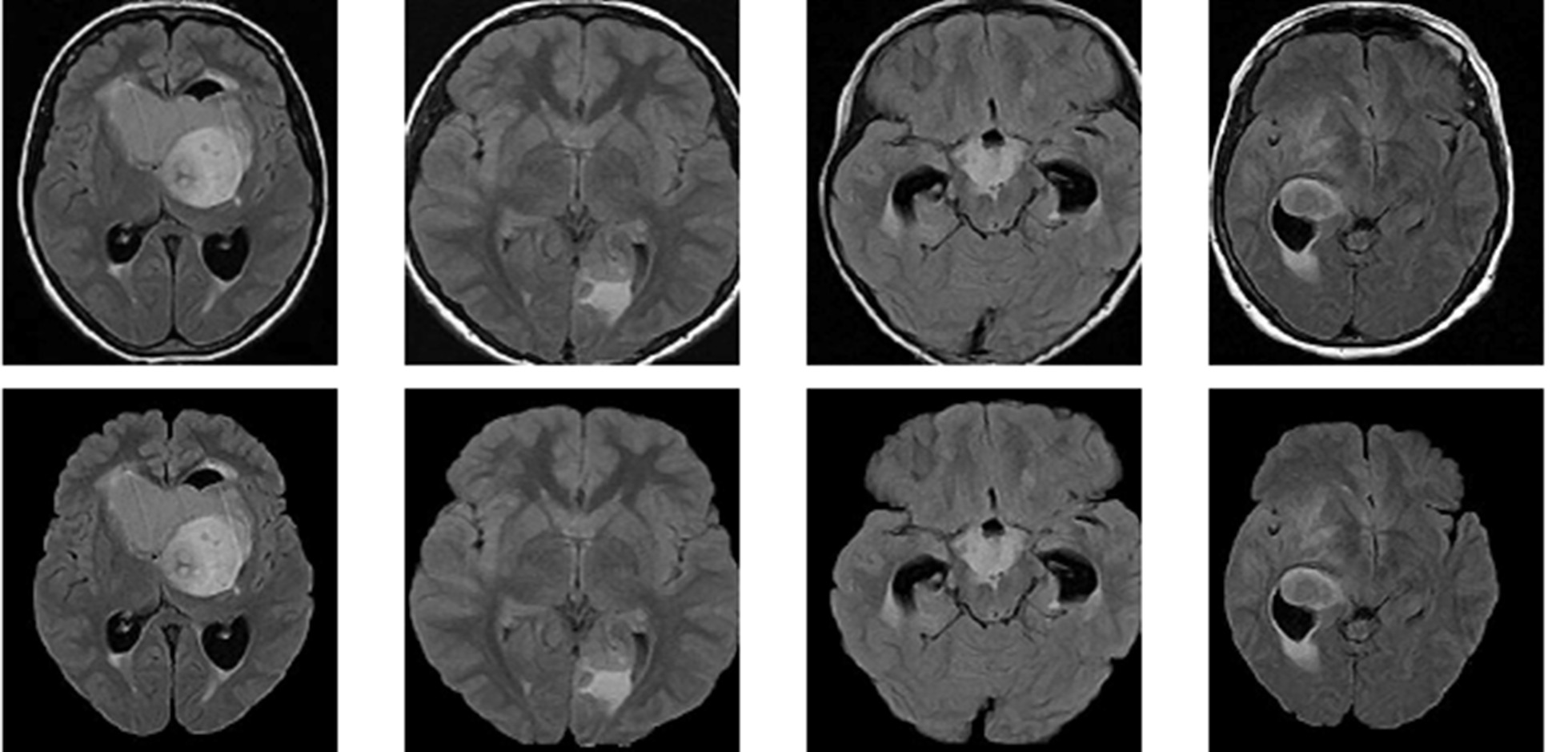
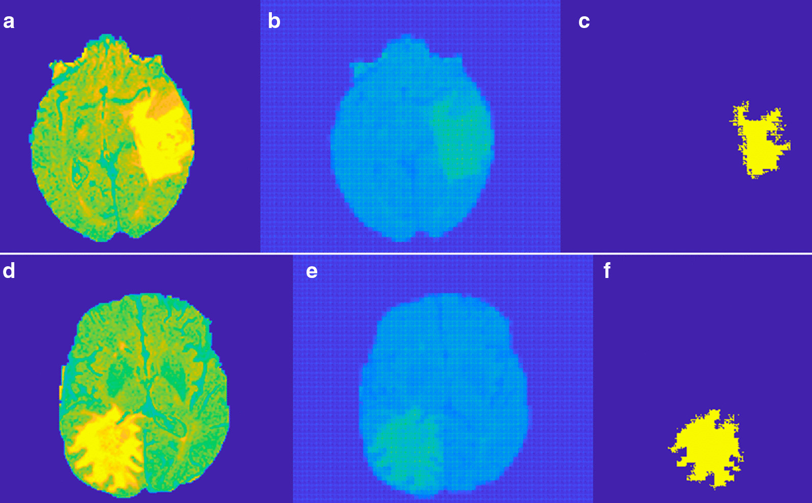
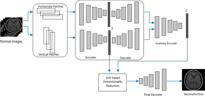
The brain tumor is the growth of abnormal cells inside the brain. These cells can be grown into malignant or benign tumors. Segmentation of tumor from MRI images using image processing techniques started decades back. Image processing based brain tumor segmentation can be divided in to three categories conventional image processing methods, Machine Learning methods and Deep Learning methods. Conventional methods lacks the accuracy in segmentation due to complex spatial variation of tumor. Machine Learning methods stand as a good alternative to conventional methods. Methods like SVM, KNN, Fuzzy and a combination of either of these provide good accuracy with reasonable processing speed. The difficulty in processing the various feature extraction methods and maintain accuracy as per the medical standards still exist as a limitation for machine learning methods. In Deep Learning features are extracted automatically in various stages of the network and maintain accuracy as per the medical standards. Huge database requirement and high computational time is still poses a problem for deep learning. To overcome the limitations specified above we propose an unsupervised dual autoencoder with latent space optimization here. The model require only normal MRI images for its training thus reducing the huge tumor database requirement. With a set of normal class data, an autoencoder can reproduce the feature vector into an output layer. This trained autoencoder works well with normal data while it fails to reproduce an anomaly to the output layer. But a classical autoencoder suffer due to poor latent space optimization. The Latent space loss of classical autoencoder is reduced using an auxiliary encoder along with the feature optimization based on singular value decomposition (SVD). The patches used for training are not traditional square patches but we took both horizontal and vertical patches to keep both local and global appearance features on the training set. An Autoencoder is applied separately for learning both horizontal and vertical patches. While training a logistic sigmoid transfer function is used for both encoder and decoder parts. SGD optimizer is used for optimization with an initial learning rate of .001 and the maximum epochs used are 4000. The network is trained in MATLAB 2018a with a processor capacity of 3.7 GHz with NVIDIA GPU and 16 GB of RAM.

RESULTS

The results are obtained using a patch size of 16 × 64, 64 × 16 for horizontal and vertical patches respectively. In Glioma images tumor is not grown from a point rather it spreads randomly. Region filling and connectivity operations are performed to get the final tumor segmentation. Overall the method segments Meningioma better than Gliomas. Three evaluation metrics are considered to measure the performance of the proposed system such as Dice Similarity Coefficient, Positive Predictive Value, and Sensitivity.

CONCLUSION

An unsupervised method for the segmentation of brain tumor from MRI images is proposed here. The proposed dual autoencoder with SVD based feature optimization reduce the latent space loss in the classical autoencoder. The proposed method have advantages in computational efficiency, no need of huge database requirement and better accuracy than machine learning methods. The method is compared Machine Learning methods Like SVM, KNN and supervised deep learning methods like CNN and commentable results are obtained.

https://cdn.ncbi.nlm.nih.gov/pmc/blobs/7db7/8117624/4fdf144d6b65/12880_2021_614_Fig6_HTML.jpg
https://cdn.ncbi.nlm.nih.gov/pmc/blobs/7db7/8117624/40059f149286/12880_2021_614_Fig1_HTML.jpg
https://cdn.ncbi.nlm.nih.gov/pmc/blobs/7db7/8117624/64197c18961f/12880_2021_614_Fig2_HTML.jpg
https://cdn.ncbi.nlm.nih.gov/pmc/blobs/7db7/8117624/68d7dccd2d60/12880_2021_614_Fig3_HTML.jpg
https://cdn.ncbi.nlm.nih.gov/pmc/blobs/7db7/8117624/91b5f2dffb8f/12880_2021_614_Fig4_HTML.jpg
https://cdn.ncbi.nlm.nih.gov/pmc/blobs/7db7/8117624/8593815c5c02/12880_2021_614_Fig5_HTML.jpg
https://cdn.ncbi.nlm.nih.gov/pmc/blobs/7db7/8117624/4fdf144d6b65/12880_2021_614_Fig6_HTML.jpg
摘要

背景

脑瘤是大脑内部异常细胞的生长。这些细胞可以长成恶性或良性肿瘤。使用图像处理技术从 MRI 图像中分割肿瘤始于几十年前。基于图像处理的脑肿瘤分割可分为三类:传统图像处理方法、机器学习方法和深度学习方法。由于肿瘤的空间变化复杂,传统方法在分割方面缺乏准确性。机器学习方法是传统方法的良好替代品。支持向量机 (SVM)、KNN、模糊等方法以及这些方法的任意组合都能以合理的处理速度提供良好的准确性。在处理各种特征提取方法和保持符合医疗标准的准确性方面仍然存在困难,这仍然是机器学习方法的一个限制。在深度学习中,特征在网络的各个阶段自动提取,并根据医疗标准保持准确性。庞大的数据库需求和高计算时间仍然是深度学习的一个问题。为了克服上述限制,我们在这里提出了一种带有潜在空间优化的无监督双自动编码器。该模型仅需要正常的 MRI 图像进行训练,从而减少了对大型肿瘤数据库的需求。使用一组正常类数据,自动编码器可以将特征向量重新生成到输出层。经过训练的自动编码器在处理正常数据时效果很好,但无法将异常情况重新生成到输出层。但是,经典自动编码器由于潜在空间优化不佳而受到影响。通过沿特征优化基于奇异值分解 (SVD) 使用辅助编码器,降低了经典自动编码器的潜在空间损失。用于训练的补丁不是传统的正方形补丁,而是我们同时使用水平和垂直补丁来保留训练集中的局部和全局外观特征。自动编码器分别用于学习水平和垂直补丁。在训练过程中,逻辑斯谛 sigmoid 传递函数用于编码器和解码器部分。使用 SGD 优化器进行优化,初始学习率为.001,最大 epoch 数为 4000。网络在具有 NVIDIA GPU 和 16GB RAM 的 3.7GHz 处理器容量的 MATLAB 2018a 中进行训练。

结果

使用 16×64 和 64×16 的补丁大小分别获得水平和垂直补丁的结果。在神经胶质瘤图像中,肿瘤不是从一个点生长的,而是随机扩散的。进行区域填充和连接操作以获得最终的肿瘤分割。总体而言,该方法对脑膜瘤的分割优于神经胶质瘤。考虑了三个评估指标来衡量所提出系统的性能,例如骰子相似系数、阳性预测值和灵敏度。

结论

本文提出了一种从 MRI 图像中分割脑肿瘤的无监督方法。所提出的基于 SVD 的特征优化双自动编码器减少了经典自动编码器中的潜在空间损失。与机器学习方法相比,所提出的方法具有计算效率高、不需要庞大的数据库需求和更高的准确性的优点。该方法与 SVM、KNN 等机器学习方法以及 CNN 等监督深度学习方法进行了比较,并获得了可评论的结果。

相似文献

1
A dual autoencoder and singular value decomposition based feature optimization for the segmentation of brain tumor from MRI images.基于双自动编码器和奇异值分解的特征优化在 MRI 图像脑部肿瘤分割中的应用。
BMC Med Imaging. 2021 May 13;21(1):82. doi: 10.1186/s12880-021-00614-3.
2
A Hybrid Approach Based on Deep CNN and Machine Learning Classifiers for the Tumor Segmentation and Classification in Brain MRI.基于深度卷积神经网络和机器学习分类器的脑 MRI 肿瘤分割与分类的混合方法。
Comput Math Methods Med. 2022 Aug 5;2022:6446680. doi: 10.1155/2022/6446680. eCollection 2022.
3
MRI-based brain tumor detection using convolutional deep learning methods and chosen machine learning techniques.基于 MRI 的脑肿瘤检测:使用卷积深度学习方法和选定的机器学习技术。
BMC Med Inform Decis Mak. 2023 Jan 23;23(1):16. doi: 10.1186/s12911-023-02114-6.
4
Automated glioma grading on conventional MRI images using deep convolutional neural networks.使用深度卷积神经网络对传统MRI图像进行自动脑胶质瘤分级
Med Phys. 2020 Jul;47(7):3044-3053. doi: 10.1002/mp.14168. Epub 2020 May 11.
5
Brain tumor segmentation using holistically nested neural networks in MRI images.MRI 图像中基于整体嵌套神经网络的脑肿瘤分割。
Med Phys. 2017 Oct;44(10):5234-5243. doi: 10.1002/mp.12481. Epub 2017 Aug 20.
6
Brain tumor classification for MRI images using dual-discriminator conditional generative adversarial network.基于双鉴别器条件生成对抗网络的 MRI 图像脑肿瘤分类。
Electromagn Biol Med. 2024 Apr 2;43(1-2):81-94. doi: 10.1080/15368378.2024.2321352. Epub 2024 Mar 10.
7
Tumor Diagnosis against Other Brain Diseases Using T2 MRI Brain Images and CNN Binary Classifier and DWT.使用T2加权磁共振成像脑图像、卷积神经网络二元分类器和离散小波变换进行肿瘤与其他脑部疾病的诊断
Brain Sci. 2023 Feb 17;13(2):348. doi: 10.3390/brainsci13020348.
8
[Fully Automatic Glioma Segmentation Algorithm of Magnetic Resonance Imaging Based on 3D-UNet With More Global Contextual Feature Extraction: An Improvement on Insufficient Extraction of Global Features].基于具有更多全局上下文特征提取的3D-UNet的磁共振成像全自动胶质瘤分割算法:对全局特征提取不足的改进
Sichuan Da Xue Xue Bao Yi Xue Ban. 2024 Mar 20;55(2):447-454. doi: 10.12182/20240360208.
9
An Intelligent Diagnosis Method of Brain MRI Tumor Segmentation Using Deep Convolutional Neural Network and SVM Algorithm.基于深度卷积神经网络和支持向量机算法的脑 MRI 肿瘤分割智能诊断方法。
Comput Math Methods Med. 2020 Jul 14;2020:6789306. doi: 10.1155/2020/6789306. eCollection 2020.
10
Semantic segmentation of cerebrospinal fluid and brain volume with a convolutional neural network in pediatric hydrocephalus-transfer learning from existing algorithms.基于卷积神经网络的小儿脑积水脑积水分割及脑容量计算——从现有算法进行迁移学习。
Acta Neurochir (Wien). 2020 Oct;162(10):2463-2474. doi: 10.1007/s00701-020-04447-x. Epub 2020 Jun 25.

引用本文的文献

1
Integrating artificial intelligence in healthcare: applications, challenges, and future directions.将人工智能整合到医疗保健中:应用、挑战及未来方向。
Future Sci OA. 2025 Dec;11(1):2527505. doi: 10.1080/20565623.2025.2527505. Epub 2025 Jul 4.
2
Performance of Convolutional Neural Network Models in Meningioma Segmentation in Magnetic Resonance Imaging: A Systematic Review and Meta-Analysis.卷积神经网络模型在磁共振成像中脑膜瘤分割的性能:系统评价与荟萃分析
Neuroinformatics. 2025 Jan;23(1):14. doi: 10.1007/s12021-024-09704-3. Epub 2024 Dec 28.
3
A systematic review on deep learning-based automated cancer diagnosis models.

本文引用的文献

1
Autoencoders for unsupervised anomaly segmentation in brain MR images: A comparative study.基于自动编码器的脑磁共振图像无监督异常分割方法的对比研究
Med Image Anal. 2021 Apr;69:101952. doi: 10.1016/j.media.2020.101952. Epub 2021 Jan 2.
2
Concatenated and Connected Random Forests With Multiscale Patch Driven Active Contour Model for Automated Brain Tumor Segmentation of MR Images.拼接连接随机森林与多尺度斑块驱动主动轮廓模型在磁共振图像的脑肿瘤自动分割中的应用。
IEEE Trans Med Imaging. 2018 Aug;37(8):1943-1954. doi: 10.1109/TMI.2018.2805821. Epub 2018 Feb 13.
3
Brain tumor segmentation with Deep Neural Networks.
基于深度学习的自动化癌症诊断模型的系统评价。
J Cell Mol Med. 2024 Mar;28(6):e18144. doi: 10.1111/jcmm.18144.
4
Automatic quadriceps and patellae segmentation of MRI with cascaded U -Net and SASSNet deep learning model.基于级联 U-Net 和 SASSNet 深度学习模型的 MRI 自动股四头肌和髌骨分割。
Med Phys. 2022 Jan;49(1):443-460. doi: 10.1002/mp.15335. Epub 2021 Nov 22.
基于深度神经网络的脑肿瘤分割。
Med Image Anal. 2017 Jan;35:18-31. doi: 10.1016/j.media.2016.05.004. Epub 2016 May 19.
4
Brain Tumor Segmentation Using Convolutional Neural Networks in MRI Images.基于 MRI 图像的卷积神经网络脑肿瘤分割。
IEEE Trans Med Imaging. 2016 May;35(5):1240-1251. doi: 10.1109/TMI.2016.2538465. Epub 2016 Mar 4.
5
Segmentation and measurement of the cortex from 3-D MR images using coupled-surfaces propagation.使用耦合表面传播从三维磁共振图像中分割和测量皮质。
IEEE Trans Med Imaging. 1999 Oct;18(10):927-37. doi: 10.1109/42.811276.