Industrial Electronics and Control Engineering Department, Faculty of Electronic Engineering, Menoufia University, Menoufia, Egypt.
Electronics and Electrical Communications Engineering Department, Faculty of Electronic Engineering, Menoufia University, Menoufia, Egypt.
PLoS One. 2021 May 20;16(5):e0251899. doi: 10.1371/journal.pone.0251899. eCollection 2021.
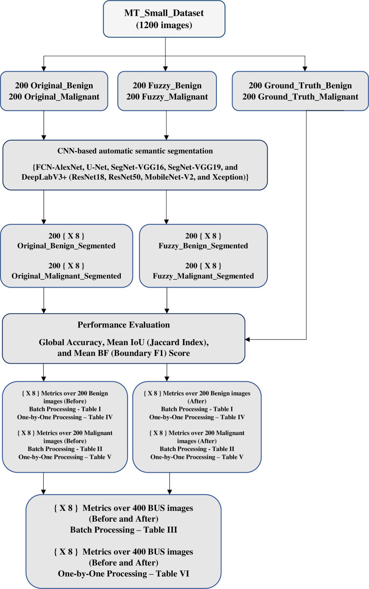
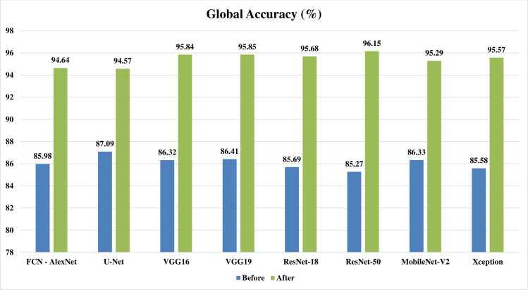
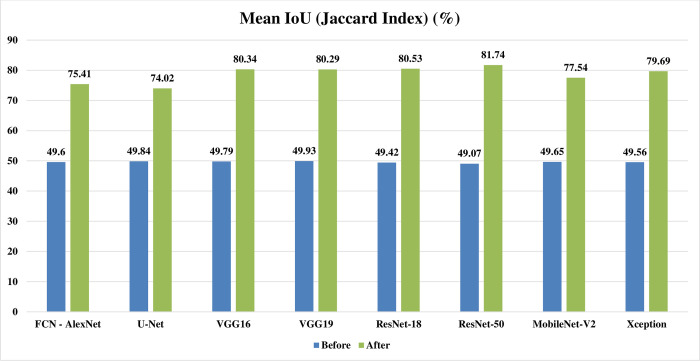
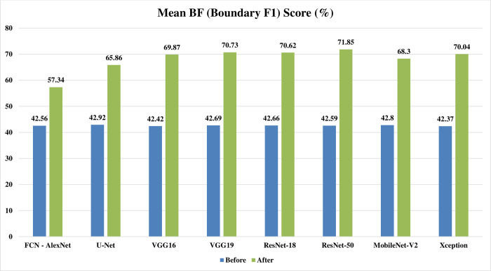
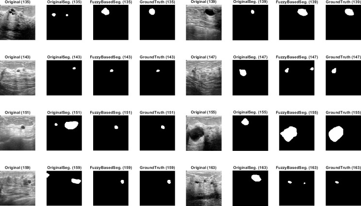
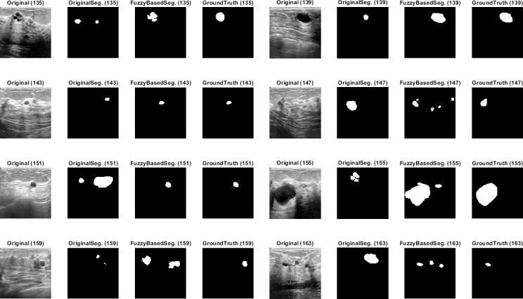
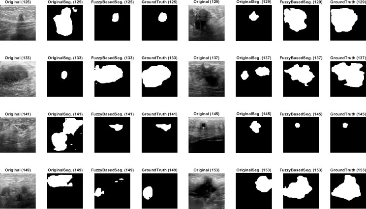
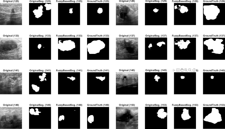
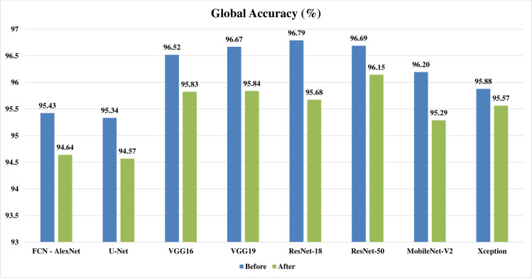
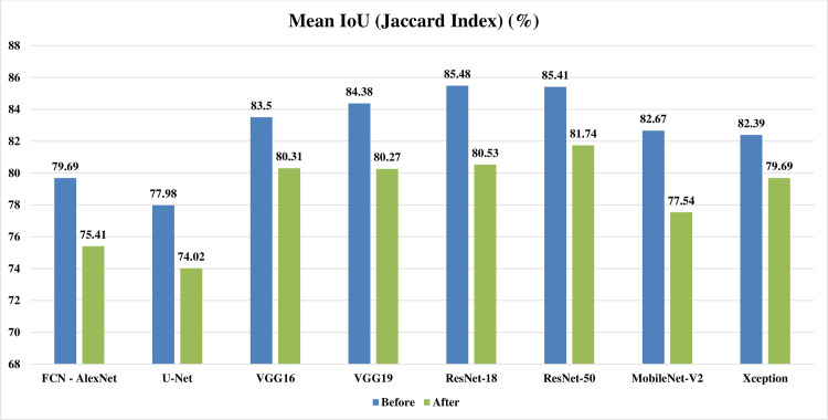
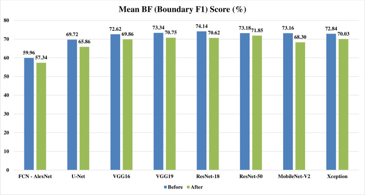
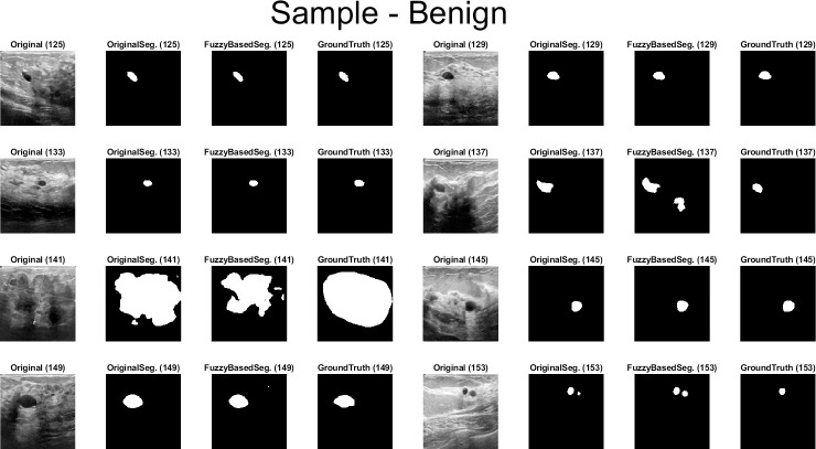
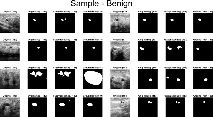
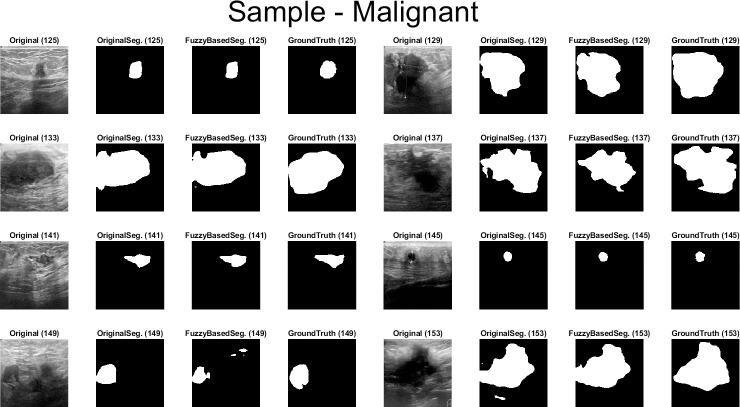
Computer aided diagnosis (CAD) of biomedical images assists physicians for a fast facilitated tissue characterization. A scheme based on combining fuzzy logic (FL) and deep learning (DL) for automatic semantic segmentation (SS) of tumors in breast ultrasound (BUS) images is proposed. The proposed scheme consists of two steps: the first is a FL based preprocessing, and the second is a Convolutional neural network (CNN) based SS. Eight well-known CNN based SS models have been utilized in the study. Studying the scheme was by a dataset of 400 cancerous BUS images and their corresponding 400 ground truth images. SS process has been applied in two modes: batch and one by one image processing. Three quantitative performance evaluation metrics have been utilized: global accuracy (GA), mean Jaccard Index (mean intersection over union (IoU)), and mean BF (Boundary F1) Score. In the batch processing mode: quantitative metrics' average results over the eight utilized CNNs based SS models over the 400 cancerous BUS images were: 95.45% GA instead of 86.08% without applying fuzzy preprocessing step, 78.70% mean IoU instead of 49.61%, and 68.08% mean BF score instead of 42.63%. Moreover, the resulted segmented images could show tumors' regions more accurate than with only CNN based SS. While, in one by one image processing mode: there has been no enhancement neither qualitatively nor quantitatively. So, only when a batch processing is needed, utilizing the proposed scheme may be helpful in enhancing automatic ss of tumors in BUS images. Otherwise applying the proposed approach on a one-by-one image mode will disrupt segmentation's efficiency. The proposed batch processing scheme may be generalized for an enhanced CNN based SS of a targeted region of interest (ROI) in any batch of digital images. A modified small dataset is available: https://www.kaggle.com/mohammedtgadallah/mt-small-dataset (S1 Data).
计算机辅助诊断 (CAD) 可辅助医生对生物医学图像进行快速、便捷的组织特征分析。提出了一种基于模糊逻辑 (FL) 和深度学习 (DL) 的方案,用于对乳腺超声 (BUS) 图像中的肿瘤进行自动语义分割 (SS)。该方案包括两个步骤:第一步是基于 FL 的预处理,第二步是基于卷积神经网络 (CNN) 的 SS。研究中利用了 8 种著名的基于 CNN 的 SS 模型。该方案的研究基于 400 例癌症 BUS 图像及其对应的 400 幅真实图像数据集。SS 过程应用于两种模式:批处理和逐个图像处理。利用了三个定量性能评估指标:全局准确率 (GA)、平均交并比 (mean intersection over union (IoU)) 和平均 BF (Boundary F1) 分数。在批处理模式下:在 400 例癌症 BUS 图像上,利用 8 种基于 CNN 的 SS 模型的平均结果,应用模糊预处理步骤后,GA 从 86.08%提高到 95.45%,mean IoU 从 49.61%提高到 78.70%,mean BF 分数从 42.63%提高到 68.08%。此外,与仅基于 CNN 的 SS 相比,分割得到的图像可以更准确地显示肿瘤区域。然而,在逐个图像处理模式下,无论在质量上还是数量上都没有提高。因此,只有在需要批量处理时,利用该方案才能有助于提高 BUS 图像中肿瘤的自动 SS。否则,在逐个图像模式下应用该方法将降低分割效率。该批量处理方案可推广到任何批量数字图像的目标感兴趣区域 (ROI) 的增强型 CNN 基于 SS。提供了一个修改后的小数据集:https://www.kaggle.com/mohammedtgadallah/mt-small-dataset(S1 数据)。