• 文献检索
  • 文档翻译
  • 深度研究
  • 学术资讯
  • Suppr Zotero 插件Zotero 插件
  • 邀请有礼
  • 套餐&价格
  • 历史记录
应用&插件
Suppr Zotero 插件Zotero 插件浏览器插件Mac 客户端Windows 客户端微信小程序
定价
高级版会员购买积分包购买API积分包
服务
文献检索文档翻译深度研究API 文档MCP 服务
关于我们
关于 Suppr公司介绍联系我们用户协议隐私条款
关注我们

Suppr 超能文献

核心技术专利:CN118964589B侵权必究
粤ICP备2023148730 号-1Suppr @ 2026

文献检索

告别复杂PubMed语法,用中文像聊天一样搜索,搜遍4000万医学文献。AI智能推荐,让科研检索更轻松。

立即免费搜索

文件翻译

保留排版,准确专业,支持PDF/Word/PPT等文件格式,支持 12+语言互译。

免费翻译文档

深度研究

AI帮你快速写综述,25分钟生成高质量综述,智能提取关键信息,辅助科研写作。

立即免费体验

远程眼科设置中可转诊视网膜病变的自动识别。

Automated Identification of Referable Retinal Pathology in Teleophthalmology Setting.

机构信息

Department of Electrical and Computer Engineering, Duke University, Durham, NC, USA.

Department of Ophthalmology, Duke University, Durham, NC, USA.

出版信息

Transl Vis Sci Technol. 2021 May 3;10(6):30. doi: 10.1167/tvst.10.6.30.

DOI:10.1167/tvst.10.6.30
PMID:34036304
原文链接:https://pmc.ncbi.nlm.nih.gov/articles/PMC8161696/
Abstract

PURPOSE

This study aims to meet a growing need for a fully automated, learning-based interpretation tool for retinal images obtained remotely (e.g. teleophthalmology) through different imaging modalities that may include imperfect (uninterpretable) images.

METHODS

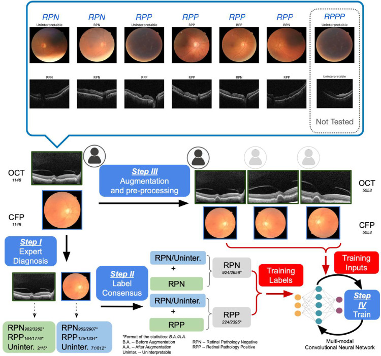
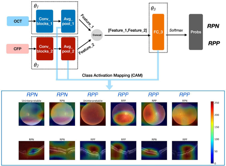
A retrospective study of 1148 optical coherence tomography (OCT) and color fundus photography (CFP) retinal images obtained using Topcon's Maestro care unit on 647 patients with diabetes. To identify retinal pathology, a Convolutional Neural Network (CNN) with dual-modal inputs (i.e. CFP and OCT images) was developed. We developed a novel alternate gradient descent algorithm to train the CNN, which allows for the use of uninterpretable CFP/OCT images (i.e. ungradable images that do not contain sufficient image biomarkers for the reviewer to conclude absence or presence of retinal pathology). Specifically, a 9:1 ratio to split the training and testing dataset was used for training and validating the CNN. Paired CFP/OCT inputs (obtained from a single eye of a patient) were grouped as retinal pathology negative (RPN; 924 images) in the absence of retinal pathology in both imaging modalities, or if one of the imaging modalities was uninterpretable and the other without retinal pathology. If any imaging modality exhibited referable retinal pathology, the corresponding CFP/OCT inputs were deemed retinal pathology positive (RPP; 224 images) if any imaging modality exhibited referable retinal pathology.

RESULTS

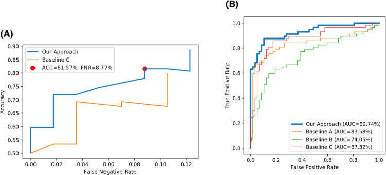
Our approach achieved 88.60% (95% confidence interval [CI] = 82.76% to 94.43%) accuracy in identifying pathology, along with the false negative rate (FNR) of 12.28% (95% CI = 6.26% to 18.31%), recall (sensitivity) of 87.72% (95% CI = 81.69% to 93.74%), specificity of 89.47% (95% CI = 83.84% to 95.11%), and area under the curve of receiver operating characteristic (AUC-ROC) was 92.74% (95% CI = 87.71% to 97.76%).

CONCLUSIONS

Our model can be successfully deployed in clinical practice to facilitate automated remote retinal pathology identification.

TRANSLATIONAL RELEVANCE

A fully automated tool for early diagnosis of retinal pathology might allow for earlier treatment and improved visual outcomes.

摘要

目的

本研究旨在满足远程(例如远程眼科)获取的视网膜图像的完全自动化、基于学习的解释工具的需求,这些图像可能包括不完美(不可解释)的图像。

方法

对 647 名糖尿病患者使用 Topcon 的 Maestro care 单元获得的 1148 份光学相干断层扫描(OCT)和眼底彩色摄影(CFP)视网膜图像进行回顾性研究。为了识别视网膜病变,我们开发了一种具有双模态输入(即 CFP 和 OCT 图像)的卷积神经网络(CNN)。我们开发了一种新的交替梯度下降算法来训练 CNN,该算法允许使用不可解释的 CFP/OCT 图像(即没有足够的图像生物标志物的图像,无法让审阅者得出是否存在视网膜病变的结论)。具体来说,使用 9:1 的比例将训练和测试数据集分开用于训练和验证 CNN。来自患者单眼的配对 CFP/OCT 输入,如果两种成像方式均无视网膜病变,或者如果一种成像方式不可解释而另一种没有视网膜病变,则将其分组为视网膜病变阴性(RPN;924 张图像)。如果任何一种成像方式显示出可参考的视网膜病变,则将相应的 CFP/OCT 输入视为视网膜病变阳性(RPP;224 张图像),如果任何一种成像方式显示出可参考的视网膜病变。

结果

我们的方法在识别病变方面达到了 88.60%(95%置信区间 [CI] = 82.76%至 94.43%)的准确率,假阴性率(FNR)为 12.28%(95% CI = 6.26%至 18.31%),召回率(敏感性)为 87.72%(95% CI = 81.69%至 93.74%),特异性为 89.47%(95% CI = 83.84%至 95.11%),接收器工作特征(ROC)曲线下面积(AUC-ROC)为 92.74%(95% CI = 87.71%至 97.76%)。

结论

我们的模型可以成功地部署在临床实践中,以促进远程自动视网膜病变识别。

翻译

这是一篇关于使用深度学习算法进行视网膜图像分析的研究报告。研究人员开发了一种基于卷积神经网络的工具,可以自动识别视网膜病变,并对其进行分类。该工具使用了来自多个医疗机构的视网膜图像数据集进行训练和验证,取得了较高的准确率和特异性。研究结果表明,这种自动化工具可以帮助医生快速、准确地诊断视网膜病变,提高诊断效率和准确性。

https://cdn.ncbi.nlm.nih.gov/pmc/blobs/3a5c/8161696/0f27af300a37/tvst-10-6-30-f003.jpg
https://cdn.ncbi.nlm.nih.gov/pmc/blobs/3a5c/8161696/9d6af7a11323/tvst-10-6-30-f001.jpg
https://cdn.ncbi.nlm.nih.gov/pmc/blobs/3a5c/8161696/870226753a35/tvst-10-6-30-f002.jpg
https://cdn.ncbi.nlm.nih.gov/pmc/blobs/3a5c/8161696/0f27af300a37/tvst-10-6-30-f003.jpg
https://cdn.ncbi.nlm.nih.gov/pmc/blobs/3a5c/8161696/9d6af7a11323/tvst-10-6-30-f001.jpg
https://cdn.ncbi.nlm.nih.gov/pmc/blobs/3a5c/8161696/870226753a35/tvst-10-6-30-f002.jpg
https://cdn.ncbi.nlm.nih.gov/pmc/blobs/3a5c/8161696/0f27af300a37/tvst-10-6-30-f003.jpg

相似文献

1
Automated Identification of Referable Retinal Pathology in Teleophthalmology Setting.远程眼科设置中可转诊视网膜病变的自动识别。
Transl Vis Sci Technol. 2021 May 3;10(6):30. doi: 10.1167/tvst.10.6.30.
2
Evaluation of a Remote Diagnosis Imaging Model vs Dilated Eye Examination in Referable Macular Degeneration.评估远程诊断成像模型与散瞳眼底检查在可转诊的黄斑变性中的应用。
JAMA Ophthalmol. 2019 Jul 1;137(7):802-808. doi: 10.1001/jamaophthalmol.2019.1203.
3
Fully automated detection of retinal disorders by image-based deep learning.基于图像的深度学习技术对视网膜疾病进行全自动检测。
Graefes Arch Clin Exp Ophthalmol. 2019 Mar;257(3):495-505. doi: 10.1007/s00417-018-04224-8. Epub 2019 Jan 4.
4
Advancing glaucoma detection with convolutional neural networks: a paradigm shift in ophthalmology.利用卷积神经网络推进青光眼检测:眼科学的范式转变。
Rom J Ophthalmol. 2023 Jul-Sep;67(3):222-237. doi: 10.22336/rjo.2023.39.
5
Optical coherence tomography for age-related macular degeneration and diabetic macular edema: an evidence-based analysis.光学相干断层扫描在年龄相关性黄斑变性和糖尿病性黄斑水肿中的应用:一项基于证据的分析。
Ont Health Technol Assess Ser. 2009;9(13):1-22. Epub 2009 Sep 1.
6
Multimodal Retinal Imaging Classification for Parkinson's Disease Using a Convolutional Neural Network.基于卷积神经网络的帕金森病多模态视网膜图像分类
Transl Vis Sci Technol. 2024 Aug 1;13(8):23. doi: 10.1167/tvst.13.8.23.
7
A 3D Deep Learning System for Detecting Referable Glaucoma Using Full OCT Macular Cube Scans.基于 OCT 全 macular 立方扫描的 3D 深度学习系统检测可致盲性青光眼
Transl Vis Sci Technol. 2020 Feb 18;9(2):12. doi: 10.1167/tvst.9.2.12.
8
Geometric Correspondence-Based Multimodal Learning for Ophthalmic Image Analysis.基于几何对应关系的眼科图像分析多模态学习
IEEE Trans Med Imaging. 2024 May;43(5):1945-1957. doi: 10.1109/TMI.2024.3352602. Epub 2024 May 2.
9
Incidence of Referable Retinal Disease in Diabetic Patients at a Primary Care Practice.基层医疗实践中糖尿病患者可转诊视网膜疾病的发病率
J Vitreoretin Dis. 2021 Oct 4;6(2):138-146. doi: 10.1177/24741264211044223. eCollection 2022 Mar-Apr.
10
Glaucoma Diagnosis with Machine Learning Based on Optical Coherence Tomography and Color Fundus Images.基于光学相干断层扫描和眼底彩色图像的机器学习青光眼诊断。
J Healthc Eng. 2019 Feb 18;2019:4061313. doi: 10.1155/2019/4061313. eCollection 2019.

引用本文的文献

1
RobOCTNet: Robotics and Deep Learning for Referable Posterior Segment Pathology Detection in an Emergency Department Population.RobOCTNet:机器人技术和深度学习在急诊科人群中用于可转诊的后节病理检测。
Transl Vis Sci Technol. 2024 Mar 1;13(3):12. doi: 10.1167/tvst.13.3.12.
2
The Present and Future of Artificial Intelligence-Based Medical Image in Diabetes Mellitus: Focus on Analytical Methods and Limitations of Clinical Use.人工智能在糖尿病医学影像中的现状与未来:聚焦分析方法及临床应用的局限性。
J Korean Med Sci. 2023 Aug 7;38(31):e253. doi: 10.3346/jkms.2023.38.e253.
3
Incidence of Referable Retinal Disease in Diabetic Patients at a Primary Care Practice.

本文引用的文献

1
Text Data Augmentation for Deep Learning.用于深度学习的文本数据增强
J Big Data. 2021;8(1):101. doi: 10.1186/s40537-021-00492-0. Epub 2021 Jul 19.
2
Automated diagnoses of age-related macular degeneration and polypoidal choroidal vasculopathy using bi-modal deep convolutional neural networks.使用双模态深度卷积神经网络进行与年龄相关的黄斑变性和息肉样脉络膜血管病变的自动诊断。
Br J Ophthalmol. 2021 Apr;105(4):561-566. doi: 10.1136/bjophthalmol-2020-315817. Epub 2020 Jun 4.
3
Multimodal Retinal Image Analysis via Deep Learning for the Diagnosis of Intermediate Dry Age-Related Macular Degeneration: A Feasibility Study.
基层医疗实践中糖尿病患者可转诊视网膜疾病的发病率
J Vitreoretin Dis. 2021 Oct 4;6(2):138-146. doi: 10.1177/24741264211044223. eCollection 2022 Mar-Apr.
4
Deep Learning Models for Segmenting Non-perfusion Area of Color Fundus Photographs in Patients With Branch Retinal Vein Occlusion.用于分割视网膜分支静脉阻塞患者彩色眼底照片无灌注区的深度学习模型
Front Med (Lausanne). 2022 Jun 30;9:794045. doi: 10.3389/fmed.2022.794045. eCollection 2022.
基于深度学习的多模态视网膜图像分析用于诊断中度干性年龄相关性黄斑变性:一项可行性研究
J Ophthalmol. 2020 Jan 13;2020:7493419. doi: 10.1155/2020/7493419. eCollection 2020.
4
DMENet: Diabetic Macular Edema diagnosis using Hierarchical Ensemble of CNNs.DMENet:基于 CNN 分层集成的糖尿病性黄斑水肿诊断。
PLoS One. 2020 Feb 10;15(2):e0220677. doi: 10.1371/journal.pone.0220677. eCollection 2020.
5
Evaluation of a Remote Diagnosis Imaging Model vs Dilated Eye Examination in Referable Macular Degeneration.评估远程诊断成像模型与散瞳眼底检查在可转诊的黄斑变性中的应用。
JAMA Ophthalmol. 2019 Jul 1;137(7):802-808. doi: 10.1001/jamaophthalmol.2019.1203.
6
Deep Learning-Based Automated Classification of Multi-Categorical Abnormalities From Optical Coherence Tomography Images.基于深度学习的光学相干断层扫描图像多类别异常自动分类
Transl Vis Sci Technol. 2018 Dec 28;7(6):41. doi: 10.1167/tvst.7.6.41. eCollection 2018 Nov.
7
From Machine to Machine: An OCT-Trained Deep Learning Algorithm for Objective Quantification of Glaucomatous Damage in Fundus Photographs.从机器到机器:一种基于 OCT 训练的深度学习算法,用于客观量化眼底照片中的青光眼损伤。
Ophthalmology. 2019 Apr;126(4):513-521. doi: 10.1016/j.ophtha.2018.12.033. Epub 2018 Dec 20.
8
Performance of Deep Learning Architectures and Transfer Learning for Detecting Glaucomatous Optic Neuropathy in Fundus Photographs.深度学习架构和迁移学习在眼底照片中检测青光眼视神经病变的性能。
Sci Rep. 2018 Nov 12;8(1):16685. doi: 10.1038/s41598-018-35044-9.
9
Fundus photograph-based deep learning algorithms in detecting diabetic retinopathy.基于眼底照片的深度学习算法在糖尿病视网膜病变检测中的应用。
Eye (Lond). 2019 Jan;33(1):97-109. doi: 10.1038/s41433-018-0269-y. Epub 2018 Nov 6.
10
The possibility of the combination of OCT and fundus images for improving the diagnostic accuracy of deep learning for age-related macular degeneration: a preliminary experiment.OCT 与眼底图像相结合提高深度学习诊断年龄相关性黄斑变性准确性的可能性:一项初步实验。
Med Biol Eng Comput. 2019 Mar;57(3):677-687. doi: 10.1007/s11517-018-1915-z. Epub 2018 Oct 22.