• 文献检索
  • 文档翻译
  • 深度研究
  • 学术资讯
  • Suppr Zotero 插件Zotero 插件
  • 邀请有礼
  • 套餐&价格
  • 历史记录
应用&插件
Suppr Zotero 插件Zotero 插件浏览器插件Mac 客户端Windows 客户端微信小程序
定价
高级版会员购买积分包购买API积分包
服务
文献检索文档翻译深度研究API 文档MCP 服务
关于我们
关于 Suppr公司介绍联系我们用户协议隐私条款
关注我们

Suppr 超能文献

核心技术专利:CN118964589B侵权必究
粤ICP备2023148730 号-1Suppr @ 2026

文献检索

告别复杂PubMed语法,用中文像聊天一样搜索,搜遍4000万医学文献。AI智能推荐,让科研检索更轻松。

立即免费搜索

文件翻译

保留排版,准确专业,支持PDF/Word/PPT等文件格式,支持 12+语言互译。

免费翻译文档

深度研究

AI帮你快速写综述,25分钟生成高质量综述,智能提取关键信息,辅助科研写作。

立即免费体验

基于解剖学信息的深度学习在对比增强心脏磁共振成像上用于瘢痕分割和临床特征提取

Anatomically informed deep learning on contrast-enhanced cardiac magnetic resonance imaging for scar segmentation and clinical feature extraction.

作者信息

Popescu Dan M, Abramson Haley G, Yu Rebecca, Lai Changxin, Shade Julie K, Wu Katherine C, Maggioni Mauro, Trayanova Natalia A

机构信息

Alliance for Cardiovascular Diagnostic and Treatment Innovation (ADVANCE), Johns Hopkins University, Baltimore, Maryland.

Department of Biomedical Engineering, Johns Hopkins University School of Medicine, Baltimore, Maryland.

出版信息

Cardiovasc Digit Health J. 2021 Nov 26;3(1):2-13. doi: 10.1016/j.cvdhj.2021.11.007. eCollection 2022 Feb.

DOI:10.1016/j.cvdhj.2021.11.007
PMID:35265930
原文链接:https://pmc.ncbi.nlm.nih.gov/articles/PMC8890075/
Abstract

BACKGROUND

Visualizing fibrosis on cardiac magnetic resonance (CMR) imaging with contrast enhancement (late gadolinium enhancement; LGE) is paramount in characterizing disease progression and identifying arrhythmia substrates. Segmentation and fibrosis quantification from LGE-CMR is intensive, manual, and prone to interobserver variability. There is an unmet need for automated LGE-CMR image segmentation that ensures anatomical accuracy and seamless extraction of clinical features.

OBJECTIVE

This study aimed to develop a novel deep learning solution for analysis of contrast-enhanced CMR images that produces anatomically accurate myocardium and scar/fibrosis segmentations and uses these to calculate features of clinical interest.

METHODS

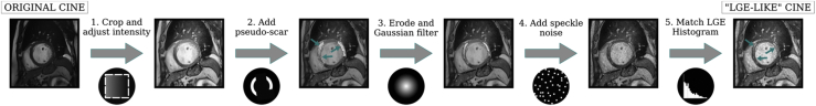
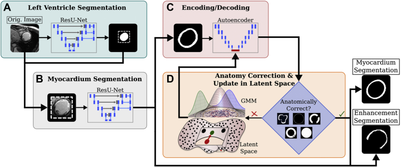
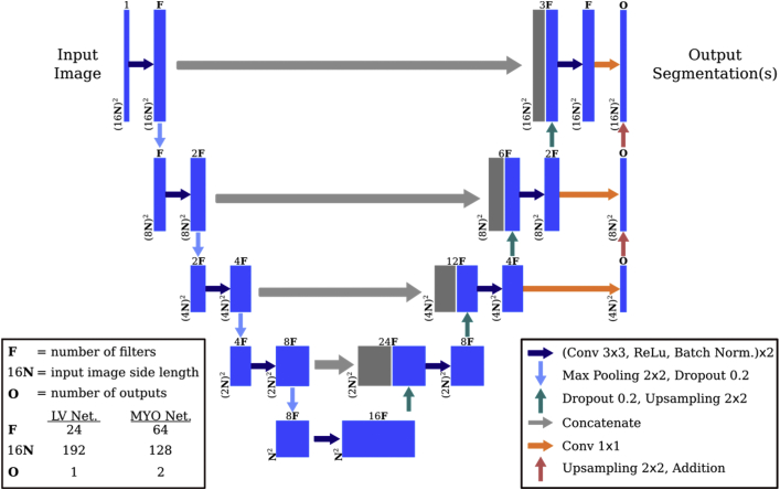
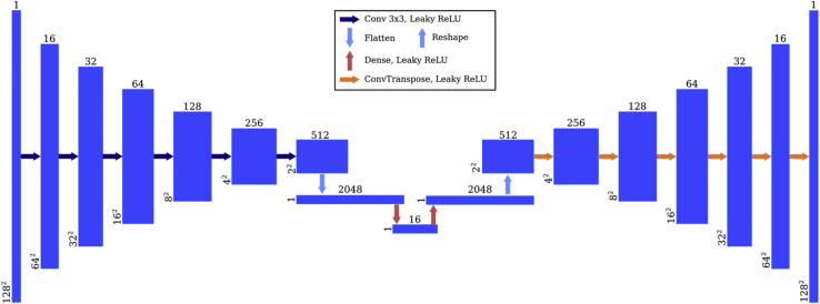
Data sources were 155 2-dimensional LGE-CMR patient scans (1124 slices) and 246 synthetic "LGE-like" scans (1360 slices) obtained from cine CMR using a novel style-transfer algorithm. We trained and tested a 3-stage neural network that identified the left ventricle (LV) region of interest (ROI), segmented ROI into viable myocardium and regions of enhancement, and postprocessed the segmentation results to enforce conforming to anatomical constraints. The segmentations were used to directly compute clinical features, such as LV volume and scar burden.

RESULTS

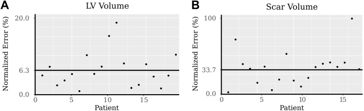
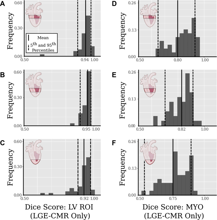
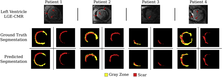
Predicted LV and scar segmentations achieved 96% and 75% balanced accuracy, respectively, and 093 and 057 Dice coefficient when compared to trained expert segmentations. The mean scar burden difference between manual and predicted segmentations was 2%.

CONCLUSION

We developed and validated a deep neural network for automatic, anatomically accurate expert-level LGE- CMR myocardium and scar/fibrosis segmentation, allowing direct calculation of clinical measures. Given the training set heterogeneity, our approach could be extended to multiple imaging modalities and patient pathologies.

摘要

背景

通过对比增强(延迟钆增强;LGE)在心脏磁共振(CMR)成像上可视化纤维化对于表征疾病进展和识别心律失常基质至关重要。从LGE-CMR进行分割和纤维化定量是密集的、手动的,并且容易出现观察者间的变异性。对于确保解剖准确性和无缝提取临床特征的自动化LGE-CMR图像分割存在未满足的需求。

目的

本研究旨在开发一种用于分析对比增强CMR图像的新型深度学习解决方案,该方案能生成解剖学上准确的心肌和瘢痕/纤维化分割,并利用这些分割来计算临床感兴趣的特征。

方法

数据来源为155例二维LGE-CMR患者扫描(1124层)和246例使用新型风格迁移算法从电影CMR获得的合成“类LGE”扫描(1360层)。我们训练并测试了一个三阶段神经网络,该网络识别左心室(LV)感兴趣区域(ROI),将ROI分割为存活心肌和增强区域,并对分割结果进行后处理以使其符合解剖学约束。这些分割用于直接计算临床特征,如LV容积和瘢痕负担。

结果

与训练有素的专家分割相比,预测的LV和瘢痕分割的平衡准确率分别达到96%和75%,Dice系数分别为0.93和0.57。手动分割和预测分割之间的平均瘢痕负担差异为2%。

结论

我们开发并验证了一种深度神经网络,用于自动、解剖学上准确的专家级LGE-CMR心肌和瘢痕/纤维化分割,允许直接计算临床指标。鉴于训练集的异质性,我们的方法可扩展到多种成像模式和患者病理情况。

https://cdn.ncbi.nlm.nih.gov/pmc/blobs/e481/8890075/fa65c6d40d35/gr8.jpg
https://cdn.ncbi.nlm.nih.gov/pmc/blobs/e481/8890075/484b1a4e8255/gr1.jpg
https://cdn.ncbi.nlm.nih.gov/pmc/blobs/e481/8890075/9c5630f2d5ff/gr2.jpg
https://cdn.ncbi.nlm.nih.gov/pmc/blobs/e481/8890075/1486bb005342/gr3.jpg
https://cdn.ncbi.nlm.nih.gov/pmc/blobs/e481/8890075/a6c7e90347df/gr4.jpg
https://cdn.ncbi.nlm.nih.gov/pmc/blobs/e481/8890075/3fd0276c0b79/gr5.jpg
https://cdn.ncbi.nlm.nih.gov/pmc/blobs/e481/8890075/e3267442c4d3/gr6.jpg
https://cdn.ncbi.nlm.nih.gov/pmc/blobs/e481/8890075/494d6e98c555/gr7.jpg
https://cdn.ncbi.nlm.nih.gov/pmc/blobs/e481/8890075/fa65c6d40d35/gr8.jpg

相似文献

1
Anatomically informed deep learning on contrast-enhanced cardiac magnetic resonance imaging for scar segmentation and clinical feature extraction.基于解剖学信息的深度学习在对比增强心脏磁共振成像上用于瘢痕分割和临床特征提取
Cardiovasc Digit Health J. 2021 Nov 26;3(1):2-13. doi: 10.1016/j.cvdhj.2021.11.007. eCollection 2022 Feb.
2
Improved Quantification of Myocardium Scar in Late Gadolinium Enhancement Images: Deep Learning Based Image Fusion Approach.基于深度学习的图像融合方法提高钆延迟增强图像中心肌瘢痕的定量评估。
J Magn Reson Imaging. 2021 Jul;54(1):303-312. doi: 10.1002/jmri.27555. Epub 2021 Feb 17.
3
Joint Deep Learning Framework for Image Registration and Segmentation of Late Gadolinium Enhanced MRI and Cine Cardiac MRI.用于延迟钆增强磁共振成像和心脏电影磁共振成像的图像配准与分割的联合深度学习框架
Proc SPIE Int Soc Opt Eng. 2021 Feb;11598. doi: 10.1117/12.2581386. Epub 2021 Feb 15.
4
Fully automated segmentation of left ventricular scar from 3D late gadolinium enhancement magnetic resonance imaging using a cascaded multi-planar U-Net (CMPU-Net).基于级联多平面 U-Net(CMPU-Net)的 3D 钆延迟增强磁共振成像左心室瘢痕的全自动分割。
Med Phys. 2020 Apr;47(4):1645-1655. doi: 10.1002/mp.14022. Epub 2020 Feb 10.
5
Deep learning for automatic volumetric segmentation of left ventricular myocardium and ischaemic scar from multi-slice late gadolinium enhancement cardiovascular magnetic resonance.基于多切片延迟钆增强心血管磁共振成像的深度学习左心室心肌和缺血性瘢痕自动容积分割
Eur Heart J Cardiovasc Imaging. 2024 May 31;25(6):829-838. doi: 10.1093/ehjci/jeae022.
6
Convolutional neural network-based approach for segmentation of left ventricle myocardial scar from 3D late gadolinium enhancement MR images.基于卷积神经网络的方法用于从 3D 晚期钆增强磁共振图像中分割左心室心肌瘢痕。
Med Phys. 2019 Apr;46(4):1740-1751. doi: 10.1002/mp.13436. Epub 2019 Feb 28.
7
Cine and late gadolinium enhancement MRI registration and automated myocardial infarct heterogeneity quantification.电影和晚期钆增强 MRI 配准及自动心肌梗死异质性定量分析。
Magn Reson Med. 2021 May;85(5):2842-2855. doi: 10.1002/mrm.28596. Epub 2020 Nov 23.
8
Evaluation of fully automated myocardial segmentation techniques in native and contrast-enhanced T1-mapping cardiovascular magnetic resonance images using fully convolutional neural networks.使用全卷积神经网络评估 native 和 contrast-enhanced T1-mapping 心血管磁共振成像中的全自动心肌分割技术。
Med Phys. 2021 Jan;48(1):215-226. doi: 10.1002/mp.14574. Epub 2020 Dec 1.
9
A deep learning pipeline for automatic analysis of multi-scan cardiovascular magnetic resonance.用于多扫描心血管磁共振自动分析的深度学习管道。
J Cardiovasc Magn Reson. 2021 Apr 26;23(1):47. doi: 10.1186/s12968-020-00695-z.
10
Development and testing of a deep learning-based strategy for scar segmentation on CMR-LGE images.基于深度学习的CMR-LGE图像瘢痕分割策略的开发与测试
MAGMA. 2019 Apr;32(2):187-195. doi: 10.1007/s10334-018-0718-4. Epub 2018 Nov 20.

引用本文的文献

1
Multimodal AI to forecast arrhythmic death in hypertrophic cardiomyopathy.用于预测肥厚型心肌病心律失常性死亡的多模态人工智能
Nat Cardiovasc Res. 2025 Jul 2. doi: 10.1038/s44161-025-00679-1.
2
Automated fibrosis segmentation from wideband post-contrast mapping in an animal model of ischemic heart disease with implantable cardioverter-defibrillators.在植入式心脏复律除颤器的缺血性心脏病动物模型中,从宽带对比后映射自动分割纤维化
Magn Reson Med. 2025 Jun;93(6):2401-2413. doi: 10.1002/mrm.30468. Epub 2025 Mar 11.
3
Progress in the Clinical Application of Artificial Intelligence for Left Ventricle Analysis in Cardiac Magnetic Resonance.

本文引用的文献

1
Cardiac segmentation on late gadolinium enhancement MRI: A benchmark study from multi-sequence cardiac MR segmentation challenge.延迟钆增强磁共振成像的心脏分割:来自多序列心脏磁共振分割挑战赛的一项基准研究。
Med Image Anal. 2022 Oct;81:102528. doi: 10.1016/j.media.2022.102528. Epub 2022 Jul 9.
2
Late-Gadolinium Enhancement Interface Area and Electrophysiological Simulations Predict Arrhythmic Events in Patients With Nonischemic Dilated Cardiomyopathy.晚期钆增强界面区和电生理模拟预测非缺血性扩张型心肌病患者的心律失常事件。
JACC Clin Electrophysiol. 2021 Feb;7(2):238-249. doi: 10.1016/j.jacep.2020.08.036. Epub 2020 Oct 29.
3
人工智能在心脏磁共振左心室分析临床应用中的进展
Rev Cardiovasc Med. 2024 Dec 19;25(12):447. doi: 10.31083/j.rcm2512447. eCollection 2024 Dec.
4
Improving the efficiency and accuracy of cardiovascular magnetic resonance with artificial intelligence-review of evidence and proposition of a roadmap to clinical translation.利用人工智能提高心血管磁共振成像的效率和准确性——证据综述及临床转化路线图建议
J Cardiovasc Magn Reson. 2024;26(2):101051. doi: 10.1016/j.jocmr.2024.101051. Epub 2024 Jun 22.
5
The beating heart: artificial intelligence for cardiovascular application in the clinic.跳动的心脏:人工智能在心血管临床应用。
MAGMA. 2024 Jul;37(3):369-382. doi: 10.1007/s10334-024-01180-9. Epub 2024 Jun 22.
6
Toward Enabling Cardiac Digital Twins of Myocardial Infarction Using Deep Computational Models for Inverse Inference.利用用于反向推理的深度计算模型实现心肌梗死的心脏数字孪生。
IEEE Trans Med Imaging. 2024 Jul;43(7):2466-2478. doi: 10.1109/TMI.2024.3367409. Epub 2024 Jul 1.
7
Cardiac Fibrosis Automated Diagnosis Based on FibrosisNet Network Using CMR Ischemic Cardiomyopathy.基于FibrosisNet网络利用心脏磁共振成像缺血性心肌病的心脏纤维化自动诊断
Diagnostics (Basel). 2024 Jan 24;14(3):255. doi: 10.3390/diagnostics14030255.
8
Computational modeling of cardiac electrophysiology and arrhythmogenesis: toward clinical translation.心脏电生理学和心律失常发生的计算建模:迈向临床转化。
Physiol Rev. 2024 Jul 1;104(3):1265-1333. doi: 10.1152/physrev.00017.2023. Epub 2023 Dec 28.
9
Twenty-five years of research in cardiac imaging in electrophysiology procedures for atrial and ventricular arrhythmias.25 年在心律失常的心房和心室电生理程序中的心脏成像研究。
Europace. 2023 Aug 25;25(8). doi: 10.1093/europace/euad183.
10
On the Analyses of Medical Images Using Traditional Machine Learning Techniques and Convolutional Neural Networks.关于使用传统机器学习技术和卷积神经网络对医学图像进行分析
Arch Comput Methods Eng. 2023;30(5):3173-3233. doi: 10.1007/s11831-023-09899-9. Epub 2023 Apr 4.
Improved Quantification of Myocardium Scar in Late Gadolinium Enhancement Images: Deep Learning Based Image Fusion Approach.
基于深度学习的图像融合方法提高钆延迟增强图像中心肌瘢痕的定量评估。
J Magn Reson Imaging. 2021 Jul;54(1):303-312. doi: 10.1002/jmri.27555. Epub 2021 Feb 17.
4
Discordance in Scar Detection Between Electroanatomical Mapping and Cardiac MRI in an Infarct Swine Model.梗死猪模型中心电图与心脏 MRI 瘢痕检测的不相符。
JACC Clin Electrophysiol. 2020 Oct 26;6(11):1452-1464. doi: 10.1016/j.jacep.2020.08.033.
5
Post-DAE: Anatomically Plausible Segmentation via Post-Processing With Denoising Autoencoders.后 DAE:基于去噪自动编码器的后处理的解剖合理分割。
IEEE Trans Med Imaging. 2020 Dec;39(12):3813-3820. doi: 10.1109/TMI.2020.3005297. Epub 2020 Nov 30.
6
Fully automated segmentation of left ventricular scar from 3D late gadolinium enhancement magnetic resonance imaging using a cascaded multi-planar U-Net (CMPU-Net).基于级联多平面 U-Net(CMPU-Net)的 3D 钆延迟增强磁共振成像左心室瘢痕的全自动分割。
Med Phys. 2020 Apr;47(4):1645-1655. doi: 10.1002/mp.14022. Epub 2020 Feb 10.
7
Three-dimensional Deep Convolutional Neural Networks for Automated Myocardial Scar Quantification in Hypertrophic Cardiomyopathy: A Multicenter Multivendor Study.三维深度学习卷积神经网络在肥厚型心肌病心肌瘢痕自动量化中的应用:一项多中心多厂家研究。
Radiology. 2020 Jan;294(1):52-60. doi: 10.1148/radiol.2019190737. Epub 2019 Nov 12.
8
Computationally guided personalized targeted ablation of persistent atrial fibrillation.计算指导下的持续性心房颤动个体化靶向消融
Nat Biomed Eng. 2019 Nov;3(11):870-879. doi: 10.1038/s41551-019-0437-9. Epub 2019 Aug 19.
9
Personalized virtual-heart technology for guiding the ablation of infarct-related ventricular tachycardia.用于指导梗死相关室性心动过速消融的个性化虚拟心脏技术。
Nat Biomed Eng. 2018 Oct;2(10):732-740. doi: 10.1038/s41551-018-0282-2. Epub 2018 Sep 3.
10
Deep learning cardiac motion analysis for human survival prediction.用于人类生存预测的深度学习心脏运动分析
Nat Mach Intell. 2019 Feb 11;1:95-104. doi: 10.1038/s42256-019-0019-2.